
The oral cavity: teeth, tongue, palate & salivary glands
On this page
Overview & boundaries
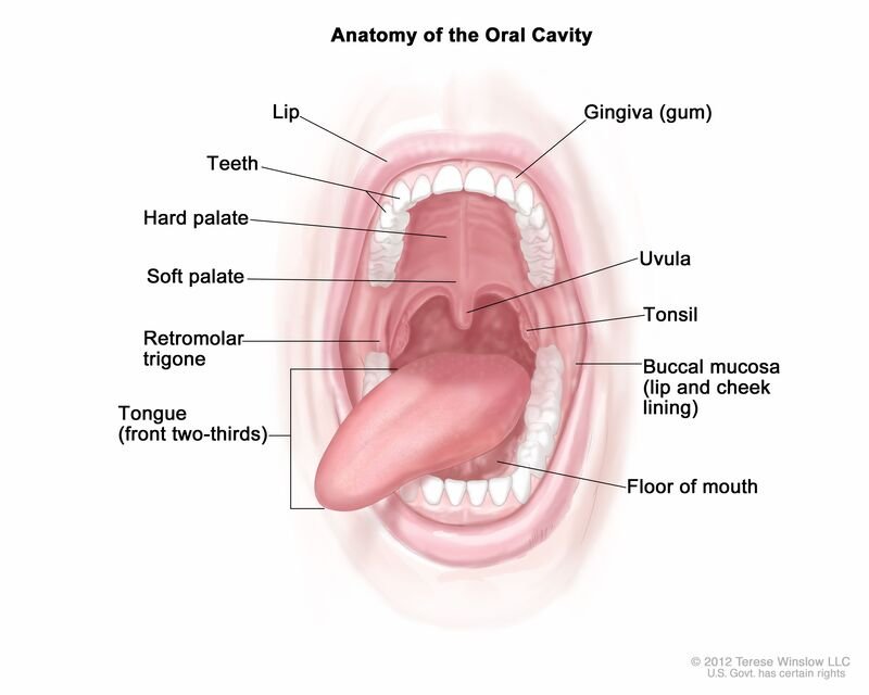
The oral cavity (mouth) represents the first division of the human digestive tract and serves secondary, indispensable roles in vocal articulation and airway patency. It extends from the labial fissure anteriorly to the isthmus of the fauces (oropharyngeal isthmus) posteriorly, where it transitions directly into the oropharynx.
Subdivisions of the Oral Cavity
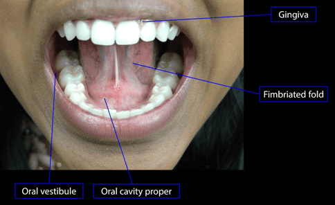
The dental arches (composed of the maxillary and mandibular alveolar processes, teeth, and overlying gingivae) subdivide the oral cavity into two anatomical compartments:
1. The Oral Vestibule (Vestibulum Oris)
The slit-like outer chamber situated between the mobile peripheral facial tissues and the rigid dental apparatus:
- External Boundary: Lined by the mucous membrane of the lips (labia) anteriorly and the cheeks (buccae) laterally, which are supported by the orbicularis oris and buccinator muscles, respectively.
- Internal Boundary: Bounded by the outer (labial and buccal) surfaces of the teeth and their supporting gums (gingivae).
- Parotid Duct Drainage: Communicates with the parotid salivary gland via Stensen’s duct, which pierces the buccinator muscle and opens onto a small mucosal elevation—the parotid papilla—situated in the vestibule opposite the crown of the maxillary second molar tooth.
- Communication: When the teeth are occluded, the vestibule communicates with the oral cavity proper through the narrow retromolar space located behind the last molar teeth on either side.
2. The Oral Cavity Proper (Cavitas Oris Propria)
The primary internal chamber occupied largely by the tongue:
- Anterior & Lateral Boundaries: Defined by the inner (lingual and palatal) margins of the maxillary and mandibular dental arcades and gingivae.
- Superior Boundary (Roof): Formed entirely by the palate (the anterior four-fifths hard palate and posterior one-fifth soft palate), which separates the mouth from the overlying nasal cavity and nasopharynx.
- Inferior Boundary (Floor): Formed by the anterior two-thirds of the tongue and the sublingual mucosal reflection, supported by a muscular hammock comprising the paired mylohyoid muscles (the oral diaphragm) reinforced superiorly by the geniohyoid muscles.
- Posterior Boundary: Communicates with the oropharynx across the isthmus of the fauces, which is bounded superiorly by the soft palate, laterally by the palatoglossal arches, and inferiorly by the dorsum of the tongue.

The palate: hard & soft
The palate forms the arched roof of the oral cavity and the floor of the nasal cavities. It is partitioned structurally into an anterior immobile bony plate (the hard palate) and a posterior dynamic fibromuscular valve (the soft palate).
1. The Hard Palate (Palatum Durum)
The hard palate occupies the anterior four-fifths (4/5) of the total palatal surface. It forms an osteomucosal partition lined inferiorly by masticatory mucosa (keratinized stratified squamous epithelium bound to underlying periosteum via a dense collagenous submucosa containing numerous mucus-secreting palatine glands).
- Bony Architecture: Formed by the union of two pairs of bones along the median and transverse palatine sutures:
- The palatine processes of the maxillae (constituting the anterior two-thirds).
- The horizontal plates of the palatine bones (constituting the posterior one-third).
- Foramina of the Hard Palate:
- Incisive Foramen (Fossa): Positioned in the anterior midline immediately behind the central incisor teeth. Transmits the terminal branches of the nasopalatine nerve (CN V2) and the sphenopalatine vessels between the nasal septum and oral mucosa.
- Greater Palatine Foramen: Located in the posterolateral aspect of the hard palate, medial to the third molar tooth. Transmits the greater palatine nerve and greater palatine artery forward onto the hard palate.
- Lesser Palatine Foramina: Situated in the pyramidal process of the palatine bone immediately posterior to the greater palatine foramen. Transmit the lesser palatine nerves and arteries posteriorly into the soft palate.
- Neurovascular Supply:
- Sensory Innervation: Mediated exclusively by branches of the maxillary division of the trigeminal nerve (CN V2). The anterior premaxillary region behind the incisors is supplied by the nasopalatine nerve; the remainder of the hard palate is innervated by the greater palatine nerve.
- Arterial Supply: Derived primarily from the greater palatine artery (a terminal branch of the descending palatine artery from the third part of the maxillary artery), which runs anteriorly in a bony groove along the lateral margin of the palate to anastomose through the incisive canal with the sphenopalatine artery.
2. The Soft Palate (Velum Palatinum)
The soft palate comprises the posterior one-fifth (1/5) of the palate. It is a mobile, dynamic fibromuscular curtain suspended from the posterior margin of the hard palate, terminating posteroinferiorly in a conical, free-hanging median projection called the uvula.
- Physiological Action: Swallowing (deglutition) and phonation require the soft palate to swing posterosuperiorly against the posterior and lateral pharyngeal walls (Passavant’s ridge). This seals the pharyngeal isthmus, completely isolating the nasopharynx from the oropharynx to prevent nasal regurgitation of liquids and solids.
- Faucial Arches: Laterally, the soft palate is continuous with two descending mucosal folds:
- Palatoglossal Fold (Anterior Pillar): Extends from the soft palate to the lateral border of the tongue, encasing the palatoglossus muscle.
- Palatopharyngeal Fold (Posterior Pillar): Extends from the soft palate to the lateral pharyngeal wall, encasing the palatopharyngeus muscle.
- The Tonsillar Sinus (Fossa): The triangular space between these two arches houses the palatine tonsil.
- Blood Supply of the Soft Palate: Receives an extensive collateral arterial anastomosis from:
- The greater and lesser palatine arteries (branches of the descending palatine artery from the maxillary artery).
- The ascending palatine artery (a direct cervical branch of the facial artery).
- The palatine branch of the ascending pharyngeal artery (from the external carotid artery).
- Innervation of the Soft Palate:
- Sensory: Conducted by the lesser palatine nerves (CN V2 via the pterygopalatine ganglion) and branches of the glossopharyngeal nerve (CN IX).
- Motor: All intrinsic and extrinsic palatal muscles are innervated by the vagus nerve (CN X) via the pharyngeal plexus, with the single exception of the tensor veli palatini, which is innervated by the mandibular division of the trigeminal nerve (CN V3).
Muscles of the palate
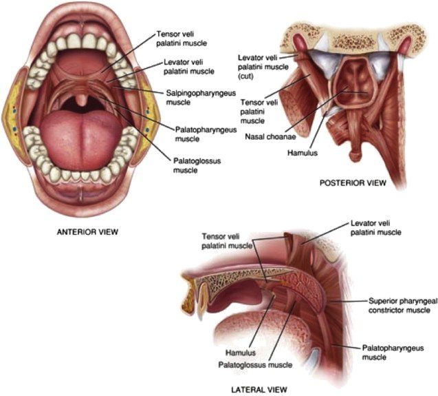
The soft palate contains five paired striated muscles that attach to and mobilize the central fibrous palatine aponeurosis (the expanded tendon of the tensor veli palatini, which anchors anteriorly to the posterior edge of the hard palate).
Scroll the table sideways on a smaller screen.
| Muscle | Proximal Attachment (Origin) | Distal Attachment (Insertion) | Innervation | Primary Mechanical Action |
|---|---|---|---|---|
| Tensor Veli Palatini | Scaphoid fossa of sphenoid, spine of sphenoid, and membranous/cartilaginous auditory (Eustachian) tube | Tendon hooks at a right angle around the pterygoid hamulus of the medial pterygoid plate to expand into the palatine aponeurosis | Mandibular nerve (CN V3) via the nerve to medial pterygoid | Tenses the soft palate horizontally; dilates and opens the auditory tube during swallowing and yawning to equalize middle ear pressure |
| Levator Veli Palatini | Inferior surface of petrous part of temporal bone (anterior to carotid canal) and cartilaginous auditory tube | Passes medially into the soft palate to insert into the superior surface of the palatine aponeurosis | Vagus nerve (CN X) via the pharyngeal plexus | Elevates the soft palate posterosuperiorly against the posterior pharyngeal wall, closing the pharyngeal isthmus |
| Palatoglossus | Oral surface of the palatine aponeurosis | Passes anteroinferiorly in the palatoglossal arch to insert into the dorsolateral border of the tongue | Vagus nerve (CN X) via the pharyngeal plexus | Elevates the root of the tongue and depresses the soft palate; acts as an oral sphincter to constrict the isthmus of the fauces |
| Palatopharyngeus | Palatine aponeurosis and posterior hard palate | Descends in the palatopharyngeal arch to insert into the posterior border of the thyroid cartilage and fibrous coat of the pharynx | Vagus nerve (CN X) via the pharyngeal plexus | Elevates the pharynx and larynx during deglutition; draws the palatopharyngeal arches together to seal off the nasopharynx |
| Musculus Uvulae | Posterior nasal spine of the palatine bones and the palatine aponeurosis | Mucous membrane envelope of the uvula | Vagus nerve (CN X) via the pharyngeal plexus | Elevates, thickens, and shortens the uvula, aiding the levator veli palatini in sealing the pharyngeal isthmus |
The tongue: parts & surfaces
The tongue (lingua/glossa) is a mobile muscular hydrostat that occupies the floor of the oral cavity and forms the anterior wall of the oropharynx. It executes mastication, bolus preparation, deglutition, vocal phonation, and taste perception (gustation).
Anatomical Subdivisions
- 1. Root (Base) of the Tongue: The attached posterior third that rests between the mandible, hyoid bone, and the posterior vertical wall of the oral cavity. It is fixed in position and anchored securely to the hyoid bone and mandible by the hyoglossus and genioglossus muscles.
- 2. Body (Corpus) of the Tongue: The anterior two-thirds of the organ situated horizontally between the root and apex. Highly mobile; its superior surface forms the anterior floor of the oral cavity proper.
- 3. Apex (Tip) of the Tongue: The pointed anterior extremity of the body that normally rests against the lingual surfaces of the mandibular incisor teeth. The body and apex demonstrate multi-axial mobility.
Surfaces and External Attachments
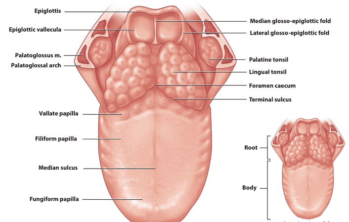
- Dorsum of the Tongue: The convex superior and posterior surface. A chevron-shaped groove called the sulcus terminalis (terminal sulcus) divides the dorsum into an anterior two-thirds (oral/presulcal part) facing superiorly into the mouth, and a posterior one-third (pharyngeal/postsulcal part) facing posteriorly into the oropharynx.
- Inferior (Ventral) Surface: The smooth mucosal surface resting on the floor of the mouth:
- Lingual Frenulum: A median vertical fold of mucous membrane connecting the underside of the tongue to the floor of the mouth. It stabilizes the organ while allowing the anterior apex to move freely.
- Deep Lingual Veins: Large, blue-tinged venous channels running on either side of the frenulum, visible through the thin, non-keratinized mucosal epithelium.
- Fimbriated Folds (Plicae Fimbriatae): Delicate, scalloped mucosal ridges lying lateral to the deep lingual veins.
- Sublingual Caruncles (Papillae): Small papillae on each side of the base of the lingual frenulum that house the terminal orifices of the submandibular (Wharton’s) ducts.
- Skeletal Attachments: The tongue is anchored by muscle slips to five surrounding structures: the hyoid bone, mandible, styloid process, soft palate, and pharyngeal wall.

Papillae & tonsillar tissue
The Sulcus Terminalis & Foramen Caecum
The dorsum of the tongue is demarcated by the V-shaped sulcus terminalis, whose apex points posteriorly toward the blind depression known as the foramen caecum.
- Embryological Significance: The foramen caecum marks the origin of the embryonic thyroglossal duct, which descends through the developing tongue and anterior neck to establish the bilobed thyroid gland. Incomplete obliteration of this pathway leads to developmental abnormalities such as thyroglossal duct cysts or an ectopic lingual thyroid.
- Developmental Dichotomy: The anterior two-thirds develops from the first pharyngeal arch (lateral lingual swellings and tuberculum impar), whereas the posterior one-third develops from the third pharyngeal arch (cranial portion of the hypopharyngeal eminence/copula). This embryological divergence explains their differing structural features, vascular supplies, and sensory innervations.
Morphology of Lingual Papillae
The mucosa of the presulcal anterior two-thirds contains four types of mucosal projections termed lingual papillae:
1. Vallate (Circumvallate) Papillae
Large, cylindrical, dome-shaped mucosal projections (numbering 8 to 12) arranged in a distinct V-shaped row immediately anterior and parallel to the sulcus terminalis:
- Structural Trench: Each papilla is surrounded by a deep circular moat-like trench into which the serous glands of von Ebner empty. Their watery secretions flush food debris from the trench, preparing the taste buds for new gustatory stimuli.
- Taste Buds & Innervation: The lateral walls of the trenches are densely populated with taste buds. Despite lying on the anterior side of the sulcus terminalis, their taste buds and mucosa are innervated by the glossopharyngeal nerve (CN IX).
2. Fungiform Papillae
Mushroom-shaped mucosal elevations with rounded, bright-red heads (due to rich capillary vascularization visible through thin, non-keratinized epithelium):
- Distribution: Scattered individually across the dorsum, concentrated predominantly along the lateral margins and apex of the tongue.
- Gustatory Role: Contain one to several functional taste buds on their apical surfaces, innervated for taste by the chorda tympani (CN VII via the lingual nerve).
3. Filiform Papillae
Minute, slender, conical, thread-like projections arranged in tightly packed, V-shaped rows parallel to the sulcus terminalis across the entire presulcal dorsum:
- Keratinization: Covered by heavily keratinized stratified squamous epithelium, which gives the tongue surface its characteristic grayish-pink, velvety texture.
- Mechanical Function: Devoid of taste buds. They provide a high-friction abrasive surface that aids in licking, moving food boluses, and food texture perception (tactile and thermal general somatic sensation via CN V3).
4. Foliate Papillae
Parallel, leaf-like mucosal folds situated along the posterolateral margins of the tongue anterior to the palatoglossal arch. While prominent and rich in taste buds in lagomorphs and other mammals, they are rudimentary in adult humans.
The Lingual Tonsil (Root of the Tongue)
The mucous membrane of the postsulcal posterior third of the tongue lacks lingual papillae. Instead, it has a cobblestone appearance produced by underlying nodular aggregates of lymphoid follicles embedded in submucosa: the lingual tonsil. Each nodule contains a central microscopic crypt opening onto the mucosal surface. Together with the palatine tonsils, tubal tonsils, and pharyngeal tonsil (adenoid), the lingual tonsil completes the protective immunological ring of the pharynx (Waldeyer’s ring).

Musculature of the tongue
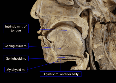
The tongue is composed almost entirely of striated skeletal muscle fibers enveloped by mucous membrane. The lingual musculature is symmetrically paired, separated along the midline by a vertical median fibrous lingual septum that merges posteriorly with the sublingual aponeurosis. The muscles are divided into two functional classes: extrinsic muscles (which alter the spatial position of the tongue) and intrinsic muscles (which alter its shape).
1. Extrinsic Muscles of the Tongue
The four paired extrinsic muscles originate from bony or cartilaginous structures outside the tongue and insert into its muscular body:
Scroll the table sideways on a smaller screen.
| Extrinsic Muscle | Proximal Origin | Distal Insertion | Innervation | Primary Mechanical Action |
|---|---|---|---|---|
| Genioglossus | Superior mental spine (genial tubercle) on the inner posterior surface of the symphysis menti of the mandible | Fans out broadly: inferior fibers attach to the body of the hyoid bone; middle fibers insert into the root; superior fibers ascend to the apex | Hypoglossal nerve (CN XII) | Protrudes the tongue out of the mouth; bilateral action depresses the central dorsum (forming a longitudinal trough for swallowing); unilateral action deviates the tongue to the contralateral side |
| Hyoglossus | Superior border of the body and entire length of the greater horn of the hyoid bone | Ascends vertically into the side of the tongue between styloglossus and the inferior longitudinal muscle | Hypoglossal nerve (CN XII) | Depresses the tongue, pulling its sides downward; assists in retraction (retrusion) |
| Styloglossus | Anterolateral surface of the styloid process of the temporal bone and the stylohyoid ligament | Runs anteroinferiorly to divide and insert into the lateral margins and inferior aspect of the tongue, interdigitating with hyoglossus | Hypoglossal nerve (CN XII) | Retracts (pulls backward) and elevates the tongue; draws the lateral borders upward to form a swallowing trough |
| Palatoglossus | Inferior oral surface of the palatine aponeurosis of the soft palate | Descends in the palatoglossal arch to insert into the dorsolateral posterolateral margins of the tongue | Vagus nerve (CN X) via the pharyngeal plexus | Elevates the posterior tongue while depressing the soft palate; closes the isthmus of the fauces to initiate swallowing |
2. Intrinsic Muscles of the Tongue
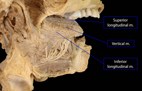
The four paired intrinsic muscles are contained entirely within the substance of the tongue without external skeletal attachments. They are oriented in three spatial planes:
Scroll the table sideways on a smaller screen.
| Intrinsic Muscle | Fiber Architecture & Topography | Innervation | Primary Mechanical Action |
|---|---|---|---|
| Superior Longitudinal | Thin longitudinal stratum lying immediately subjacent to the dorsal mucous membrane, running from the median fibrous septum and submucosa of the root forward to the lateral margins and apex | Hypoglossal nerve (CN XII) | Curls the apex and lateral margins of the tongue upward (dorsally), making the dorsum concave; shortens (retrudes) the tongue |
| Inferior Longitudinal | Narrow cylindrical longitudinal fascicle situated on the ventral aspect of the tongue between the genioglossus and hyoglossus, extending from the root of the tongue to the apex | Hypoglossal nerve (CN XII) | Curls the apex of the tongue downward (ventrally), making the dorsum convex; shortens and retrudes the tongue |
| Transverse | Horizontal fibers that arise from the median fibrous lingual septum and radiate laterally through the lingual substance to insert into the submucosa of the lateral margins | Hypoglossal nerve (CN XII) | Narrows the tongue and increases its vertical height; pushes the tongue forward (elongates and protrudes) |
| Vertical | Vertical fibers that extend from the dorsal submucosal fibrous layer to the ventral submucosa along the lateral borders of the tongue, intersecting with transverse fibers | Hypoglossal nerve (CN XII) | Flattens and broadens the tongue; depresses the margins against the floor of the mouth |
Innervation of the tongue
Because the tongue derives from multiple embryonic pharyngeal arches (first, second, third, and fourth arches) along with migrating occipital somites, its adult innervation comprises motor, general somatic sensory, and special sensory (taste) fibers.
1. Motor Innervation
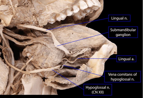
All eight paired muscles of the tongue (four intrinsic, four extrinsic) are innervated by the hypoglossal nerve (CN XII), with the single exception of the palatoglossus, which is innervated by the vagus nerve (CN X) via the pharyngeal plexus.
2. Sensory Innervation (General Somatic Sensation & Taste)
A. Anterior Two-Thirds (Presulcal Body & Apex)
- General Sensation (Pain, Touch, Temperature): Mediated by the lingual nerve, a terminal branch of the mandibular division of the trigeminal nerve (CN V3). Cell bodies reside in the trigeminal (semilunar) ganglion.
- Special Sensation (Taste): Mediated by the chorda tympani (a branch of the facial nerve, CN VII). The chorda tympani hitches a ride with the lingual nerve in the infratemporal fossa to distribute to fungiform papillae taste buds. Primary sensory cell bodies reside in the geniculate ganglion of CN VII.
B. Posterior One-Third (Postsulcal Root & Vallate Papillae)
- Dual Sensation (General & Taste): Both general somatic sensation (tactile, thermal, pain) and special sensory taste are conducted by the glossopharyngeal nerve (CN IX) via its lingual branches. This encompasses the root of the tongue, the palatoglossal folds, and the circumvallate (vallate) papillae. Primary sensory cell bodies reside in the inferior (petrosal) ganglion of CN IX.
C. Posterior Root & Epiglottic Region
- Valleculae and Epiglottis: The posterior root of the tongue adjacent to the epiglottic valleculae receives general sensation and taste from the internal laryngeal branch of the vagus nerve (CN X). Sensory cell bodies reside in the inferior vagal (nodose) ganglion.
Scroll the table sideways on a smaller screen.
| Tongue Region | Motor Efferent | General Somatosensory (GSA) | Special Sensory / Taste (SVA) |
|---|---|---|---|
| Anterior 2/3 (Body & Apex) | Hypoglossal nerve (CN XII) (Palatoglossus by CN X) |
Lingual nerve (from Mandibular CN V3) | Chorda tympani (from Facial CN VII via lingual nerve) |
| Posterior 1/3 (Root & Vallate Papillae) | Hypoglossal nerve (CN XII) | Glossopharyngeal nerve (CN IX) | Glossopharyngeal nerve (CN IX) |
| Vallecular & Epiglottic Area | Vagus nerve (CN X via pharyngeal plexus) | Internal laryngeal nerve (CN X) | Internal laryngeal nerve (CN X) |
Blood supply & lymphatics
Arterial Supply
The primary vascular source for the tongue is the lingual artery, supplemented by minor contributions from the tonsillar and ascending palatine branches of the facial artery, and the ascending pharyngeal artery.
- Course of the Lingual Artery: Arises from the anterior aspect of the external carotid artery within the carotid triangle, opposite the tip of the greater horn of the hyoid bone. It loops upward over the middle pharyngeal constrictor, passes deep to the posterior border of the hyoglossus muscle, and courses forward through the sublingual compartment.
- Branching Pattern:
- Suprahyoid Artery: Courses along the upper border of the hyoid bone, supplying the attached infra- and suprahyoid musculature.
- Dorsal Lingual Arteries (2–3 branches): Arise deep to hyoglossus, ascending vertically to supply the posterior one-third (root) of the tongue, palatoglossal arch, palatine tonsil, and epiglottis. Crucial Surgical Note: The dorsal lingual arteries are prevented from communicating across the midline by the median fibrous lingual septum.
- Sublingual Artery: Arises at the anterior border of hyoglossus; courses between mylohyoid and genioglossus to supply the sublingual salivary gland, mylohyoid muscle, and adjacent alveolar mucosa.
- Deep Lingual Artery (Ranine Artery): The terminal continuation of the lingual artery. Ascends tortuously between the genioglossus and inferior longitudinal muscles near the ventral surface to supply the anterior body. Near the apex of the tongue, the left and right deep lingual arteries communicate across the midline through terminal anastomoses.
Venous Drainage
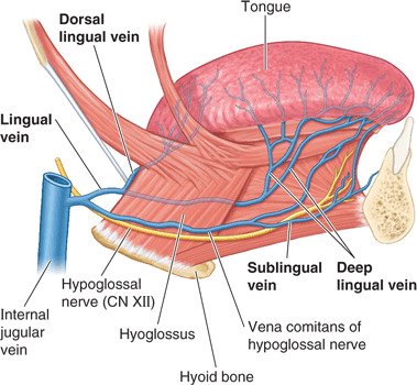
Venous blood from the tongue drains via two networks that ultimately empty into the internal jugular vein (IJV), either directly or via the common facial/lingual trunk:
- 1. Deep Lingual Veins: Begin near the apex on the ventral surface and run posteriorly beside the lingual frenulum. They pass over the lateral surface of the hyoglossus muscle (accompanied by the hypoglossal nerve) to join the sublingual vein. In elderly individuals, the sublingual veins often become visibly dilated and tortuous (lingual varicosities / caviar tongue), which is a normal benign age-related change.
- 2. Dorsal Lingual Veins: Accompany the dorsal lingual branches of the lingual artery deep to hyoglossus, draining the posterior root and dorsal margins.
Lymphatic Drainage: Four Distinct Clinical Routes
The lymphatic drainage of the tongue is clinically important due to the propensity of squamous cell carcinomas (SCC) of the tongue to metastasize early to deep cervical lymph nodes. While most lymphatic channels follow the venous system, central and apical lymphatics take independent, crossing courses:

The Four Lymphatic Drainage Routes
- Route 1: Root of the Tongue (Posterior 1/3): Lymphatic vessels cross the midline and drain bilaterally into the superior deep cervical lymph nodes, particularly the jugulodigastric node. Cancers of the base of the tongue carry a high frequency of bilateral cervical nodal metastasis.
- Route 2: Medial Body of the Tongue: Vessels from the central dorsal gutter drain directly and bilaterally across the median septum, passing through the mylohyoid to reach the inferior deep cervical lymph nodes (specifically the jugulo-omohyoid node).
- Route 3: Lateral Parts of the Body (Lateral Margins): Channels drain unilaterally (ipsilaterally) into the submandibular lymph nodes. Secondary drainage passes from submandibular nodes into the superior and inferior deep cervical chains.
- Route 4: Apex and Lingual Frenulum: Vessels drain inferiorly into the submental lymph nodes. Crucially, the medial portion drains bilaterally to both submental basins, followed by secondary drainage to the jugulo-omohyoid nodes.
Teeth: structure & classes
Teeth (dentes) are specialized, mineralized hard-tissue organs anchored within the alveolar sockets (alveoli dentales) of the maxillae and mandible by a specialized fibrous gomphosis joint.
Primary Physiological Functions
- Mastication: Cutting (incising), tearing, crushing, and grinding food particles, mixing them with saliva to form a digestible bolus.
- Periodontal Integrity: Maintaining the bone height of the alveolar ridges through physiological masticatory mechanical loading.
- Speech Articulation: Serving as critical contact surfaces for the tongue and lips to produce labiodental (e.g., “f”, “v”) and linguodental (e.g., “th”) phonetic sounds.
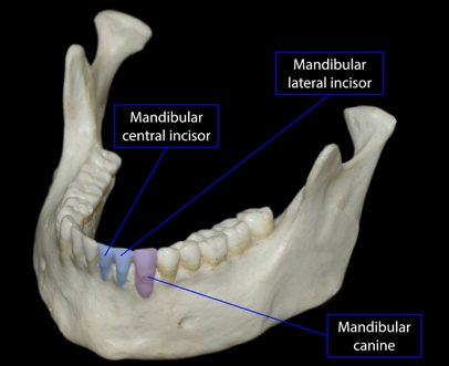
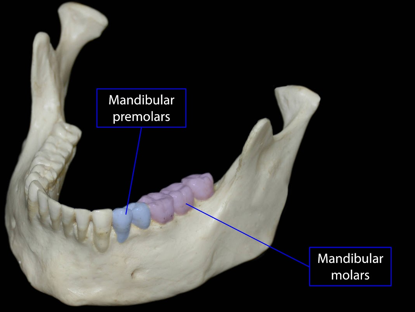
Anatomical Components of a Tooth
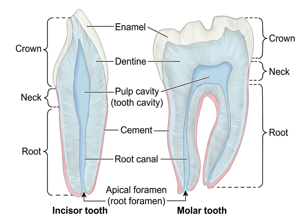
- 1. Crown (Corona Dentis): The visible anatomical segment projecting above the gingival margin into the oral cavity, capped by crystalline enamel.
- 2. Neck (Cervix Dentis): The slightly constricted transitional boundary zone at the cementoenamel junction (CEJ) where the enamel-covered crown meets the cementum-covered root.
- 3. Root (Radix Dentis): The subgingival portion embedded within the bony tooth socket (alveolus). It is covered by a thin layer of bone-like tissue called cementum and anchored to the alveolar bone by a collagenous ligament: the periodontal ligament (PDL).
- Maxillary molars: Typically possess three roots (one palatal, two buccal).
- Mandibular molars: Typically possess two roots (one mesial, one distal).
- Maxillary first premolar: Frequently possesses a bifid or double root; all other premolars generally have a single root.
Internal Histological Architecture
- 1. Enamel: The hardest biological substance in the human body, composed of 96% to 97% inorganic mineralized hydroxyapatite crystals arranged in hexagonal prisms. It forms an acellular, non-regenerative cap protecting the coronal dentin. Synthesized by epithelial ameloblasts during odontogenesis, which are lost when the tooth erupts.
- 2. Dentin (Dentine): A mineralized, bonelike tissue (70% inorganic, 20% organic collagen matrix, 10% water) forming the primary structural bulk of the tooth. It is traversed by microscopic dentinal tubules that contain the long cytoplasmic processes of odontoblasts. Odontoblast cell bodies line the peripheral wall of the pulp cavity, remaining metabolically active throughout life to produce secondary and tertiary (reparative) dentin.
- 3. Dental Pulp: The loose, gelatinous, non-mineralized connective tissue core filling the central pulp cavity (coronal pulp chamber) and extending down the roots as the root canals. It contains an extensive neurovascular network, sensory nerve endings (transmitting sharp pain via A-delta fibers and dull aching pain via C-fibers), and lymphatics, which enter and exit through the microscopic apical foramen at the root apex.
Primary vs. Permanent Dentition Sets
1. Deciduous (Primary / Milk) Teeth
The temporary childhood dentition (erupting between 6 months and 2.5 years of age). Consists of 20 teeth in total (5 per quadrant):
- Per Quadrant: Two incisors (central, lateral), one canine, and two deciduous molars.
- Clinical Rule: There are no premolars in the deciduous dentition. The deciduous molars are replaced during late childhood by the permanent premolars.
2. Permanent (Secondary / Adult) Teeth
The adult dentition (erupting between 6 years and 18–25 years of age). Consists of 32 teeth in total (8 per quadrant):
- Per Quadrant: Two incisors (central, lateral), one canine, two premolars, and three molars (including the third molar or “wisdom tooth”).
Scroll the table sideways on a smaller screen.
| Tooth Class | Coronal Morphology & Cusps | Root Architecture | Primary Masticatory Function |
|---|---|---|---|
| Incisors (Central & Lateral) | Chisel-shaped crown with a sharp, straight incisal edge | Single conical root | Cutting and incising food |
| Canines (Cuspids) | Robust, pointed crown with a single prominent conical cusp | Single, exceptionally long and strong root (forms canine eminence) | Piercing and tearing coarse food materials |
| Premolars (Bicuspids) | Broad occlusal surface displaying two distinct cusps (one buccal, one lingual) | Single root (except the maxillary first premolar, which is typically bifid) | Crushing and grinding food |
| Molars (1st, 2nd, 3rd) | Broad rhomboid or trapezoid occlusal table bearing 3 to 5 grinding cusps | Multi-rooted: Maxillary molars have 3 roots; Mandibular molars have 2 roots | Heavy pulverization and grinding of food |
Vessels & nerves of teeth & gums
Arterial Supply & Venous Drainage of Teeth
- Arteries: All dental arteries arise from branches of the maxillary artery (a terminal branch of the external carotid artery):
- Maxillary Teeth: Supplied by the posterior superior alveolar artery (from the third part of the maxillary artery for molars/premolars) and the anterior and middle superior alveolar arteries (branches of the infraorbital artery for canine and incisors).
- Mandibular Teeth: Supplied by the inferior alveolar artery (from the first part of the maxillary artery), which enters the mandibular canal through the mandibular foramen and sends dental branches into each root apex.
- Veins: Alveolar veins accompany their named arterial counterparts, emerging from dental sockets to drain into the pterygoid venous plexus in the infratemporal fossa.
- Lymphatics: Lymphatic vessels from both maxillary and mandibular teeth and their surrounding periodontal tissues drain primarily to the submandibular lymph nodes, with minor apical drainage to submental nodes.
Innervation of the Teeth and Gingivae
Dental innervation is provided entirely by branches of the trigeminal nerve (CN V), dividing along the maxillary and mandibular arcades:
Scroll the table sideways on a smaller screen.
| Arcade / Structure | Innervation of the Teeth (Pulps) | Innervation of Outer (Buccal/Labial) Gingiva | Innervation of Inner (Palatal/Lingual) Gingiva |
|---|---|---|---|
| Maxillary Dental Arcade |
• Anterior superior alveolar nerve (Incisors & Canine) • Middle superior alveolar nerve (Premolars & mesiobuccal root of 1st molar) • Posterior superior alveolar nerve (Molars) (All derived from Maxillary CN V2) |
• Labial gingiva: Infraorbital nerve and anterior superior alveolar nerve • Buccal gingiva: Posterior superior alveolar nerve |
• Anterior palatal gingiva: Nasopalatine nerve (behind incisors/canines) • Posterior palatal gingiva: Greater palatine nerve (adjacent to premolars/molars) |
| Mandibular Dental Arcade | • Inferior alveolar nerve (via its dental plexus, derived from Mandibular CN V3); divides terminally into the incisive nerve (incisors/canine) and mental nerve |
• Anterior labial gingiva: Mental nerve (from inferior alveolar) • Posterior buccal gingiva: Long buccal nerve (direct branch of CN V3) |
• Lingual gingiva along the entire mandibular dental arch: Lingual nerve (from Mandibular CN V3) |
Salivary glands
Saliva is synthesized and secreted by three pairs of major salivary glands (parotid, submandibular, and sublingual) alongside hundreds of microscopic minor salivary glands dispersed throughout the submucosa of the lips, cheeks, palate, and tongue.
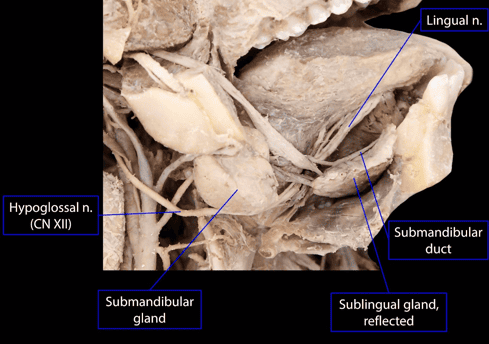
1. The Submandibular Gland
The submandibular gland is a large, mixed (predominantly serous, partially mucous) salivary gland occupying the submandibular triangle of the anterior cervical triangle. It is encapsulated by the investing layer of deep cervical fascia.
- Morphological Lobes: The gland is shaped like the letter “C,” folded around the free posterior border of the mylohyoid muscle:
- Superficial Portion: The larger lobe, situated superficial to the mylohyoid within the submandibular fossa of the inner mandible. Its inferolateral surface is covered by cervical skin, platysma, and cervical fascia, crossed by the marginal mandibular branch of the facial nerve.
- Deep Portion: Extends forward into the floor of the mouth, lying in the interval between the hyoglossus and styloglossus muscles medially and the mylohyoid muscle laterally. It is positioned between the lingual nerve above and the hypoglossal nerve below.
- Wharton’s Duct (Submandibular Duct): Approximately 5 cm in length. Emerges from the anterior end of the deep portion of the gland and courses forward between the mylohyoid and hyoglossus muscles.
Surgical Relationship: As it travels forward, the lingual nerve loops around the duct, crossing first lateral, then inferior (underneath), and finally medial to the duct as it ascends into the tongue substance.
Terminal Orifice: The duct continues forward between the sublingual gland and genioglossus to terminate at the summit of the sublingual caruncle (papilla) at the base of the lingual frenulum. - Parasympathetic Secretomotor Pathway:
- Preganglionic parasympathetic cell bodies reside in the superior salivatory nucleus of the pons.
- Fibers exit in the facial nerve (CN VII) via the nervus intermedius, enter the middle ear within the chorda tympani, and join the lingual nerve in the infratemporal fossa.
- Preganglionic fibers synapse within the submandibular ganglion (which hangs suspended from the lingual nerve on the hyoglossus muscle).
- Short postganglionic parasympathetic secretomotor fibers enter the submandibular gland parenchyma directly to stimulate watery serous secretion.

2. The Sublingual Gland
The sublingual gland is the smallest of the three major salivary glands. It is predominantly mucous in character, almond-shaped, and situated in the sublingual space beneath the oral floor mucosa.
- Topographical Relations: Lies directly beneath the mucous membrane of the mouth, elevating the sublingual fold (plica sublingualis). It is bounded laterally by the sublingual fossa of the mandible, medially by the genioglossus muscle and submandibular duct, and inferiorly by the mylohyoid muscle.
- Duct System (Ducts of Rivinus & Bartholin): Does not possess a single dominant drainage conduit. Instead, it empties through 8 to 20 small short ducts (ducts of Rivinus) along the crest of the sublingual fold. A few anterior ducts may unite to form the greater sublingual duct (duct of Bartholin), which empties directly into the submandibular duct.
- Parasympathetic Innervation: Driven by postganglionic parasympathetic secretomotor fibers originating from the submandibular ganglion, which reach the gland either directly or by hitchhiking along the distal branches of the lingual nerve.
Clinical correlations
1. Sialolithiasis (Salivary Calculi)
Over 80% to 90% of salivary gland stones form within the submandibular gland and Wharton’s duct. This high clinical incidence is governed by three anatomical and physiological factors:
- Antigravity Duct Trajectory: Wharton’s duct courses upward and forward around the posterior margin of mylohyoid, causing salivary stasis during recumbency.
- High Calcium & Mucin Composition: Submandibular secretions are more viscous, richer in mucin proteins, and have higher concentrations of calcium and phosphate salts than parotid saliva.
- Alkaline pH: Favors the precipitation of calcium phosphate stones.
Presentation: Painful, postprandial swelling of the submandibular triangle (“mealtime syndrome”). Transoral palpation along the floor of the mouth often reveals a calculus trapped in the distal duct.
2. Ludwig’s Angina
A rapidly spreading, life-threatening cellulitis of the floor of the mouth that involves the submandibular, sublingual, and submental spaces bilaterally. It typically arises from an odontogenic apical abscess of the mandibular second or third molar teeth, whose root apices extend inferiorly below the mylohyoid line into the submandibular space.
Clinical Presentation: Brawny suprahyoid swelling, elevated and displaced tongue (causing acute mechanical airway compromise), trismus, fever, and stridor. Management requires immediate airway security (often awake fiberoptic or surgical tracheostomy) and surgical decompression.
3. Lingual Nerve Injury During Oral Surgery
Because the lingual nerve loops around Wharton’s duct and lies adjacent to the lingual cortical plate near the mandibular third molar, it is vulnerable to traction or transection during surgical extraction of impacted lower wisdom teeth or transoral submandibular duct sialolithotomies.
Clinical Consequence: Loss of both general sensation (numbness) and taste (ageusia) over the anterior two-thirds of the tongue on the ipsilateral side.
4. Ankyloglossia (Tongue-Tie)
A congenital developmental anomaly in which an excessively short, thick, or anteriorly displaced lingual frenulum anchors the apex of the tongue to the floor of the mouth and mandibular gingiva. It restricts tongue protrusion, interfering with infant latch during breastfeeding and later contributing to speech articulation difficulties (especially lingual consonants: “t”, “d”, “l”). Managed via frenotomy (surgical division of the frenulum).
Quick recap
References
The following authoritative anatomical reference works were verified in compiling this curriculum study note:
- Moore’s Clinically Oriented Anatomy: Dalley, A.F. and Agur, A.M.R. (2022) Moore’s Clinically Oriented Anatomy. 9th edn. Philadelphia: Wolters Kluwer. [Chapter 8: Head – Oral Cavity, Teeth, Tongue, and Palate].
- Gray’s Anatomy: Standring, S. (2021) Gray’s Anatomy: The Anatomical Basis of Clinical Practice. 42nd edn. London: Elsevier. [Section 4: Head and Neck; Chapter 30: Oral Cavity and Salivary Glands].
- Last’s Anatomy: Sinnatamby, C.S. (2011) Last’s Anatomy: Regional and Applied. 12th edn. Edinburgh: Churchill Livingstone. [Chapter 6: Head and Neck and Spine – Mouth and Tongue].
- Ten Cate’s Oral Histology: Nanci, A. (2018) Ten Cate’s Oral Histology: Development, Structure, and Function. 9th edn. St. Louis: Elsevier. [Chapter 8: Dentin-Pulp Complex; Chapter 12: Salivary Glands].
- Netter’s Atlas of Human Anatomy: Netter, F.H. (2019) Atlas of Human Anatomy. 7th edn. Philadelphia: Elsevier. [Plates 60–66: Oral Cavity, Tongue, Teeth, and Salivary Glands].