Groups of Muscles
Comprehensive anatomical study of the major muscle groups of the human body, detailing their origin, insertion, nerve supply, and action. This guide is organized by anatomical region to facilitate clinical reasoning and surgical planning.
1. Introduction
The human body contains over 600 skeletal muscles. These are organized into functional groups based on their anatomical location and the movements they produce. Muscles are named according to specific criteria:
- Size: e.g., gluteus maximus.
- Shape: e.g., deltoid.
- Location: e.g., tibialis anterior.
- Number of origins: e.g., biceps (two heads), triceps (three heads).
- Direction of fibers: e.g., rectus abdominis (straight).
- Action: e.g., flexor digitorum.
2. Muscles of the Head and Neck
2.1 Muscles of Facial Expression
These muscles are unique because they insert into the skin rather than onto bone. They are all innervated by the Facial nerve (Cranial Nerve VII).
| Muscle | Origin | Insertion | Action |
|---|---|---|---|
| Frontalis | Epicranial aponeurosis | Skin of eyebrows and forehead | Raises eyebrows, wrinkles forehead |
| Orbicularis oculi | Medial orbital margin, lacrimal sac | Skin around orbit | Closes eyelids |
| Orbicularis oris | Maxilla, mandible, surrounding muscles | Lips | Closes and protrudes lips |
| Buccinator | Maxilla, mandible, pterygomandibular raphe | Orbicularis oris | Compresses cheek (blowing, sucking) |
| Platysma | Fascia of deltoid and pectoralis major | Mandible, skin of lower face | Depresses mandible, tenses neck skin |
Bell's Palsy
Facial nerve palsy results in the paralysis of the muscles of facial expression on the affected side. Patients typically present with an inability to close the eye, a drooping corner of the mouth, and an inability to wrinkle the forehead.
2.2 Muscles of Mastication
These muscles move the mandible and are all innervated by the Mandibular division of the Trigeminal nerve (CN V3).
| Muscle | Origin | Insertion | Action |
|---|---|---|---|
| Masseter | Zygomatic arch | Lateral surface of mandibular ramus and angle | Elevates mandible (closes jaw) |
| Temporalis | Temporal fossa | Coronoid process of mandible | Elevates and retracts mandible |
| Medial pterygoid | Medial surface of lateral pterygoid plate | Medial surface of mandibular ramus and angle | Elevates and protrudes mandible |
| Lateral pterygoid | Lateral surface of lateral pterygoid plate, greater wing of sphenoid | Neck of mandibular condyle, articular disc of TMJ | Protrudes and depresses mandible (opens mouth) |
3. Muscles of the Trunk
3.1 Anterior Abdominal Wall Muscles
This group consists of three flat muscles and one vertical strap-like muscle. They function to protect viscera and increase intra-abdominal pressure. All are innervated by the Thoracoabdominal nerves (T7-T12); the internal oblique and transversus also receive supply from L1.
| Muscle | Origin | Insertion | Action |
|---|---|---|---|
| Rectus abdominis | Pubic symphysis, pubic crest | Xiphoid process, costal cartilages 5-7 | Flexes trunk, compresses abdomen |
| External oblique | External surfaces of ribs 5-12 | Iliac crest, pubic tubercle, linea alba | Compresses abdomen, flexes and rotates trunk |
| Internal oblique | Iliac crest, inguinal ligament, thoracolumbar fascia | Ribs 10-12, linea alba, pubis | Compresses abdomen, flexes and rotates trunk |
| Transversus abdominis | Iliac crest, inguinal ligament, ribs 7-12 | Linea alba, pubic crest | Compresses abdomen ("corset" muscle) |
3.2 Intercostal Muscles
Essential for respiration, located in the intercostal spaces and innervated by the Intercostal nerves.
- External intercostal: Elevates ribs (Inspiration).
- Internal intercostal: Depresses ribs (Expiration).
- Innermost intercostal: Assists in expiration.
4. Muscles of the Upper Limb
4.1 Shoulder (Glenohumeral Joint) Muscles
These muscles provide stability to the most mobile joint in the body.
| Muscle | Origin | Insertion | Nerve Supply | Action |
|---|---|---|---|---|
| Deltoid | Clavicle, acromion, spine of scapula | Deltoid tuberosity of humerus | Axillary n. (C5-C6) | Abducts, flexes, and extends arm |
| Supraspinatus | Supraspinous fossa | Greater tubercle of humerus | Suprascapular n. (C5-C6) | Initiates abduction (first 15°) |
| Infraspinatus | Infraspinous fossa | Greater tubercle of humerus | Suprascapular n. (C5-C6) | Laterally rotates arm |
| Teres minor | Lateral border of scapula | Greater tubercle of humerus | Axillary n. (C5-C6) | Laterally rotates arm |
| Subscapularis | Subscapular fossa | Lesser tubercle of humerus | Subscapular nn. (C5-C7) | Medially rotates arm |
4.2 Anterior Arm Muscles
These muscles act primarily as flexors and are all supplied by the Musculocutaneous nerve.
- Biceps brachii: Flexes elbow, supinates forearm, weakly flexes shoulder.
- Brachialis: The primary flexor of the elbow.
- Coracobrachialis: Flexes and adducts the arm.
5. Muscles of the Lower Limb
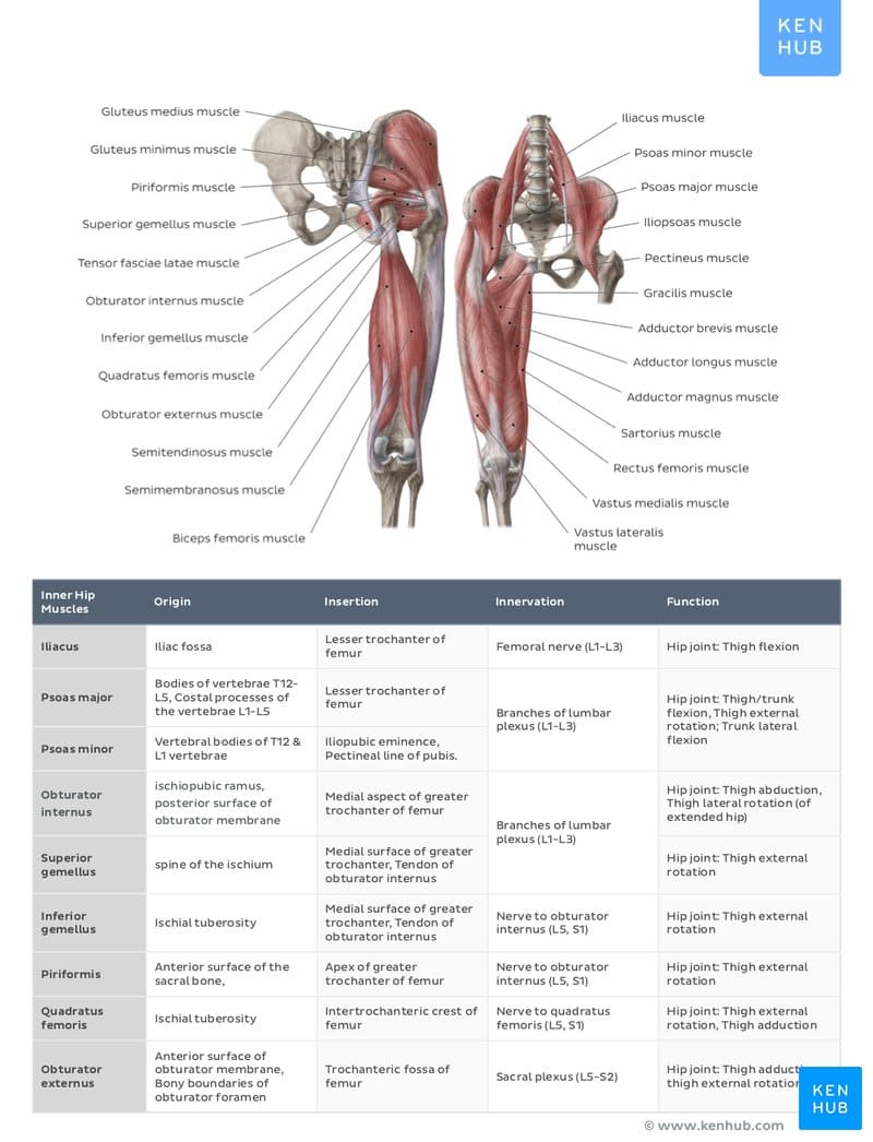
5.1 Anterior Thigh Muscles (Quadriceps Femoris)
Four muscles sharing a common insertion on the tibial tuberosity via the patellar ligament. All are supplied by the Femoral nerve (L2-L4).
- Rectus femoris: Extends knee and flexes hip.
- Vastus lateralis: Extends knee.
- Vastus medialis: Extends knee, stabilizes patella.
- Vastus intermedius: Extends knee.
5.2 Posterior Thigh Muscles (Hamstrings)
Responsible for hip extension and knee flexion, innervated by the Sciatic nerve.
| Muscle | Origin | Insertion | Action |
|---|---|---|---|
| Biceps femoris | Ischial tuberosity (long), Linea aspera (short) | Head of fibula, lateral tibial condyle | Extends hip, flexes knee, laterally rotates leg |
| Semitendinosus | Ischial tuberosity | Proximal medial tibia (Pes anserinus) | Extends hip, flexes knee, medially rotates leg |
| Semimembranosus | Ischial tuberosity | Posterior medial condyle of tibia | Extends hip, flexes knee, medially rotates leg |
6. Clinical Relevance
The SITS muscles (Supraspinatus, Infraspinatus, Teres minor, Subscapularis) are common sites of injury. Supraspinatus tears are the most frequent, often occurring due to impingement under the acromion.
- Radial Nerve Injury: Results in wrist drop due to paralysis of extensors.
- Femoral Nerve Palsy: Results in an inability to extend the knee and a loss of the patellar reflex.
- Sciatic Nerve Injury: Can result in foot drop if the common fibular division is affected.
Injury to CN VII paralyzes all facial muscles on the affected side (Bell's Palsy). This must be distinguished from a stroke (Upper Motor Neuron lesion), which spares the forehead because the forehead receives bilateral cortical innervation.
Summary Key Points
- Facial expression muscles are unique for inserting into skin (CN VII).
- Mastication muscles move the mandible (CN V3).
- The Rotator cuff provides essential stability to the glenohumeral joint.
- The Quadriceps extend the knee; Hamstrings flex the knee and extend the hip.
- Understanding nerve supply is required to predict motor deficits after trauma.
