Functions and Types of Muscles
A comprehensive anatomical and physiological study of skeletal, smooth, and cardiac muscle tissue, detailing their structural characteristics, functional roles, and clinical significance in medical practice.
1. Introduction
Muscle tissue is a specialized tissue designed for contraction and is responsible for nearly all movement in the human body. Beyond locomotion, it performs vital roles in stability, heat generation, and the internal transport of substances. There are three distinct histological types: Skeletal, Smooth, and Cardiac.
2. Functions of Muscle Tissue
- Movement: Skeletal muscles produce voluntary movement by pulling on bones at joints (locomotion, facial expression). Smooth muscle handles involuntary movements like peristalsis and vascular constriction. Cardiac muscle provides rhythmic contractions to pump blood.
- Maintenance of Posture: Continuous contraction of skeletal muscles (specifically in the back, neck, and legs) maintains body position against gravity. These muscles are rich in Type I (slow-twitch) fibers.
- Joint Stabilization: Muscles provide dynamic stability by compressing joints during contraction. The rotator cuff muscles are a prime example of stabilizers for the glenohumeral joint.
- Heat Production: Muscle contraction is an exothermic process. Skeletal muscle accounts for approximately 85% of heat production during physical activity. Shivering is a mechanism of rapid, involuntary contraction to prevent hypothermia.
- Protection and Support: Abdominal muscles protect viscera, and pelvic floor muscles support pelvic organs.
Muscle tissue is not just for movement; it is a metabolic engine that generates heat and a structural component that stabilizes the skeletal framework and protects internal organs.
3. Skeletal Muscle
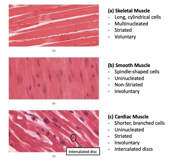
Skeletal muscle constitutes approximately 40% of total body weight. It is characterized as striated voluntary muscle.
3.1 Structure and Connective Tissue
Skeletal muscle fibers are long, cylindrical, multinucleated cells with nuclei located peripherally. They range from 10 to 100 micrometers in diameter.
- Striations: Alternating light and dark bands caused by the arrangement of actin (thin) and myosin (thick) filaments within sarcomeres.
- Epimysium: Surrounds the entire muscle.
- Perimysium: Surrounds bundles of fibers called fascicles.
- Endomysium: Surrounds individual muscle fibers.
3.2 Fiber Types
| Feature | Type I (Slow-twitch) | Type II (Fast-twitch) |
|---|---|---|
| Metabolism | Oxidative (Aerobic) | Glycolytic (Anaerobic) |
| Fatigue Resistance | High | Low |
| Color | Red (High myoglobin) | White/Pale (Low myoglobin) |
| Specialization | Endurance, posture | Explosive, short-duration power |
4. Smooth Muscle
Smooth muscle is found in the walls of hollow organs and blood vessels. It is non-striated and involuntary.
4.1 Structure and Control
Cells are spindle-shaped (fusiform) with a single central nucleus. They lack sarcomeres; instead, actin and myosin are anchored to dense bodies scattered throughout the cytoplasm and cell membrane.
4.2 Types of Smooth Muscle
- Single-unit (Visceral): Cells are connected by gap junctions and contract as a coordinated unit. Found in the stomach, intestines, and uterus.
- Multi-unit: Cells are not electrically connected and contract independently. Found in the iris of the eye, ciliary body, and arrector pili muscles.
5. Cardiac Muscle
Forms the myocardium. It combines features of both skeletal (striated) and smooth (involuntary) muscle.
5.1 Intercalated Discs
Cardiac muscle cells (cardiomyocytes) are branched and connected by intercalated discs, which contain:
- Desmosomes: Mechanical attachments.
- Gap Junctions: Allow rapid electrical coupling for synchronized contraction of the myocardium.
5.2 Unique Properties
- Automaticity: The ability to generate spontaneous action potentials (SA and AV nodes).
- Rhythmicity: Regular, repeating patterns of contraction.
- Refractory Period: A long refractory period prevents tetanic contraction, ensuring the heart has time to fill between beats.
6. Comparison of Muscle Types
| Feature | Skeletal Muscle | Cardiac Muscle | Smooth Muscle |
|---|---|---|---|
| Striations | Present | Present | Absent |
| Nuclei | Multiple, peripheral | Single/Double, central | Single, central |
| Control | Voluntary (Somatic) | Involuntary (Autonomic) | Involuntary (Autonomic) |
| Regeneration | Limited (Satellite cells) | None (Scar tissue forms) | Capable |
7. Clinical Relevance
Muscular Dystrophies
Inherited disorders like Duchenne muscular dystrophy (dystrophin gene mutation) lead to progressive skeletal muscle degeneration and severe weakness beginning in childhood.
Hypertension
Increased smooth muscle tone (vasoconstriction) in arteriole walls increases peripheral resistance. Medications like calcium channel blockers work by reducing this contraction.
Myocardial Infarction (MI)
Ischemic death of cardiomyocytes results in irreversible damage. Because cardiac muscle cannot regenerate, the area is replaced by non-contractile fibrous scar tissue, potentially leading to heart failure.
Asthma
Smooth muscle hyperreactivity in the bronchi leads to bronchospasm. Beta-2 agonists (bronchodilators) are used to relax this smooth muscle and open the airways.
Skeletal muscle has some regenerative capacity via satellite cells, but cardiac muscle lacks this ability, making heart injuries permanent. Smooth muscle is the only type capable of significant hyperplasia and regeneration.
