Definition of Key Terms: Muscular System
Comprehensive anatomical study of the fundamental terminology of the muscular system, focusing on the hierarchical organization of skeletal muscle, the functional concepts of origin and insertion, and clinical applications for diagnosis and rehabilitation.
1. Introduction
Understanding the fundamental terminology of the muscular system is essential for clinical practice. The terms muscle, origin, and insertion form the foundation upon which all knowledge of muscle anatomy, physiology, and pathology is built. These concepts enable the clinician to predict muscle action, identify sites of injury, and plan appropriate rehabilitation strategies.
2. Definition of Muscle
A muscle is a soft tissue structure composed of elongated cells (muscle fibers) that have the specialized ability to contract and produce force. Muscle tissue is one of the four primary tissue types in the human body. It is responsible for movement, maintenance of posture, joint stabilization, heat production, and organ protection.
Histological Classification
Muscles are classified into three distinct histological types:
- Skeletal Muscle: Typically attached to bones via tendons; under voluntary control.
- Smooth Muscle: Found in the walls of hollow organs and blood vessels; operates involuntarily.
- Cardiac Muscle: Forms the walls of the heart; functions involuntarily.
2.1 Structural Organization of a Skeletal Muscle
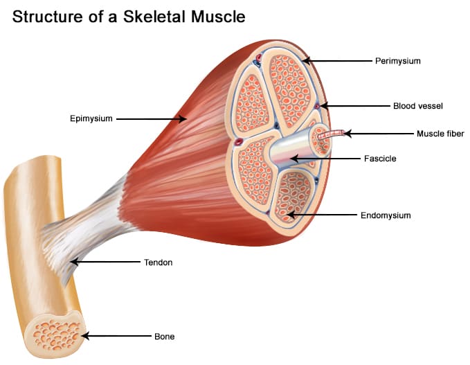
A skeletal muscle exhibits a hierarchical organization from the gross to the microscopic level:
- Muscle (Whole Organ): The complete muscle is surrounded by a dense irregular connective tissue sheath called the epimysium. This layer protects the muscle from friction against surrounding structures.
- Fascicle: The muscle is internally divided into bundles of muscle fibers called fascicles. Each fascicle is surrounded by the perimysium, which contains blood vessels and nerves.
- Muscle Fiber (Muscle Cell): Individual cells within the fascicle, surrounded by the endomysium, a delicate layer of areolar connective tissue.
- Myofibril: Thread-like structures within each fiber containing contractile proteins (actin and myosin) arranged in units called sarcomeres.
2.2 Muscle Attachments
Muscles attach to bones through two primary structures:
- Tendon: A tough, cord-like band of dense regular connective tissue. Tendons are highly resistant to tension and efficiently transmit contractile force to the skeleton.
- Aponeurosis: A broad, flat sheet of connective tissue that serves the same function as a tendon.
- A muscle is a contractile tissue composed of fibers organized into fascicles.
- The epimysium, perimysium, and endomysium surround the whole muscle, fascicles, and fibers, respectively.
- Tendons connect muscles to bones and transmit force.
3. Definition of Origin
The origin of a muscle is the attachment site that remains relatively fixed during muscle contraction. It serves as the anchor point from which the muscle pulls.
3.1 Characteristics of the Origin
- Proximal Location: Usually located on the bone closer to the axial skeleton. (Example: Biceps brachii origin is on the scapula).
- Greater Stability: The bone at the origin site is typically larger and more stable, providing a firm anchor for contraction.
- Multiple Origins: Some muscles have more than one. These are named by the number of heads: bi- (2), tri- (3), or quadri- (4).
4. Definition of Insertion
The insertion of a muscle is the attachment site that moves during muscle contraction. It is typically the distal attachment, farther from the trunk or center of the body.
4.1 Characteristics of the Insertion
- Distal Location: Usually located on the bone farther from the axial skeleton. (Example: Gastrocnemius insertion is on the calcaneus).
- Greater Mobility: The bone at the insertion site is more mobile, allowing the muscle to produce movement at the joint.
- Reversibility of Action: Roles can reverse depending on the movement. In a pull-up, the insertion (hand) is fixed, and the origin (trunk) moves toward the insertion.
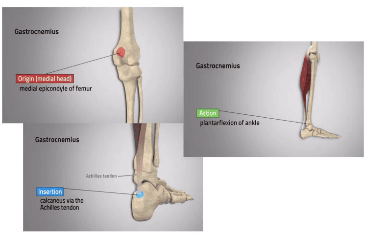
Origin and insertion are functional concepts rather than absolute anatomical designations, as their roles can reverse based on stabilizing forces or gravity.
5. Related Anatomical Terms
- Muscle Belly: The thick, central contractile portion of the muscle located between the origin and insertion. It is the part that bulges or shortens during contraction.
- Tendon: Primarily composed of parallel collagen fibers. They are relatively avascular and contain Golgi tendon organs to detect tension and prevent injury.
- Ligament: Dense regular connective tissue connecting bone to bone. Their function is to stabilize joints and limit excessive movement.
- Fascicle: A bundle of muscle fibers surrounded by perimysium. Arrangements include parallel, fusiform, pennate, circular, and convergent.
- Muscle Fiber: Multi-nucleated cells containing myofibrils. They range from 10 to 100 micrometers in diameter.
6. Summary Table: Key Terms
| Term | Definition | Clinical Relevance |
|---|---|---|
| Muscle | Contractile tissue composed of fibers producing force. | Target of rehabilitation; site of strains/tears. |
| Origin | The relatively fixed, proximal attachment. | Less commonly injured; avulsions may occur. |
| Insertion | The movable, distal attachment. | Common site of tendinitis and avulsion fractures. |
| Tendon | Connective tissue connecting muscle to bone. | Site of tendon rupture. |
| Ligament | Connective tissue connecting bone to bone. | Site of sprains; crucial for stability. |
| Muscle Belly | The thick, contractile central portion. | Site of muscle strains and contusions. |
7. Clinical Relevance
Understanding muscle origins and insertions has direct clinical applications:
- Muscle Testing: Knowledge of attachments allows clinicians to test strength by placing a joint at a mechanical disadvantage (e.g., abducting the arm against resistance to test the deltoid).
- Injection Sites: Intramuscular injections are administered into the muscle belly to avoid major nerves and vessels.
- Avulsion Fractures: Occur when a tendon or ligament pulls a fragment of bone away. Common at the tibial tuberosity (patellar ligament insertion) or ischial tuberosity (hamstring origin).
- Surgical Approaches: Surgeons must plan incisions to preserve muscle function and avoid important neurovascular structures based on attachment maps.
- The origin is fixed/proximal; the insertion is movable/distal.
- Avulsion fractures commonly occur at muscle insertion sites.
- Knowledge of attachments is essential for surgical planning and injection technique.
