Congenital Malformations
A Comprehensive Medical Reference covering Congenital Malformations, Causes, Critical Periods, and Clinical Management.
1. INTRODUCTION
A congenital malformation is defined as a structural abnormality present at birth that results from a disturbance in normal embryonic or fetal development. These abnormalities may affect any organ system and range from minor cosmetic defects to severe, life-threatening conditions. Congenital malformations are a leading cause of infant mortality and childhood morbidity worldwide, including in Uganda.
The study of congenital malformations (teratology) is essential for clinical medicine practitioners. Understanding the timing, mechanisms, and causes of developmental disruptions enables prevention, early detection, and appropriate management. Many congenital malformations are preventable through prenatal care, nutritional supplementation, and avoidance of teratogenic exposures.
2. EPIDEMIOLOGY
Globally, congenital anomalies affect approximately 3-6% of all live births. The World Health Organization estimates that approximately 240,000 newborns die worldwide within 28 days of birth each year due to congenital anomalies. In low- and middle-income countries, including Uganda, the burden is compounded by limited access to prenatal screening, surgical services, and specialized neonatal care.
The most common congenital malformations include congenital heart defects, neural tube defects, cleft lip and palate, clubfoot, and Down syndrome. The prevalence varies by geographic region, genetic background, maternal nutrition, and environmental exposures.
In Uganda, neural tube defects and cleft lip/palate are among the most frequently encountered major congenital anomalies in clinical practice.
3. CAUSES OF CONGENITAL MALFORMATIONS
The causes of congenital malformations are traditionally categorized into genetic, environmental, and multifactorial etiologies. However, in many cases, the exact cause remains unknown.
3.1 Genetic Causes
- Chromosomal abnormalities: Involve alterations in chromosome number or structure. Aneuploidy (abnormal chromosome number) includes trisomies such as Down syndrome (trisomy 21), Edwards syndrome (trisomy 18), and Patau syndrome (trisomy 13). Monosomy X (Turner syndrome) and Klinefelter syndrome (XXY) are sex chromosome aneuploidies. Structural abnormalities include deletions, duplications, inversions, and translocations.
- Single gene mutations: Follow Mendelian inheritance patterns. Autosomal dominant disorders include achondroplasia and Marfan syndrome. Autosomal recessive disorders include cystic fibrosis and sickle cell disease. X-linked disorders include hemophilia and Duchenne muscular dystrophy.
- Polygenic inheritance: Involves multiple genes contributing to a phenotype. Many common malformations, including cleft lip with or without cleft palate and congenital heart defects, demonstrate polygenic inheritance with environmental interaction.
3.2 Environmental Causes (Teratogens)
Teratogens are environmental agents that cause structural or functional abnormalities in the developing embryo or fetus. The effect of a teratogen depends on the timing of exposure, dose, duration, and genetic susceptibility of the embryo.
3.2.1 Infectious Teratogens
- Rubella virus: Infection during the first trimester causes congenital rubella syndrome, characterized by cataracts, congenital heart defects (patent ductus arteriosus, pulmonary stenosis), sensorineural deafness, and microcephaly. Vaccination has dramatically reduced incidence in countries with immunization programs.
- Cytomegalovirus (CMV): The most common congenital viral infection. It causes microcephaly, intracranial calcifications, chorioretinitis, sensorineural hearing loss, and hepatosplenomegaly.
- Toxoplasma gondii: Infection causes chorioretinitis, intracranial calcifications, hydrocephalus, and seizures. Transmission occurs through ingestion of undercooked meat or contact with cat feces.
- Zika virus: Infection during pregnancy causes microcephaly and other severe brain abnormalities. The virus is transmitted by Aedes mosquitoes, which are endemic in parts of Uganda.
- Syphilis (Treponema pallidum): Causes congenital syphilis with manifestations including hepatosplenomegaly, rash, anemia, jaundice, and bone abnormalities. Late manifestations include Hutchinson teeth, saddle nose, and interstitial keratitis.
3.2.2 Chemical and Drug Teratogens
- Alcohol: A potent teratogen. Fetal alcohol spectrum disorders (FASD) include fetal alcohol syndrome (FAS) characterized by growth retardation, facial dysmorphism (short palpebral fissures, smooth philtrum, thin upper lip), and central nervous system abnormalities. No safe level of alcohol consumption during pregnancy has been established.
- Antiepileptic drugs: Such as valproic acid and carbamazepine increase the risk of neural tube defects, craniofacial anomalies, and cardiac defects. Folic acid supplementation reduces the risk in women taking antiepileptic medications.
- Thalidomide: A sedative used in the 1950s-60s, caused severe limb reduction defects (phocomelia). It remains a powerful example of drug teratogenicity and has been repurposed for treatment of leprosy and multiple myeloma with strict pregnancy prevention programs.
- Retinoids: (isotretinoin, etretinate) cause craniofacial, cardiac, thymic, and central nervous system defects. Isotretinoin is absolutely contraindicated in pregnancy.
- Warfarin: Causes fetal warfarin syndrome (nasal hypoplasia, stippled epiphyses, central nervous system abnormalities) when used in the first trimester.
3.2.3 Physical Teratogens
- Ionizing radiation: Exposure during pregnancy, particularly in the first trimester, increases the risk of microcephaly, growth retardation, and intellectual disability. The risk is dose-dependent, with diagnostic radiation typically below the teratogenic threshold.
- Hyperthermia: Maternal fever >38.9°C or hot tub use in the first trimester is associated with neural tube defects, microcephaly, and facial clefts.
3.2.4 Maternal Disease States
- Maternal diabetes mellitus: (Both pregestational and gestational) increases the risk of congenital heart defects, neural tube defects, caudal regression syndrome, and macrosomia. Strict glycemic control before conception and during early pregnancy significantly reduces risk.
- Maternal phenylketonuria (PKU): Causes microcephaly, growth retardation, congenital heart defects, and intellectual disability when maternal phenylalanine levels are elevated during pregnancy.
- Maternal hypothyroidism: Is associated with impaired neurodevelopment and may increase the risk of congenital anomalies.
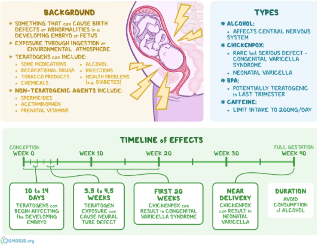
4. CRITICAL PERIODS OF DEVELOPMENT
The concept of critical periods is fundamental to understanding teratogenesis. A critical period is the specific time during development when an organ system is most susceptible to teratogenic insult. Exposure to a teratogen during an organ's critical period is most likely to produce a malformation of that organ.
The embryonic period (weeks 3-8 post-fertilization) is the most vulnerable period for teratogenic effects because all major organ systems begin development during this time.
- Pre-implantation period (weeks 1-2): Is characterized by the 'all-or-none' principle: teratogenic exposure typically causes either embryonic death or no effect, as the cells are still pluripotent.
- Fetal period (week 9 to birth): Is less susceptible to major structural malformations but remains vulnerable to functional abnormalities and growth disturbances.
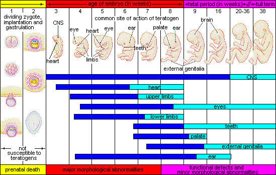
4.1 Critical Periods by Organ System
| Organ System | Critical Period | Common Teratogenic Effects |
|---|---|---|
| Central nervous system | Weeks 3-5 (neurulation) | Neural tube defects, microcephaly |
| Heart | Weeks 4-9 | Septal defects, tetralogy of Fallot |
| Upper limbs | Weeks 4-7 | Limb reduction defects, polydactyly |
| Lower limbs | Weeks 4-8 | Clubfoot, limb reduction defects |
| Palate | Weeks 6-9 | Cleft palate |
| Lip | Weeks 5-7 | Cleft lip |
| External genitalia | Weeks 7-12 (male) | Hypospadias, ambiguous genitalia |
| Teeth | Weeks 8-20 | Enamel hypoplasia, missing teeth |
| Ear | Weeks 4-9 | Microtia, hearing loss |
| Eye | Weeks 4-8 | Microphthalmia, cataracts |
5. COMMON CONGENITAL MALFORMATIONS
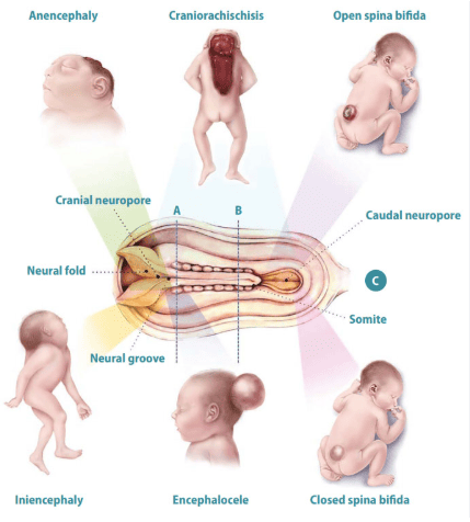
5.1 Neural Tube Defects (NTDs)
Neural tube defects result from failure of the neural tube to close completely during neurulation (weeks 3-4). They are among the most common and serious congenital malformations of the central nervous system. The incidence varies globally, with higher rates reported in certain regions.
- Anencephaly: The most severe NTD, characterized by absence of the cranial vault and cerebral hemispheres. It results from failure of the anterior neuropore to close. The condition is incompatible with life; affected infants are either stillborn or die shortly after birth.
- Spina bifida: Results from failure of the posterior neuropore to close. Spina bifida occulta is the mildest form, with a small vertebral defect covered by skin and often asymptomatic. Spina bifida cystica (meningocele and myelomeningocele) involves protrusion of the meninges or spinal cord through the vertebral defect. Myelomeningocele causes paralysis, sensory loss, bladder and bowel dysfunction, and hydrocephalus.
- Encephalocele: A herniation of brain tissue and meninges through a skull defect, most commonly in the occipital region. Surgical repair is possible, but neurological outcomes vary.
Folic acid supplementation (400 micrograms daily) before conception and during the first trimester reduces the risk of NTDs by 50-70%. Food fortification programs have successfully reduced NTD incidence in many countries.
5.2 Cleft Lip and Cleft Palate
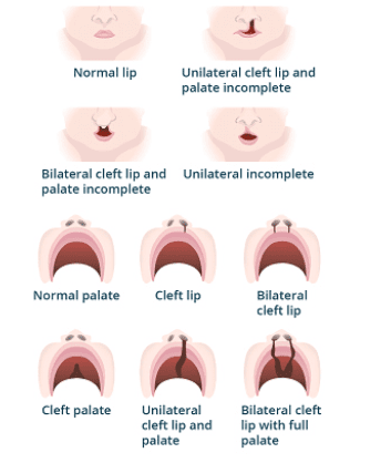
Cleft lip and cleft palate are among the most common craniofacial malformations worldwide. They result from failure of fusion of the facial processes during embryonic development (weeks 5-9).
- Cleft lip occurs when the maxillary process fails to fuse with the medial nasal elevation.
- Cleft palate occurs when the palatal shelves fail to fuse at the midline.
Cleft lip may be unilateral or bilateral and may occur with or without cleft palate. Isolated cleft palate without cleft lip has a different embryological origin and genetic association. The malformation causes feeding difficulties, speech impairment, dental problems, and psychosocial challenges.
Surgical repair of cleft lip is typically performed at 3-6 months of age, and cleft palate repair at 9-18 months. Multidisciplinary care including speech therapy, orthodontics, and psychological support is essential. Risk factors include genetic predisposition (polygenic inheritance), maternal smoking, alcohol consumption, anticonvulsant use, and folic acid deficiency.
5.3 Congenital Heart Defects
Congenital heart defects (CHDs) are the most common type of congenital malformation, affecting approximately 8 per 1,000 live births. They result from disturbances in cardiac development during weeks 4-9 of gestation.
- Ventricular septal defect (VSD): Is the most common CHD, involving a defect in the interventricular septum. Small defects may close spontaneously; large defects cause heart failure and require surgical repair.
- Atrial septal defect (ASD): Involves a defect in the interatrial septum. It may be asymptomatic in childhood but can cause pulmonary hypertension and heart failure in adulthood if unrepaired.
- Tetralogy of Fallot: Consists of four defects: VSD, pulmonary stenosis, overriding aorta, and right ventricular hypertrophy. It causes cyanosis and requires surgical correction.
- Patent ductus arteriosus (PDA): Is the persistence of the fetal ductus arteriosus after birth. In premature infants, it may close with medical management (indomethacin); persistent PDA requires surgical or catheter closure.
- Coarctation of the aorta: Is a narrowing of the aortic arch, typically distal to the left subclavian artery. It causes hypertension in the upper extremities and weak pulses in the lower extremities.
5.4 Limb Defects
Congenital limb defects result from disturbances in limb bud development (weeks 4-8). They range from minor digital abnormalities to complete limb absence.
- Clubfoot (talipes equinovarus): Is the most common congenital limb defect, affecting approximately 1 in 1,000 live births. The foot is twisted inward and downward. Treatment involves the Ponseti method (serial casting and manipulation), with excellent outcomes when initiated early.
- Polydactyly (extra digits) and syndactyly (fused digits) are common digital anomalies. They may occur as isolated findings or as part of a syndrome. Surgical correction is typically performed in infancy.
- Amelia (complete absence of a limb) and phocomelia (short, flipper-like limbs) are severe limb reduction defects. Thalidomide is a well-known cause of phocomelia.
5.5 Down Syndrome (Trisomy 21)
Down syndrome is the most common chromosomal abnormality, occurring in approximately 1 in 700 live births. It is caused by trisomy of chromosome 21 (nondisjunction during meiosis). Risk increases with advanced maternal age.
Characteristic features include intellectual disability, hypotonia, flat facial profile, upslanting palpebral fissures, epicanthal folds, single transverse palmar crease, and congenital heart defects (AV septal defect, VSD). Associated conditions include hypothyroidism, leukemia, and early-onset Alzheimer disease.
Prenatal screening includes first-trimester combined screening (nuchal translucency, maternal serum PAPP-A and free beta-hCG) and second-trimester quadruple screening. Definitive diagnosis requires chorionic villus sampling or amniocentesis.
6. PREVENTION AND MANAGEMENT
6.1 Primary Prevention
Primary prevention aims to prevent the occurrence of congenital malformations. Key strategies include:
- Folic acid supplementation — 400 micrograms daily beginning before conception and continuing through the first trimester reduces NTD risk by 50-70%.
- Rubella immunization — ensuring immunity before pregnancy.
- Diabetes management — optimizing glycemic control before conception.
- Avoidance of teratogens — alcohol, tobacco, illicit drugs, and unnecessary medications.
- Genetic counseling — for couples with known genetic conditions or family history of congenital anomalies.
6.2 Secondary Prevention
Secondary prevention involves early detection through prenatal screening and diagnosis. Ultrasound screening at 11-14 weeks (nuchal translucency) and 18-22 weeks (anomaly scan) can detect many major malformations. Maternal serum screening identifies pregnancies at increased risk for chromosomal abnormalities and NTDs. Non-invasive prenatal testing (NIPT) analyzes cell-free fetal DNA in maternal blood to screen for common aneuploidies with high accuracy.
6.3 Management of Affected Infants
Management requires a multidisciplinary approach. Surgical correction is indicated for many structural defects including cleft lip/palate, CHDs, NTDs, and limb defects. Medical management addresses associated complications such as heart failure, infections, and endocrine dysfunction. Rehabilitation services including physiotherapy, occupational therapy, and speech therapy optimize functional outcomes. Psychosocial support for families is essential, particularly in resource-limited settings.
6.4 Public Health Considerations in Uganda
In Uganda, challenges in congenital malformation management include limited access to prenatal ultrasound, shortage of pediatric surgeons and specialized centers, high cost of surgical care, and limited newborn screening programs. Community health education on folic acid supplementation, rubella vaccination, and avoidance of alcohol during pregnancy is critical. Integration of congenital anomaly surveillance into existing health information systems would improve data quality and inform policy.
7. SUMMARY TABLE: COMMON CONGENITAL MALFORMATIONS
| Malformation | Embryological Basis | Critical Period | Key Features | Prevention/Management |
|---|---|---|---|---|
| Anencephaly | Failure of anterior neuropore closure | Week 3-4 | Absent cranial vault, incompatible with life | Folic acid supplementation |
| Spina bifida | Failure of posterior neuropore closure | Week 3-4 | Vertebral defect, neurological deficits | Folic acid; surgical repair |
| Cleft lip | Failure of maxillary-medial nasal fusion | Week 5-7 | Unilateral/bilateral lip defect | Surgical repair at 3-6 months |
| Cleft palate | Failure of palatal shelf fusion | Week 6-9 | Oro-nasal communication | Surgical repair at 9-18 months |
| VSD | Septal development disturbance | Week 4-9 | Left-to-right shunt, heart failure | Surgical closure if large |
| Clubfoot | Limb bud development disturbance | Week 4-8 | Inward, downward foot deformity | Ponseti method (casting) |
| Down syndrome | Trisomy 21 (nondisjunction) | Pre-conception | Intellectual disability, characteristic facies, CHD | Screening; supportive care |
| Fetal alcohol syndrome | Alcohol teratogenicity | Throughout pregnancy | Growth retardation, facial dysmorphism, CNS defects | Alcohol abstinence |
8. KEY POINTS
- Congenital malformations are structural abnormalities present at birth resulting from disturbed embryonic or fetal development.
- Causes include genetic factors (chromosomal abnormalities, single gene mutations), environmental teratogens, and multifactorial inheritance.
- The embryonic period (weeks 3-8) is the most vulnerable time for teratogenic effects because all major organ systems develop during this period.
- Common teratogens include alcohol, antiepileptic drugs, retinoids, rubella virus, CMV, toxoplasmosis, and ionizing radiation.
- Neural tube defects result from failure of neural tube closure; folic acid supplementation reduces risk by 50-70%.
- Cleft lip and palate result from failure of facial process fusion; surgical repair is highly effective with multidisciplinary care.
- Congenital heart defects are the most common malformations; many require surgical correction in infancy.
- Primary prevention through folic acid, rubella vaccination, diabetes control, and avoidance of teratogens is the most effective strategy.
- In Uganda, limited access to prenatal screening and specialized surgical care remains a significant challenge in managing congenital anomalies.
REFERENCES
1. Moore KL, Persaud TVN, Torchia MG. The Developing Human: Clinically Oriented Embryology. 11th Edition. Elsevier; 2020.
2. Sadler TW. Langman's Medical Embryology. 14th Edition. Wolters Kluwer; 2019.
3. World Health Organization. Congenital anomalies: Fact Sheet. WHO; 2023. https://www.who.int/news-room/fact-sheets/detail/birth-defects
4. Centers for Disease Control and Prevention. Birth Defects: Data and Statistics. CDC; 2023. https://www.cdc.gov/ncbddd/birthdefects/data.html
5. Uganda Ministry of Health. Annual Health Sector Performance Report 2022/2023. Government of Uganda; 2023.
6. Brent RL. Environmental causes of human congenital malformations: the pediatrician's role in dealing with these complex clinical problems caused by a multiplicity of environmental and genetic factors. Pediatrics. 2004;113(4 Suppl):957-968.
7. Mitchell LE, Adzick NS, Melchionne J, et al. Spina bifida. Lancet. 2004;364(9448):1885-1895.
8. Mossey PA, Little J. Epidemiology of orofacial clefts: an international perspective. In: Wyszynski DF, ed. Cleft Lip and Palate: From Origin to Treatment. Oxford University Press; 2002:127-158.
