Skin Appendages
Comprehensive study notes on the structure and functions of hair, nails, and exocrine glands. This guide explores the developmental origins, anatomical components, and clinical pathologies of the integumentary system's specialized structures.
1. OVERVIEW
Skin appendages are specialized structures derived from the epidermis and dermis that extend from the skin surface. They include hair, nails, and exocrine glands (eccrine, apocrine, and sebaceous). Each appendage has a distinct structure, developmental origin, and physiological function. Together, they enhance the protective, sensory, and regulatory capabilities of the integumentary system.
2. HAIR
Hair is a filamentous biomaterial composed primarily of keratin. It is present over most of the body surface, with the exception of the palms, soles, lips, glans penis, and labia minora. The average adult has approximately 5 million hair follicles.
2.1 Structure of the Hair Follicle
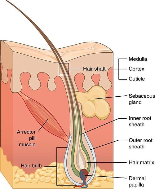
The hair follicle is an invagination of the epidermis into the dermis (and sometimes hypodermis) that produces the hair shaft. Key components include:
- Hair shaft: The visible portion above the skin surface, composed of three concentric layers: the medulla (central core), cortex (middle layer containing melanin and keratin), and cuticle (outer protective layer of overlapping keratinized cells).
- Hair root: The portion of the hair below the skin surface, anchored within the follicle.
- Hair bulb: The expanded base of the follicle containing the hair matrix—actively dividing cells that produce the hair shaft.
- Dermal papilla: A projection of connective tissue and capillaries at the base of the bulb that provides nutrition and regulates hair growth.
- Inner and outer root sheaths: Epithelial layers that surround and support the hair shaft during growth.
- Arrector pili muscle: A smooth muscle attached to the follicle that contracts to cause piloerection (goosebumps).
- Sebaceous gland: Associated with most hair follicles, secreting sebum into the follicular canal.
2.2 Hair Growth Cycle
Each hair follicle undergoes a cyclic pattern of growth and rest, independent of neighboring follicles. The cycle consists of three phases:
- Anagen (growth phase): Lasts 2–7 years for scalp hair. The matrix cells divide rapidly, and the hair shaft elongates approximately 1 cm per month. At any given time, 85–90% of scalp hairs are in anagen.
- Catagen (transitional phase): Lasts 2–3 weeks. Cell division ceases, the follicle shrinks, and the dermal papilla separates from the bulb. Approximately 1% of hairs are in catagen.
- Telogen (resting phase): Lasts 2–4 months. The hair remains anchored but does not grow. At the end of telogen, the hair is shed (exogen), and a new anagen hair begins to grow. Approximately 10–15% of hairs are in telogen.
2.3 Functions of Hair
- Protection: Scalp hair shields against UV radiation and mechanical trauma. Eyelashes and eyebrows protect the eyes from sweat, dust, and light. Nasal hairs filter airborne particles.
- Thermoregulation: Body hair traps air, providing insulation. Arrector pili contraction enhances this effect in mammals.
- Sensation: Hair follicles are richly innervated, detecting light touch and air movement.
- Social and sexual signaling: Hair distribution, color, and style convey age, sex, health status, and cultural identity.
Normal hair loss (shedding) is approximately 50–100 hairs per day. Excessive shedding (>150 hairs/day) or patchy hair loss indicates pathological alopecia requiring investigation.
3. NAILS
Nails are hard, keratinized plates on the dorsal surface of the distal phalanges. They protect the fingertips, enhance fine motor manipulation, and serve as a window to systemic health.
3.1 Structure of the Nail Unit
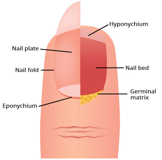
- Nail plate: The visible, translucent, keratinized structure composed of hard keratin (as opposed to the soft keratin of skin).
- Nail matrix (germinal matrix): The proximal portion beneath the proximal nail fold where nail plate cells divide and keratinize. The lunula (half-moon) is the visible distal portion of the matrix.
- Nail bed: The epithelium beneath the nail plate, containing parallel longitudinal blood vessels that give nails their pink color.
- Nail folds: The skin folds (proximal, lateral, distal) that surround and protect the nail plate margins.
- Eponychium (cuticle): The thin layer of stratum corneum that extends from the proximal nail fold onto the nail plate.
- Hyponychium: The thickened stratum corneum beneath the free edge of the nail that seals the subungual space.
- Nail root: The proximal portion of the nail plate embedded beneath the proximal nail fold.
3.2 Nail Growth
- Fingernails grow at approximately 3 mm per month; toenails at 1 mm per month.
- Complete replacement of a fingernail takes 4–6 months; a toenail takes 12–18 months.
- Growth rate is influenced by age, nutrition, season (faster in summer), and systemic disease.
3.3 Functions of Nails
- Protection: Shields the distal phalanges from trauma and enhances tactile discrimination.
- Manipulation: Acts as a rigid counterforce to the fingertip pad, enabling fine grasping and scratching.
- Diagnostic window: Nail changes (clubbing, spooning, pitting, discoloration) reflect systemic and dermatological diseases.
Beau lines (transverse grooves) indicate temporary arrest of nail matrix activity due to systemic illness, chemotherapy, or severe fever. They move distally as the nail grows, providing a timeline of the insult.
4. GLANDS
The skin contains three types of exocrine glands: eccrine sweat glands, apocrine sweat glands, and sebaceous glands. Each has a distinct structure, distribution, secretion mechanism, and physiological role.
4.1 Eccrine Sweat Glands
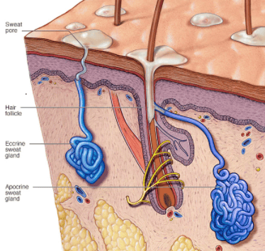
- Structure: Simple, coiled, tubular glands distributed over the entire body surface (2–4 million total).
- Secretory portion: Located in the deep dermis or superficial hypodermis; composed of clear cells (water secretion), dark cells (glycoprotein secretion), and myoepithelial cells (contraction to expel sweat).
- Duct: A straight channel lined by stratified cuboidal epithelium that reabsorbs NaCl; opens onto the skin surface via a sweat pore.
- Secretion: Watery, hypotonic sweat (pH 4.0–6.8) containing water, NaCl, urea, lactate, ammonia, and antimicrobial peptides (dermcidin).
- Function: Primary mechanism of thermoregulation via evaporative cooling. Also contributes to excretion and acid mantle maintenance.
4.2 Apocrine Sweat Glands
- Structure: Large, coiled glands that open into hair follicles in the axilla, areola, perianal region, and anogenital area.
- Development: Become functional at puberty under androgen stimulation.
- Secretion: Viscous, protein- and lipid-rich fluid (milky white) that is initially odorless.
- Odor: Bacterial decomposition of apocrine sweat produces characteristic body odor.
- Function: Pheromone secretion (possible role in social/sexual signaling); minimal thermoregulatory role.
4.3 Sebaceous Glands
- Structure: Simple, branched acinar glands associated with hair follicles (pilosebaceous unit) or existing independently (e.g., lips, glans penis, labia minora).
- Mechanism: Holocrine secretion—entire glandular cells disintegrate to release their contents.
- Secretion: Sebum—a complex lipid mixture of triglycerides, wax esters, squalene, and cholesterol esters.
- Function: Lubricates skin and hair, maintains stratum corneum hydration and flexibility, provides antimicrobial fatty acids, and transports fat-soluble antioxidants (vitamin E) to the skin surface.
- Activity: Increases at puberty under androgen stimulation; decreases with aging.
5. CLINICAL RELEVANCE
- Hirsutism: Excessive terminal hair growth in women in androgen-dependent areas (face, chest, abdomen), often due to polycystic ovary syndrome (PCOS) or adrenal disorders.
- Onychomycosis: Fungal infection of the nail plate and bed, causing thickening, discoloration, and crumbling.
- Hidradenitis suppurativa: Chronic inflammatory condition of apocrine gland-bearing skin (axilla, groin) characterized by painful nodules, abscesses, and sinus tracts.
- Acne vulgaris: Disorder of the pilosebaceous unit involving sebaceous gland hyperplasia, follicular hyperkeratinization, bacterial colonization (Cutibacterium acnes), and inflammation.
6. SUMMARY TABLE
| Appendage | Type/Structure | Distribution | Secretion | Primary Function |
|---|---|---|---|---|
| Hair | Keratinized filament; follicle with bulb, matrix | Most body surface (except palms, soles, lips) | Sebum (via associated glands) | Protection, thermoregulation, sensation |
| Nails | Hard keratin plate; matrix, bed, folds | Dorsal distal phalanges | None | Protection, manipulation, diagnostic indicator |
| Eccrine Glands | Coiled tubular glands | Entire body surface (2–4 million) | Watery, hypotonic sweat | Thermoregulation, excretion, anti-infective |
| Apocrine Glands | Large coiled glands | Axilla, areola, perianal, anogenital | Protein-rich, viscous fluid | Pheromone signaling; minimal thermoregulation |
| Sebaceous Glands | Branched acinar (holocrine) | Associated with hair follicles; face, scalp, trunk | Sebum (lipids, squalene, wax esters) | Lubrication, hydration, antimicrobial defense |
Conclusion: Skin appendages—hair, nails, and glands—are integral components of the integumentary system. Understanding their normal structure and function is essential for recognizing and managing disorders of these appendages.
