Structure of the Skin
Comprehensive medical notes on the integumentary system, detailing the histology, physiology, and clinical anatomy of the epidermis, dermis, and hypodermis.
1. INTRODUCTION
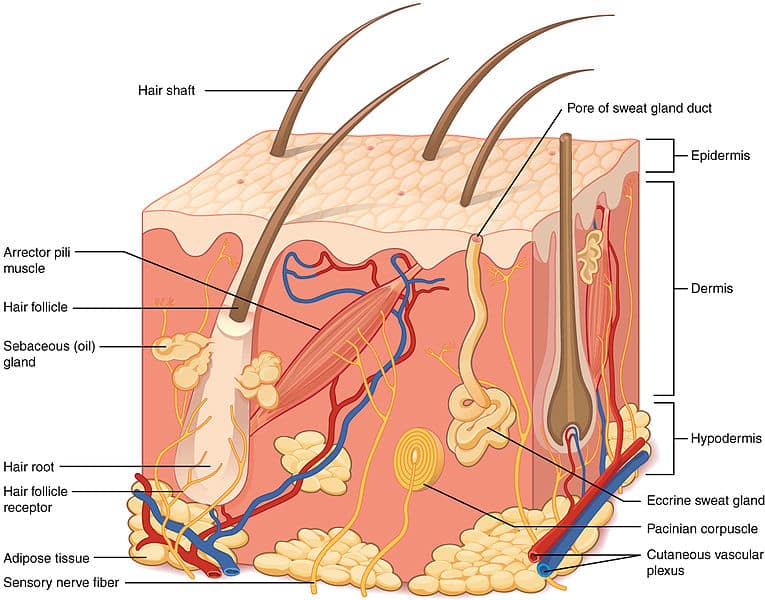
The integumentary system is the largest organ system of the human body, comprising the skin and its appendages (hair, nails, and glands). The skin alone covers approximately 1.5–2.0 m² in adults and accounts for about 15% of total body weight. It serves as the primary interface between the internal environment and the external world, performing critical protective, regulatory, and sensory functions.
The skin consists of two principal tissues: the epidermis (superficial epithelial layer) and the dermis (deeper connective tissue layer). Beneath the dermis lies the hypodermis (subcutaneous tissue), which is not technically part of the skin but is functionally integrated with it.
2. THE EPIDERMIS
The epidermis is the outermost layer of the skin, composed of stratified squamous keratinized epithelium. It is avascular and derives its nutrition by diffusion from the underlying dermis. The epidermis ranges in thickness from 0.05 mm (thin skin) to 1.5 mm (thick skin, e.g., palms and soles). It is continuously renewed through cell division in the basal layer, with complete turnover occurring approximately every 28–40 days.
2.1 Layers of the Epidermis
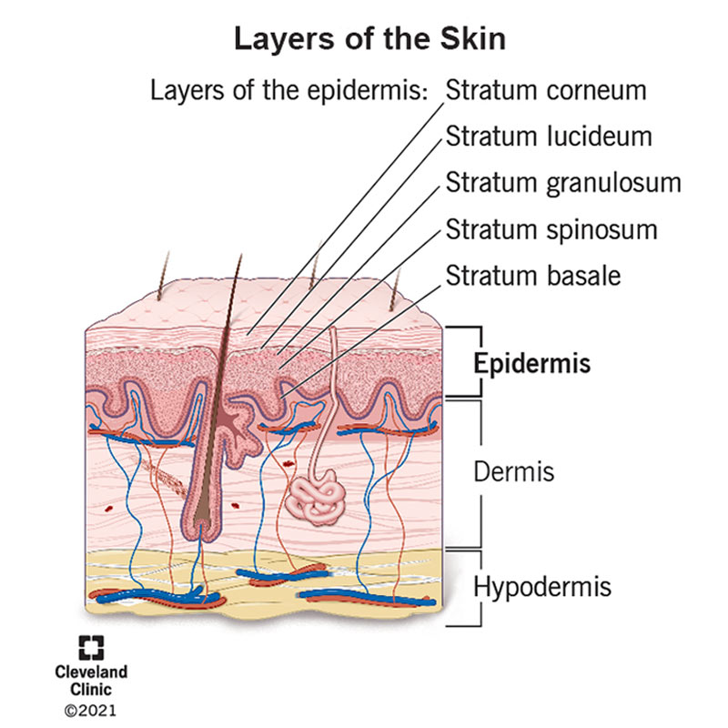
From superficial to deep, the epidermis consists of five distinct layers:
Stratum Corneum (Horny Layer)
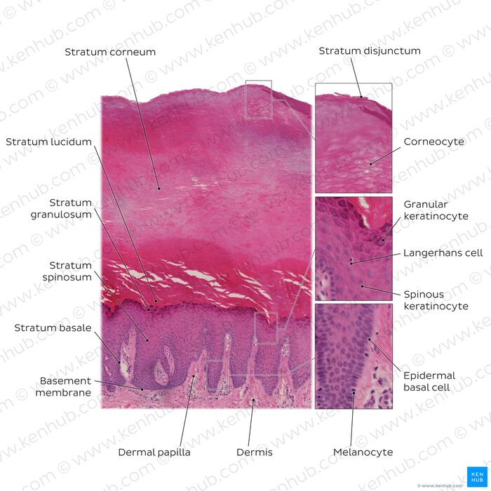
- Outermost layer, consisting of 15–30 layers of flattened, anucleate keratinized cells (corneocytes).
- Cells are filled with keratin and surrounded by a lipid-rich extracellular matrix.
- Provides the primary barrier against water loss, pathogens, and environmental toxins.
- Sheds continuously via desquamation; replaced by cells from deeper layers.
Stratum Lucidum (Clear Layer)
- Found only in thick skin (palms, soles, digits).
- Consists of 3–5 layers of flattened, dead keratinocytes with indistinct cell boundaries.
- Cells contain eleidin, a transparent substance derived from keratohyalin.
Stratum Granulosum (Granular Layer)
- Composed of 3–5 layers of keratinocytes containing prominent keratohyalin granules.
- Lamellar granules (Odland bodies) release lipids that form the waterproof barrier.
- Cells in this layer undergo apoptosis; nuclei and organelles begin to degenerate.
Stratum Spinosum (Prickle Cell Layer)
- Consists of 8–10 layers of polyhedral keratinocytes connected by desmosomes.
- Cells appear spiny under microscopy due to shrinkage artifacts around desmosomes.
- Contains Langerhans cells (dendritic antigen-presenting cells of the immune system).
- Site of initial keratin filament synthesis.
Stratum Basale (Basal Layer)
- Deepest epidermal layer, resting on the basement membrane.
- Contains a single layer of cuboidal to columnar stem cells (basal cells) that divide mitotically.
- Contains melanocytes (produce melanin) and Merkel cells (tactile sensation).
- Anchored to the basement membrane via hemidesmosomes.
2.2 Cell Types in the Epidermis
- Keratinocytes (90%): Produce keratin; form the structural framework.
- Melanocytes (5–10%): Synthesize melanin from tyrosine; transfer melanin to keratinocytes via cytocrine secretion.
- Langerhans cells (3–5%): Dendritic cells involved in immune surveillance and antigen presentation.
- Merkel cells (<1%): Mechanoreceptors associated with sensory nerve endings; release neurotransmitters.
The stratum corneum is the principal barrier to percutaneous drug absorption. Transdermal drug delivery systems exploit this property by using chemical enhancers or microneedles to bypass this layer.
3. THE DERMIS
The dermis is the thick, connective tissue layer beneath the epidermis. It provides structural support, nutrition, and sensory innervation to the epidermis. The dermis ranges from 1–4 mm in thickness and contains blood vessels, lymphatics, nerves, glands, and hair follicles.
3.1 Papillary Dermis
- Superficial layer, directly beneath the epidermis.
- Composed of loose areolar connective tissue with fine collagen (type III) and elastic fibers.
- Contains dermal papillae—finger-like projections that interdigitate with epidermal rete ridges.
- Rich in capillary loops, free nerve endings, and Meissner corpuscles (tactile receptors).
3.2 Reticular Dermis
- Deeper, thicker layer making up approximately 80% of dermal thickness.
- Composed of dense irregular connective tissue with thick collagen bundles (type I and III) and elastic fibers.
- Contains larger blood vessels, lymphatic vessels, nerve bundles, and skin appendages.
- Contains Pacinian corpuscles (pressure receptors) and Ruffini endings (stretch receptors).
3.3 Components of the Dermis
- Collagen fibers (70% of dermal dry weight): Provide tensile strength and resistance to tearing.
- Elastic fibers: Allow the skin to stretch and recoil; deficient in cutis laxa.
- Ground substance: Gel-like matrix of glycosaminoglycans (hyaluronic acid, dermatan sulfate) that hydrates the tissue.
- Fibroblasts: Principal cells that synthesize collagen, elastin, and ground substance.
- Mast cells: Release histamine and other mediators in allergic and inflammatory responses.
- Macrophages: Phagocytic cells involved in tissue repair and immune defense.
The dermo-epidermal junction (basement membrane) is critical for skin integrity. Genetic defects in hemidesmosomal proteins (e.g., collagen VII in dystrophic epidermolysis bullosa) result in blistering diseases.
4. THE HYPODERMIS (SUBCUTANEOUS TISSUE)
The hypodermis is the layer of loose connective tissue and adipose tissue beneath the dermis. It is not technically part of the skin but is functionally integrated with it.
4.1 Structure
- Composed primarily of adipocytes (fat cells) arranged in lobules separated by fibrous septa.
- Contains loose areolar connective tissue with collagen and elastic fibers.
- Thickness varies widely (1–10 cm) depending on body site, nutritional status, and sex.
4.2 Functions
- Energy storage: Adipose tissue is the body's primary energy reserve.
- Thermal insulation: Reduces heat loss and maintains core body temperature.
- Mechanical cushioning: Protects underlying structures from mechanical trauma.
- Anchorage: Attaches the skin to underlying fascia and muscles.
- Endocrine function: Adipose tissue secretes leptin, adiponectin, and other hormones.
5. THE DERMO-EPIDERMAL JUNCTION
The dermo-epidermal junction is a specialized basement membrane zone that anchors the epidermis to the dermis. It consists of three layers:
- Lamina lucida (clear layer): Contains laminin-332 and integrins that attach basal keratinocytes.
- Lamina densa (dense layer): Contains type IV collagen, laminin, and nidogen.
- Sublamina densa: Contains anchoring fibrils (type VII collagen), microfibrils, and dermal collagen.
Hemidesmosomes on basal keratinocytes connect to the lamina lucida via integrins, while anchoring fibrils secure the basement membrane to the dermis. Disruption of this junction results in blistering disorders.
6. CLINICAL RELEVANCE
| Condition | Pathophysiological Basis |
|---|---|
| Psoriasis | Accelerated epidermal turnover (3–5 days) results in thickened plaques with silvery scales. |
| Epidermolysis bullosa | Genetic defects in basement membrane proteins cause skin fragility and blistering. |
| Scleroderma | Excessive collagen deposition in the dermis leads to skin hardening and fibrosis. |
| Lipodystrophy | Loss of subcutaneous fat, leading to metabolic abnormalities. |
| Decubitus ulcers | Prolonged pressure damages the dermo-epidermal junction and underlying tissues. |
7. SUMMARY
The skin is a complex, multilayered organ with distinct structural and functional zones. The epidermis provides the primary barrier, the dermis offers structural support and vascularization, and the hypodermis provides insulation and cushioning. Understanding the normal anatomy of these layers is essential for diagnosing and managing dermatological conditions.
Skin Layer Summary Table
| Layer | Location | Principal Components | Key Functions |
|---|---|---|---|
| Epidermis | Outermost | Keratinocytes, melanocytes, Langerhans cells, Merkel cells | Barrier protection, UV filtration, immune surveillance, sensation |
| Dermis | Middle | Collagen, elastin, fibroblasts, blood vessels, nerves, glands | Structural support, nutrition, sensation, thermoregulation |
| Hypodermis | Deepest | Adipocytes, loose connective tissue | Energy storage, insulation, cushioning, anchorage |
