
The pharynx: subdivisions, wall architecture, musculature, deglutition & neurovascular supply
On this page
Overview & boundaries
The pharynx (from the Greek phárynx, meaning throat) is a funnel-shaped, fibromuscular passage situated behind the nasal cavities, the oral cavity, and the larynx. It functions as a shared anatomical crossroads where the respiratory pathway (airway) and the digestive pathway (foodway) intersect. Air inhaled through the nose travels downward and forward into the larynx, while ingested food and liquids propelled from the mouth pass downward and backward into the esophagus.

Anatomical Extent & Dimensions
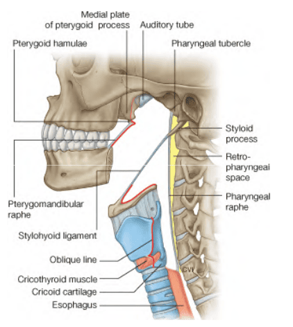
- Superior Boundary: Attached to the external base of the skull, extending across the basiocciput, petrous temporal bones, and medial pterygoid plates.
- Inferior Boundary: Terminates at the inferior border of the cricoid cartilage anteriorly, corresponding to the lower border of the sixth cervical vertebra (C6) posteriorly, where it continues directly as the cervical esophagus.
- Length & Tapering Caliber: Spans approximately 12 to 14 cm in length in adults. It is widest at its cranial skull base attachment (measuring approximately 3.5 cm transversely) and gradually narrows down to its narrowest point at the pharyngoesophageal junction (measuring approximately 1.5 cm), forming the primary physiological constriction of the alimentary tract.
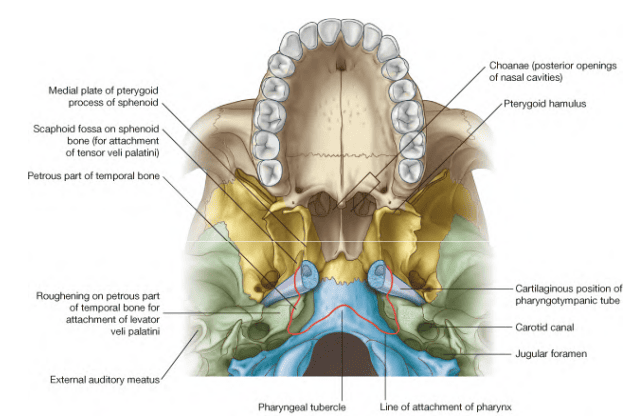
Skull Base Attachment Line
The cranial attachment of the pharynx follows an unbroken line across the basicranium on either side of the midline:
- Begins centrally at the pharyngeal tubercle on the basilar part of the occipital bone (basiocciput).
- Passes transversely across the petrous portion of the temporal bone, lying immediately anterior to the carotid canal and jugular foramen.
- Follows the cartilaginous portion of the pharyngotympanic (auditory / Eustachian) tube along the sphenopetrosal groove.
- Reaches the posterior border of the medial pterygoid plate of the sphenoid bone.
- Descends along the posterior margin of the medial pterygoid plate to the pterygoid hamulus, continuing onto the pterygomandibular raphe.
Layers of the pharyngeal wall
The pharyngeal wall is composed of five distinct concentric tissue layers arranged from inside (luminal surface) to outside:
The Five Concentric Layers
- 1. Mucous Membrane (Internal Lining): Lined by specialized epithelia continuous with the communicating cavities:
- Respiratory Epithelium: Ciliated pseudostratified columnar epithelium with goblet cells lines the nasopharynx.
- Protective Squamous Epithelium: Non-keratinized stratified squamous epithelium lines the oropharynx and laryngopharynx to resist mechanical abrasion during swallowing.
- 2. Submucosa: A loose vascular layer containing mixed seromucous glands, autonomic nerve terminals, and extensive collections of mucosa-associated lymphoid tissue (MALT).
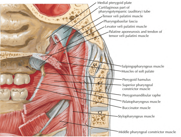
- 3. Pharyngobasilar Fascia (Fibrous Layer): A thick, tough fibrous membrane that lines the inner surface of the pharyngeal constrictor muscles. It provides structural integrity, anchors the pharyngeal wall to the cranial base, and fills the muscular defects where skeletal muscle is absent (notably the Sinus of Morgagni). It thickens posteriorly in the midline to blend with the median pharyngeal raphe.
- 4. Muscular Layer: Composed of striated skeletal muscle arranged into two distinct strata:
- An external circular layer comprising the superior, middle, and inferior pharyngeal constrictors.
- An internal longitudinal layer comprising the stylopharyngeus, palatopharyngeus, and salpingopharyngeus muscles.
- 5. Buccopharyngeal Fascia (External Sheath): A thin, fibroelastic fascial layer covering the outer surface of the muscular layer. Anteriorly, it is continuous with the fascia covering the buccinator muscle across the pterygomandibular raphe; superiorly, it blends into the skull base; inferiorly, it continues onto the adventitia of the esophagus. It houses the capillary and venous components of the pharyngeal plexus.
Posterior Relations & The Retropharyngeal Space
- Posterior Structures: The posterior pharyngeal wall rests against the cervical spine, separated by the prevertebral fascia, which encloses the prevertebral muscles (longus colli and longus capitis) and the cervical sympathetic trunk.
- The Retropharyngeal Space: A loose, potential fascial space filled with areolar tissue situated between the buccopharyngeal fascia anteriorly and the prevertebral fascia (specifically the alar layer) posteriorly.
Physiological Role: Provides a low-friction sliding plane that permits unrestricted elevation, depression, and lateral expansion of the pharynx and larynx during swallowing and neck flexion.
Clinical Hazard: Serves as a pathway for the spread of infection (“danger space”), allowing retropharyngeal abscesses to descend directly through the superior mediastinum into the posterior mediastinum.
The nasopharynx (epipharynx)
The nasopharynx is the uppermost division of the pharyngeal cavity, functioning exclusively as part of the respiratory tract. Unlike the oropharynx and laryngopharynx, its walls are rigid and non-collapsible, ensuring an open airway for nasal breathing.

Boundaries & Communications
- Superior Boundary (Roof): Formed by the sloping body of the sphenoid bone and the basilar part of the occipital bone.
- Inferior Boundary (Floor): Demarcated by the horizontal upper surface of the soft palate. Communicates with the oropharynx via the pharyngeal isthmus (closed during swallowing by elevation of the soft palate against Passavant’s ridge).
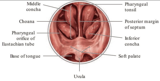
- Anterior Boundary: Communicates directly with the nasal cavities via the paired choanae (posterior internal nares). The choanae are separated in the midline by the rigid posterior free border of the bony nasal septum (formed by the vomer).
- Posterior Boundary: Slopes downward over the anterior arch of the atlas (C1) and the body of the axis (C2).
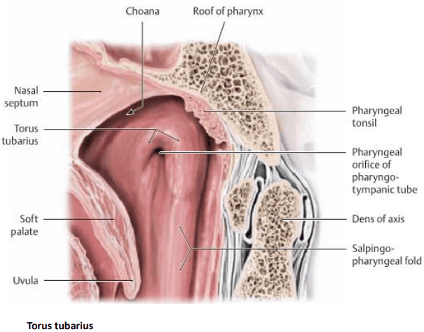
Anatomical Features of the Lateral Wall
- Pharyngeal Orifice of the Auditory Tube: A triangular or oval opening situated 1 to 1.5 cm behind the posterior end of the inferior nasal concha. The orifice points downward and forward, connecting the nasopharynx to the middle ear (tympanic cavity).
- Torus Tubarius: A prominent, hood-like cartilaginous elevation that overhangs the upper and posterior borders of the tubal orifice. It is produced by the projecting medial end of the cartilaginous auditory tube.
- Salpingopharyngeal Fold: A vertical mucosal fold descending from the posterior aspect of the torus tubarius into the lateral pharyngeal wall. It encloses the underlying salpingopharyngeus muscle.
- Salpingopalatine Fold: A smaller anterior fold running from the torus tubarius to the soft palate, overlying fibers of the tensor veli palatini.
- Torus Levatorius: A subtle ridge lying between the two folds, produced by the underlying levator veli palatini muscle as it enters the soft palate.
- Pharyngeal Recess (Fossa of Rosenmüller): A deep, slit-like lateral pouch situated immediately behind the torus tubarius and salpingopharyngeal fold.
Clinical Importance: The fossa of Rosenmüller is the most common site of origin for nasopharyngeal carcinoma (NPC). Due to its proximity to the petrous apex, tumors spreading from this recess can erode the foramen lacerum to invade the cavernous sinus, causing cranial nerve palsies (CN III, IV, V1, V2, and VI).
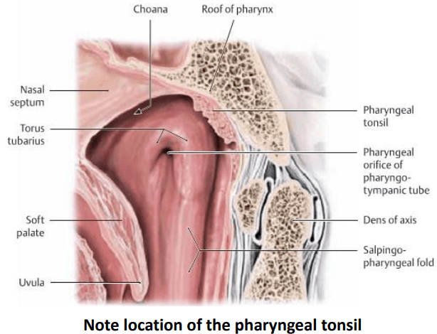
The Pharyngeal Tonsil (Adenoids)
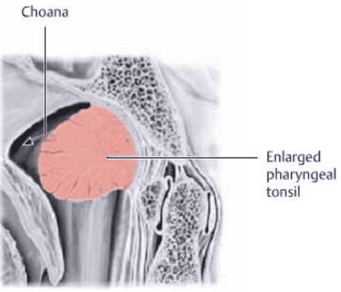
The pharyngeal tonsil is a median mass of subepithelial mucosa-associated lymphoid tissue (MALT) embedded within the mucous membrane at the junction between the roof and posterior wall of the nasopharynx.
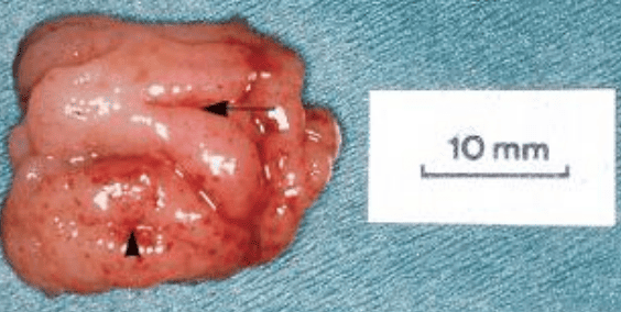
- Morphology: When fully developed in early childhood, it forms a truncated pyramid whose apex points anteriorly toward the nasal septum and whose base rests on the roof and posterior nasopharyngeal wall. Its mucosal surface features 4 to 6 prominent longitudinal ridges separated by deep clefts.
- Growth Kinetics: The pharyngeal tonsil is present at birth, undergoes rapid hyperplasia during early immune development, and reaches its maximal physiological size at approximately 5 years of age. Between 8 and 10 years of age, it undergoes progressive physiological involution, leaving minimal lymphoid remnants by adulthood.
The oropharynx (mesopharynx)
The oropharynx extends from the soft palate superiorly to the superior border of the epiglottis inferiorly. It communicates anteriorly with the oral cavity, serving both respiratory and digestive functions.
Boundaries & Architecture
- Superior Boundary (Roof): The inferior surface of the soft palate and the mobile uvula.
- Inferior Boundary (Floor): Formed by the posterior one-third of the tongue (pharyngeal part of the tongue, bearing the nodular lingual tonsil) and the interval between the tongue and epiglottis (glossoepiglottic folds and valleculae).
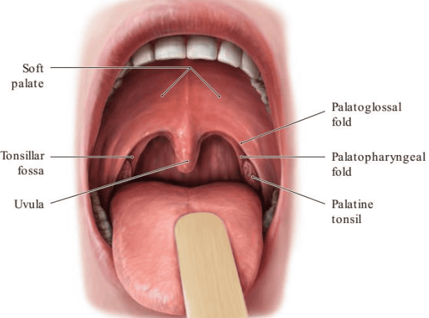
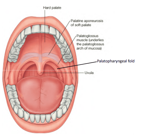
- Anterior Boundary: Communicates with the oral cavity through the oropharyngeal isthmus (isthmus of the fauces). This boundary is marked on either side by the palatoglossal fold (anterior pillar of the fauces), which encloses the palatoglossus muscle.
- Posterior Boundary: Formed by the mucosa covering the superior and middle pharyngeal constrictors, resting against the body of the axis (C2) and the third cervical vertebra (C3).
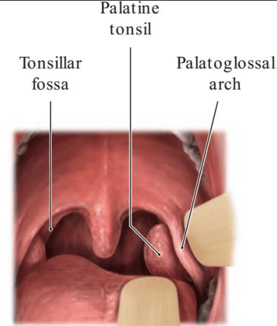
- Lateral Wall: Marked by the divergence of two vertical mucosal folds: the anterior palatoglossal arch and the posterior palatopharyngeal arch (posterior pillar, enclosing the palatopharyngeus muscle). Between these pillars lies the triangular tonsillar fossa (tonsillar sinus), which houses the palatine tonsil.
The Palatine Tonsil: Anatomy & Bed Relations
The palatine tonsil (commonly referred to simply as “the tonsil”) is an oval collection of lymphoid tissue situated in the submucosa of the lateral oropharyngeal wall. It does not completely fill the tonsillar sinus; a small crescentic depression remains above its superior pole, termed the supratonsillar fossa.
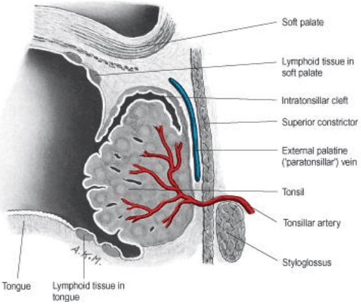
- Epithelial Crypts: The medial free surface is covered by non-keratinized stratified squamous epithelium invaginating to form 12 to 15 tonsillar crypts. The largest and deepest, the crypta magna (intratonsillar cleft), represents the embryological remnant of the second pharyngeal pouch.
- The Tonsillar Hemicapsule: The lateral deep surface is covered by a specialized, dense fibrous sheet called the hemicapsule, which separates the lymphoid tissue from the underlying muscular bed.
- The Peritonsillar Bed & Surgical Relations: Lateral to the fibrous capsule lies the loose areolar tissue of the peritonsillar space. Moving laterally from the capsule:
- The external palatine (paratonsillar) vein descends from the soft palate across the tonsillar bed; it is the primary culprit behind severe, immediate postoperative hemorrhage following tonsillectomy.
- The superior pharyngeal constrictor and styloglossus muscles form the muscular floor of the bed.
- The glossopharyngeal nerve (CN IX) courses anteriorly across the lower part of the bed, supplying sensory fibers to the tonsil and posterior tongue.
- The tortuous facial artery and its tonsillar branch lie deep to the superior constrictor.
- The internal carotid artery lies approximately 2.0 to 2.5 cm posterolateral to the tonsillar bed, safe from injury during dissection provided the surgical plane remains medial to the superior constrictor.

The laryngopharynx (hypopharynx)
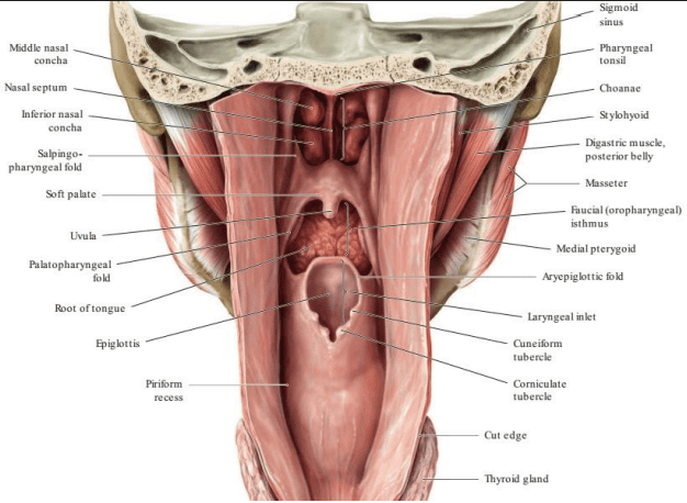
The laryngopharynx is the most caudal subdivision of the pharynx. It lies posterior to the laryngeal inlet and posterior laryngeal wall, spanning from the level of the superior border of the epiglottis down to the inferior border of the cricoid cartilage (C6 vertebral level).
Boundaries & Muscular Framework
- Superior Boundary: The plane passing through the upper border of the epiglottis and pharyngoepiglottic folds.
- Inferior Boundary: The inferior margin of the cricoid cartilage, where its muscular coat becomes continuous with the circular muscular wall of the esophagus.
- Anterior Wall: Features the elevated laryngeal inlet (aditus laryngis), flanked by the aryepiglottic folds, and the posterior aspects of the arytenoid and cricoid cartilages.
- Posterior & Lateral Walls: Formed by the overlapping sheets of the middle and inferior pharyngeal constrictor muscles, supported internally by the pharyngobasilar fascia and resting posteriorly against the prevertebral fascia covering C4, C5, and C6 vertebrae.

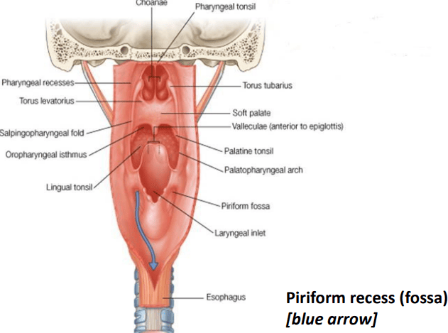
The Piriform Recess (Piriform Fossa)
The piriform recess is a deep, pear-shaped mucosal gutter situated on either side of the elevated laryngeal entrance:
- Medial Boundary: Formed by the sloping aryepiglottic fold and the lateral surfaces of the arytenoid and cricoid cartilages.
- Lateral Boundary: Formed by the inner mucosal surface of the thyroid cartilage and the overlying thyrohyoid membrane.
- Physiological Action: The paired recesses act as anatomical lateral bypass channels. During swallowing, food boluses and liquid streams are diverted around the raised laryngeal inlet into the piriform gutters, preventing aspiration before entering the esophageal lumen.
- Submucosal Neurovascular Hazard: Deep to the thin mucous membrane lining the floor of the recess run the internal laryngeal nerve (the primary sensory branch of the superior laryngeal nerve, CN X) and the superior laryngeal artery.
Clinical Hazard: Sharp swallowed foreign bodies (e.g., fine fish bones or chicken fragments) frequently lodge in the piriform recess. Attempts to retrieve them under direct vision or instrumentation carry a risk of puncturing the mucosa and injuring the internal laryngeal nerve, which causes ipsilateral loss of the protective cough reflex above the vocal cords and increases the risk of aspiration pneumonia.
External circular constrictors
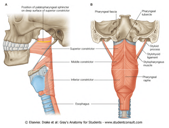
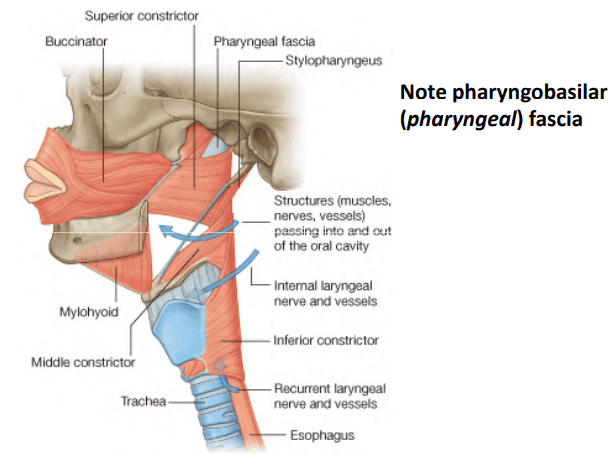
The external muscular coat of the pharynx consists of three paired, fan-shaped skeletal muscles whose fibers are predominantly circular: the superior, middle, and inferior pharyngeal constrictors.
The “Stacked Flower Pot” Arrangement & Pharyngeal Raphe
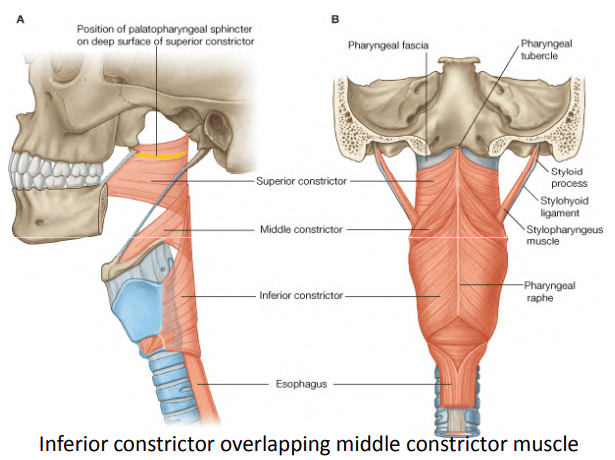
- Telescoping Overlap: The constrictor muscles overlap one another from below upward like a stack of nesting flower pots: the inferior constrictor overlaps the middle constrictor, which in turn overlaps the superior constrictor.
- The Median Pharyngeal Raphe: The muscle fibers from either side sweep posteromedially to interdigitate in the posterior midline at a vertical fibrous band: the median pharyngeal raphe. This raphe is anchored superiorly to the pharyngeal tubercle of the occipital bone and extends inferiorly to the level of the cricopharyngeus muscle, where it blends with the posterior wall of the esophagus.

1. Inferior Pharyngeal Constrictor Muscle
The thickest and most powerful of the three constrictors, originating from two cartilages of the larynx:
- Thyropharyngeus Part: Arises from the oblique line of the thyroid cartilage and a tendinous band crossing the cricothyroid muscle. Its fibers fan posteromedially, ascending obliquely to overlap the lower portion of the middle constrictor before inserting into the pharyngeal raphe.
- Cricopharyngeus Part: Arises from the lateral surface of the cricoid cartilage. Its fibers pass horizontally backward around the junction of the pharynx and esophagus without inserting into the raphe, forming an unbroken muscular ring. It functions as the physiological upper esophageal sphincter (UES), remaining tonically contracted at rest to prevent air entry into the stomach during respiration and prevent esophagopharyngeal reflux.
2. Middle Pharyngeal Constrictor Muscle
A fan-shaped muscle sheet originating from the hyoid apparatus:
- Origin: Arises from the entire length of the greater horn (cornu) and the lesser horn of the hyoid bone, as well as the lower portion of the stylohyoid ligament.
- Fiber Divergence: Its fibers diverge widely as they pass backward:
- The inferior fibers descend obliquely deep to the inferior constrictor.
- The middle fibers course horizontally to the raphe.
- The superior fibers ascend steeply to overlap the outer surface of the superior constrictor.

3. Superior Pharyngeal Constrictor Muscle
The thinnest and most cranial constrictor, arising from a continuous line of skeletal and ligamentous landmarks:
- Origin:
- The lower part of the posterior border of the medial pterygoid plate and the pterygoid hamulus.
- The pterygomandibular raphe (a tendinous band extending from the pterygoid hamulus to the posterior end of the mylohyoid line of the mandible, serving as the common junction between the buccinator muscle anteriorly and the superior constrictor posteriorly).
- The posterior end of the mylohyoid line of the mandible.
- The side of the root of the tongue.
- Insertion & The Sinus of Morgagni: The fibers curve backward to insert into the median pharyngeal raphe, with the uppermost fibers inserting directly into the pharyngeal tubercle of the skull base.
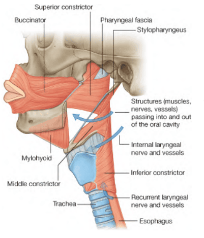
The Crescentic Defect: Because the free upper border of the superior constrictor does not reach the cranial base except in the posterior midline, a crescentic gap is left on either side: the Sinus of Morgagni. Here, the pharyngeal wall consists entirely of the dense pharyngobasilar fascia.
Structures Traversing the Gap: The auditory tube, the levator veli palatini muscle, the tensor veli palatini tendon, and the ascending palatine artery pass through this space into the nasopharynx.


Internal longitudinal elevators
Lying internal to the circular constrictor muscles is a longitudinal muscle stratum composed of three paired muscles: the stylopharyngeus, palatopharyngeus, and salpingopharyngeus. Their primary function is to elevate, shorten, and widen the pharynx and larynx during swallowing and phonation.

1. Stylopharyngeus Muscle
- Origin: Arises from the medial aspect of the base of the styloid process of the temporal bone.
- Course: Descends anteromedially through the neck, passing between the internal and external carotid arteries. It enters the pharyngeal wall by slipping through the gap between the superior and middle pharyngeal constrictors, accompanied by the glossopharyngeal nerve (CN IX), which curves around its lateral and posterior border.
- Insertion: Its fibers fan out deep to the middle and inferior constrictors to insert into the superior and posterior borders of the thyroid cartilage and blend into the lateral pharyngeal wall.
- Unique Innervation: Stylopharyngeus is the only skeletal muscle in the human body innervated by the glossopharyngeal nerve (CN IX).


2. Palatopharyngeus Muscle
- Origin: Arises from the soft palate by two heads separated by the levator veli palatini: an anterior head from the posterior border of the hard palate and palatine aponeurosis, and a posterior head from the aponeurosis.
- Course: Descends posterolaterally, creating the prominent submucosal ridge of the palatopharyngeal arch (posterior faucial pillar).
- Insertion: Inserts into the posterior border of the thyroid cartilage and the fibrous coat of the pharynx.
- Innervation: Innervated by the pharyngeal branch of the vagus nerve (CN X) via the pharyngeal plexus.
3. Salpingopharyngeus Muscle
- Origin: A slender, delicate muscular slip arising from the inferior border of the cartilaginous part of the auditory tube in the nasopharynx.
- Course: Descends vertically within the lateral wall of the pharynx, creating the prominent salpingopharyngeal fold of mucous membrane.
- Insertion: Converges onto and blends directly with the deep fibers of the palatopharyngeus muscle to insert into the pharyngeal wall.
- Dual Action: In addition to elevating the pharynx during swallowing, contraction of the salpingopharyngeus pulls down on the cartilaginous auditory tube, opening the pharyngeal orifice of the Eustachian tube to equalize atmospheric pressure between the nasopharynx and the middle ear cavity during swallowing and yawning.
- Innervation: Innervated by the pharyngeal branch of the vagus nerve (CN X) via the pharyngeal plexus.

Innervation & blood supply
The Pharyngeal Plexus
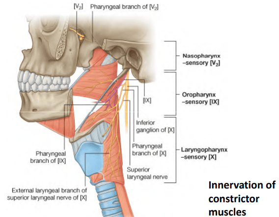
Neurovascular control of the pharyngeal wall is coordinated by the pharyngeal plexus, an autonomic and somatic neural network situated within the buccopharyngeal fascia on the posterolateral surface of the middle pharyngeal constrictor muscle.
Constituents & Functional Modalities of the Plexus
- 1. Motor Limb: Supplied by the pharyngeal branch of the vagus nerve (CN X), carrying cranial accessory (CN XI) motor fibers derived from the nucleus ambiguus in the medulla.
Muscles Supplied: Innervates all external circular constrictors (superior, middle, inferior) and all internal longitudinal muscles (palatopharyngeus, salpingopharyngeus)—with the sole exception of the stylopharyngeus. The inferior constrictor receives additional motor fibers from the external laryngeal and recurrent laryngeal branches of CN X. - 2. Sensory Limb: Supplied primarily by the pharyngeal branch of the glossopharyngeal nerve (CN IX).
Territorial Breakdown of Pharyngeal Sensation:- Nasopharynx: Sensory innervation is mediated predominantly by the pharyngeal branch of the maxillary nerve (V2) emerging from the pterygopalatine ganglion, alongside minor contributions from CN IX around the tubal orifice.
- Oropharynx: Innervated entirely by the glossopharyngeal nerve (CN IX) (supplying general somatic sensation and taste to the posterior 1/3 of the tongue, the tonsillar fossa, and the fauces; serves as the afferent limb of the gag reflex).
- Laryngopharynx: Innervated by the internal laryngeal branch of the vagus nerve (CN X) superiorly, and the recurrent laryngeal nerve (CN X) inferiorly near the cricopharyngeal junction.
- 3. Sympathetic (Vasomotor) Limb: Provided by postganglionic sympathetic fibers arising from the superior cervical sympathetic ganglion, mediating vasoconstriction across the pharyngeal vascular tree.
Arterial Supply & Venous Drainage
- Arterial Supply: Derives from branches of the external carotid artery:
- Ascending Pharyngeal Artery: Arises directly from the external carotid; ascends vertically along the pharyngobasilar fascia to supply the constrictors and nasopharynx.
- Ascending Palatine & Tonsillar Arteries: Branches of the facial artery; the tonsillar artery pierces the superior constrictor to supply the palatine tonsil.
- Descending Palatine & Sphenopalatine Arteries: Branches of the maxillary artery supplying the nasopharynx and soft palate.
- Dorsal Lingual Branches: Arise from the lingual artery to supply the oropharyngeal floor.
- Superior & Inferior Thyroid Arteries: Supply the laryngopharynx via their superior and inferior laryngeal branches.
- Venous Drainage: Blood drains into an extensive submucosal and external pharyngeal venous plexus situated within the buccopharyngeal fascia. The plexus drains into the internal jugular vein directly or via the facial and lingual veins. Crucially, the external palatine (paratonsillar) vein descends from the soft palate across the tonsillar hemicapsule to pierce the superior constrictor before entering the plexus.
- Lymphatic Drainage: Lymph from the pharynx drains primarily into the retropharyngeal lymph nodes (situated in the retropharyngeal space) and directly into the deep cervical lymph nodes along the internal jugular vein, particularly the jugulodigastric node (the “tonsillar node,” enlarged and tender in acute palatine tonsillitis).
Waldeyer’s ring & lymphoid tissue
Waldeyer’s tonsillar ring is an interrupted circular band of immunologically active mucosa-associated lymphoid tissue (MALT) encircling the entrance of the respiratory and alimentary tracts within the pharyngeal wall.

Anatomical Components of Waldeyer’s Ring
- Pharyngeal Tonsil (Adenoid): Situated superiorly in the midline within the roof and posterior wall of the nasopharynx.
- Tubal Tonsils (Tonsils of Gerlach): Submucosal lymphoid aggregates situated on the lateral walls of the nasopharynx surrounding the pharyngeal opening of the auditory tubes, extending onto the salpingopharyngeal folds.
- Palatine Tonsils: The largest components, situated laterally on either side of the oropharynx between the palatoglossal and palatopharyngeal arches.
- Lingual Tonsil: Diffuse nodular lymphoid aggregates covering the dorsal submucosa of the posterior one-third of the tongue.
- Accessory Aggregates: Interconnecting lateral pharyngeal lymphoid bands along the salpingopharyngeal folds and scattered lymphoid follicles across the posterior pharyngeal wall.
Immunological Role & Clinical Protection
Waldeyer’s ring acts as an immunological sentinel system. Positioned at the portal of entry for inspired air and ingested food, it samples inhaled and ingested antigens, initiating the production of secretory immunoglobulin A (sIgA) and facilitating clonal B-cell and T-cell expansion. This local immune response helps protect the lower respiratory organs and gastrointestinal tract from descending microbial infections.
Biomechanics of swallowing (deglutition)
Because the larynx is situated directly in the pathway of the digestive tract, deglutition requires precise neuromuscular coordination to propel the food bolus into the esophagus while temporarily sealing off the lower and upper airways.
The Three Phases of Swallowing
Phase 1: Voluntary (Oral) Phase
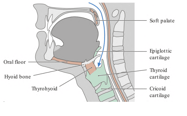
Masticated food is formed into a cohesive bolus on the dorsum of the tongue. The anterior tongue tip presses against the hard palate, and the intrinsic and extrinsic tongue muscles (particularly styloglossus and hyoglossus, innervated by CN XII) roll the bolus backward into the oropharynx through the oropharyngeal isthmus. Once the bolus contacts the sensory receptors of the posterior tongue, palatoglossal arches, and posterior oropharyngeal wall, the voluntary phase terminates and triggers the involuntary swallowing reflex.
Phase 2: Involuntary Pharyngeal Phase
A rapid reflex action (lasting approximately 1 second) controlled by the swallowing center in the medullary reticular formation. Respiration is reflexively inhibited. The bolus is propelled through the pharynx while four coordinated mechanisms prevent food from entering the airway:
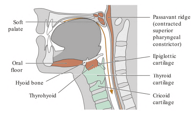
- Velopharyngeal Closure (Upper Airway Protection): The soft palate is tensed by the tensor veli palatini (CN V3) and elevated by the levator veli palatini (CN X). Simultaneously, the uppermost horizontal fibers of the superior pharyngeal constrictor contract, creating a transverse mucosal shelf on the posterior wall known as Passavant’s ridge (palatopharyngeal sphincter). The elevated soft palate meets Passavant’s ridge, closing the pharyngeal isthmus and preventing food or liquids from regurgitating into the nasopharynx or nasal cavities.
- Laryngeal Elevation & Lower Airway Sealing: The hyoid bone and larynx are pulled upward and forward beneath the base of the tongue by the suprahyoid oral floor muscles (mylohyoid [CN V3], anterior belly of digastric [CN V3], and geniohyoid [C1 via CN XII]) and the thyrohyoid muscle (C1 via CN XII), assisted by the internal pharyngeal elevators (stylopharyngeus, palatopharyngeus, salpingopharyngeus).
Epiglottic Action: As the larynx ascends, the epiglottis is retroflexed backward over the laryngeal inlet, and the aryepiglottic folds approximate, sealing the lower airway. - Bolus Deflection: The descending bolus splits around the laryngeal inlet, coursing through the bilateral piriform recesses into the hypopharynx.
- Peristaltic Pharyngeal Transit: The circular pharyngeal constrictor muscles contract in rapid serial craniocaudal sequence (superior → middle → inferior), stripping the bolus downward toward the esophagus.
Phase 3: Involuntary Esophageal Phase
As the bolus reaches the lower laryngopharynx, the tonically active cricopharyngeus muscle (upper esophageal sphincter) reflexively relaxes, allowing the bolus to enter the proximal esophagus. Once the bolus clears the sphincter, the cricopharyngeus contracts to prevent regurgitation. Coordinated primary peristaltic waves driven by the circular and longitudinal esophageal musculature (under vagal autonomic control) propel the bolus down into the stomach.
The gag reflex & neurology
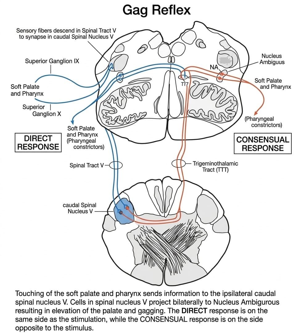
The gag reflex (pharyngeal reflex) is an involuntary, protective cephalic reflex designed to prevent foreign bodies from entering the pharynx, larynx, and respiratory tract. It is elicited clinically by lightly touching the mucosa of the posterior oropharyngeal wall, palatoglossal arch, or posterior one-third of the tongue with a spatula or cotton swab.
Neuroanatomical Arc of the Gag Reflex
- Afferent (Sensory) Limb: Mediated by the glossopharyngeal nerve (CN IX). General somatic afferent (GSA) fibers travel from the stimulated oropharyngeal mucosa through the superior and inferior glossopharyngeal ganglia into the medulla oblongata. These fibers descend in the spinal trigeminal tract to synapse upon second-order sensory neurons within the caudal spinal trigeminal nucleus (nucleus caudalis).
- Central Interneuronal Transmission: Interneurons in the spinal trigeminal nucleus project bilaterally via trigeminothalamic pathways and the reticular formation to synapse upon motor neurons within the nucleus ambiguus of both medullary hemispheres.
- Efferent (Motor) Limb: Mediated by the vagus nerve (CN X). Special visceral efferent (SVE) fibers emerge from the nucleus ambiguus, exiting the medulla via the postolivary sulcus to supply the striated pharyngeal musculature and soft palate via the pharyngeal plexus.
- Normal Reflex Response: Triggers bilateral contraction of the pharyngeal constrictor muscles accompanied by brisk, symmetrical elevation of the soft palate and backward pulling of the tongue (“gagging”):
- Direct Response: Elevation of the palate and pharyngeal contraction on the side of stimulation.
- Consensual Response: Simultaneous elevation of the palate and pharyngeal contraction on the opposite, non-stimulated side.
Clinical Neuro-Localization of Gag Reflex Lesions
Examining the symmetry of soft palate elevation and comparing unilateral versus consensual responses allows precise clinical localization of cranial nerve lesions:
1. Unilateral Glossopharyngeal Nerve (CN IX) Lesion (Sensory Deficit)
- Stimulation of the Damaged Side: Touching the pharyngeal wall on the affected side elicits zero response (neither direct nor consensual), because the sensory afferent arc is interrupted.
- Stimulation of the Normal Side: Touching the normal side elicits a completely normal, symmetrical bilateral response (both direct and consensual), because its sensory afferents are intact and project bilaterally to both intact vagal motor nuclei.
2. Unilateral Vagus Nerve (CN X) Lesion (Motor Deficit)
- Sensation Remains Intact: The patient feels the touch normally on both sides of the oropharynx because CN IX is functioning bilaterally.
- Asymmetrical Motor Response: Because the motor fibers supplying one side of the pharyngeal and palatal muscles are paralyzed, only the muscles on the healthy side contract. The soft palate elevates asymmetrically, and the uvula pulls toward the normal (intact) side, away from the side of the lesion.
- Consistency Across Sides: This asymmetric deviation toward the normal side occurs regardless of which side of the pharynx is touched, because sensory signals from either side activate the intact contralateral vagal nucleus.
3. Combined Unilateral CN IX and CN X Lesions
- Stimulation of the Damaged Side: Produces no response at all (no direct or consensual movement) due to sensory afferent block.
- Stimulation of the Normal Side: Elicits a unilateral motor response only on the normal side, with the palate deviating to that side. No consensual response is seen on the damaged side due to the motor efferent block.
Clinical pathologies & surgery
1. Adenoid Hypertrophy & Adenoidectomy
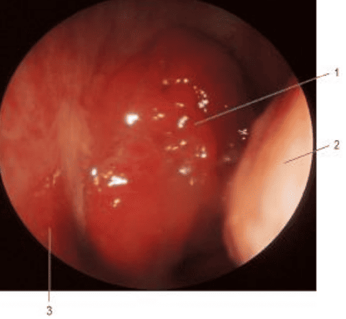
- Pathophysiology: Chronic, recurrent upper respiratory tract infections or allergic rhinitis trigger pathological hyperplasia of the pharyngeal tonsil in preschool children, encroaching on the posterior choanae.
- Clinical Manifestations:
- Nasal Obstruction & Obligate Mouth Breathing: Loud snoring, obstructive sleep apnea (OSA), daytime somnolence, and feeding difficulties in infants.
- Adenoid Facies: Long-standing mouth breathing leads to chronic craniofacial remodeling: an elongated face, dull expression, retrognathic mandible, narrow pinched nostrils, high-arched palate, and crowded maxillary incisors.
- Chronic Secretory Otitis Media (Glue Ear): Enlarged adenoids compress the pharyngeal orifice of the auditory tube, preventing ventilation and drainage of the middle ear. Negative middle ear pressure draws fluid into the tympanic cavity, causing conductive hearing loss, speech delay, and recurrent acute otitis media.
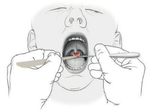
- Clinical Evaluation: Evaluated using posterior rhinoscopy (reflecting light into a small, heated mirror angled behind the soft palate) or flexible fiberoptic transnasal nasopharyngoscopy.
- Adenoidectomy: Transoral or transnasal surgical curettage/coblation performed to clear nasopharyngeal obstruction and restore Eustachian tube drainage.
2. Acute Tonsillitis & Peritonsillar Abscess (Quinsy)
- Acute Tonsillitis: Acute infection of the palatine tonsils (commonly caused by viral pathogens or Group A beta-hemolytic Streptococcus).
Clinical Features: Severe sore throat, odynophagia (painful swallowing), fever, halitosis, and referred otalgia (ear pain mediated by the shared sensory distribution of the glossopharyngeal nerve [CN IX] via its tympanic branch, Jacobson’s nerve).
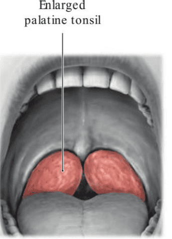
Physical Signs: Symmetrically enlarged, hyperemic palatine tonsils with purulent white-yellow exudate filling the openings of the tonsillar crypts (follicular tonsillitis). In severe cases, marked tonsillar enlargement (“kissing tonsils”) narrows the oropharyngeal outlet, producing mechanical dysphagia and stertorous breathing. - Peritonsillar Abscess (Quinsy): Occurs when infection spreads through the tonsillar capsule into the loose areolar tissue of the peritonsillar space.
Clinical Presentation: Severe unilateral throat pain, high fever, trismus (inability to open the mouth due to irritation of the adjacent medial pterygoid muscle), a muffled “hot potato” voice, and marked deviation of the uvula toward the contralateral, unaffected side. Requires urgent needle aspiration or incision and drainage alongside antibiotic therapy.
3. Tonsillectomy: Indications & Hemorrhagic Hazards
- Clinical Indications:
- Recurrent episodes of acute tonsillitis meeting paradise criteria (e.g., ≥7 episodes in one year, ≥5 episodes per year for two consecutive years, or ≥3 episodes per year for three consecutive years).
- Obstructive sleep apnea or severe dysphagia secondary to tonsillar hypertrophy.
- Peritonsillar abscess unresponsive to drainage, or recurrent quinsy.
- Suspected unilateral tonsillar malignancy (e.g., lymphoma or squamous cell carcinoma) requiring excisional biopsy.
- Post-Tonsillectomy Hemorrhage:
- Primary Hemorrhage (First 24 Hours): Arises from unligated vessels within the tonsillar bed. The primary source is usually the external palatine (paratonsillar) vein crossing the lateral wall of the bed, or the tonsillar artery (branch of the facial artery) entering the lower pole.
- Secondary Hemorrhage (Postoperative Days 5 to 10): Occurs when the healing fibrin slough covering the raw muscular bed detaches prematurely, often secondary to local bacterial infection.
4. Killian’s Dehiscence & Zenker’s Diverticulum
Between the oblique thyropharyngeus fibers and the horizontal cricopharyngeus fibers of the inferior pharyngeal constrictor lies an anatomical zone of muscular weakness: Killian’s dehiscence (Killian’s triangle).
- Pathophysiology: Incoordination during swallowing—such as failure of the cricopharyngeus to relax while the thyropharyngeus contracts forcefully—generates high intraluminal pressures in the hypopharynx.
- Zenker’s Diverticulum (Pharyngeal Pouch): High pressures cause the mucosa and submucosa to herniate posteriorly through Killian’s dehiscence, creating a false pulsion diverticulum.
- Clinical Presentation: Progressively enlarging pouch in elderly patients presenting with halitosis (from retained, decomposing food), dysphagia, regurgitation of undigested food, gurgling in the neck while swallowing, and aspiration pneumonia.
Comprehensive summary tables
Scroll the table sideways on a smaller screen.
| Muscle | Layer & Orientation | Origin Attachment | Insertion Attachment | Primary Innervation | Primary Functional Action |
|---|---|---|---|---|---|
| Superior Constrictor | External Circular | Pterygoid hamulus, pterygomandibular raphe, mylohyoid line of mandible, side of tongue | Median pharyngeal raphe and pharyngeal tubercle of basiocciput | Vagus nerve (CN X via pharyngeal plexus) | Constricts upper pharynx; forms Passavant’s ridge for velopharyngeal closure |
| Middle Constrictor | External Circular | Greater and lesser horns of hyoid bone, lower stylohyoid ligament | Median pharyngeal raphe | Vagus nerve (CN X via pharyngeal plexus) | Constricts pharynx during swallowing, stripping bolus downward |
| Inferior Constrictor | External Circular | Oblique line of thyroid cartilage (thyropharyngeus); side of cricoid cartilage (cricopharyngeus) | Median pharyngeal raphe; cricopharyngeus forms an unbroken horizontal ring | Vagus nerve (CN X via plexus, plus external and recurrent laryngeal nerves) | Constricts lower pharynx; cricopharyngeus acts as upper esophageal sphincter (UES) |
| Stylopharyngeus | Internal Longitudinal | Medial surface of styloid process of temporal bone | Superior and posterior borders of thyroid cartilage; lateral pharyngeal wall | Glossopharyngeal nerve (CN IX) — Sole muscular target | Elevates, shortens, and widens pharynx and larynx during swallowing |
| Palatopharyngeus | Internal Longitudinal | Hard palate and palatine aponeurosis of soft palate | Posterior border of thyroid cartilage; pharyngeal wall | Vagus nerve (CN X via pharyngeal plexus) | Elevates pharynx and larynx; pulls palatopharyngeal arches together |
| Salpingopharyngeus | Internal Longitudinal | Inferior border of cartilaginous auditory tube | Blends with palatopharyngeus into the lateral pharyngeal wall | Vagus nerve (CN X via pharyngeal plexus) | Elevates pharynx; dilates and opens pharyngeal orifice of Eustachian tube |
Scroll the table sideways on a smaller screen.
| Feature | Nasopharynx (Epipharynx) | Oropharynx (Mesopharynx) | Laryngopharynx (Hypopharynx) |
|---|---|---|---|
| Vertebral Levels | Skull base to C1 / C2 | C2 to C3 | C4 to C6 |
| Superior Boundary | Sphenoid and basiocciput | Soft palate | Superior border of epiglottis |
| Inferior Boundary | Soft palate (pharyngeal isthmus) | Superior border of epiglottis | Inferior border of cricoid cartilage (C6) |
| Anterior Opening | Choanae into nasal cavities | Oropharyngeal isthmus into oral cavity | Laryngeal inlet into larynx |
| Epithelial Lining | Ciliated pseudostratified columnar (respiratory) | Non-keratinized stratified squamous | Non-keratinized stratified squamous |
| Dominant Sensory Nerve | Pharyngeal branch of V2 (maxillary) | Glossopharyngeal nerve (CN IX) | Internal and recurrent laryngeal nerves (CN X) |
| Key Anatomical Landmarks | Torus tubarius, Eustachian orifice, fossa of Rosenmüller, adenoid | Palatine tonsils, faucial arches, lingual tonsil, supratonsillar fossa | Piriform recesses, aryepiglottic folds, Killian’s dehiscence |
Scroll the table sideways on a smaller screen.
| Site of Lesion | Stimulation of Damaged Side | Stimulation of Normal Side | Key Diagnostic Pearl |
|---|---|---|---|
| Unilateral CN IX (Sensory Afferent Deficit) | No response (no direct or consensual movement) | Normal bilateral response (symmetrical elevation of palate) | Loss of sensation on damaged side; normal response on stimulating healthy side. |
| Unilateral CN X (Motor Efferent Deficit) | Asymmetrical elevation: palate pulls to normal side; uvula deviates away from lesion | Asymmetrical elevation: palate pulls to normal side; uvula deviates away from lesion | Sensation intact bilaterally; palate elevates only on intact side regardless of side touched. |
| Combined CN IX & X (Complete Unilateral Deficit) | No response at all | Unilateral response only on normal side (no consensual response) | Abolished sensation and motor response on damaged side; palate pulls to intact side on testing normal side. |
Quick recap
References
The following authoritative medical anatomy and clinical textbooks support this curriculum note:
- Gray’s Anatomy: Standring, S. (2021) Gray’s Anatomy: The Anatomical Basis of Clinical Practice. 42nd edn. London: Elsevier. [Section 4: Head and Neck; Chapter 33: Pharynx].
- Clinically Oriented Anatomy: Moore, K.L., Dalley, A.F. and Agur, A.M.R. (2018) Clinically Oriented Anatomy. 8th edn. Philadelphia: Wolters Kluwer. [Chapter 8: Head; Pharynx and Deglutition].
- Last’s Anatomy: Sinnatamby, C.S. (2011) Last’s Anatomy: Regional and Applied. 12th edn. Edinburgh: Churchill Livingstone. [Chapter 6: Head and Neck and Spine; Pharynx].
- Scott-Brown’s Otorhinolaryngology: Watkinson, J.C. and Clarke, R.W. (2018) Scott-Brown’s Otorhinolaryngology and Head and Neck Surgery. 8th edn. Boca Raton: CRC Press. [Volume 3: Head and Neck Surgery; Chapter 15: Benign and Malignant Disorders of the Nasopharynx and Oropharynx].
- Snell’s Clinical Anatomy by Regions: Splittgerber, R. (2019) Snell’s Clinical Anatomy by Regions. 10th edn. Philadelphia: Wolters Kluwer. [Chapter 11: The Head and Neck; The Pharynx].