
Thyroid and parathyroid glands: surgical anatomy, embryology & clinical correlations
On this page
Overview & cervical fascia
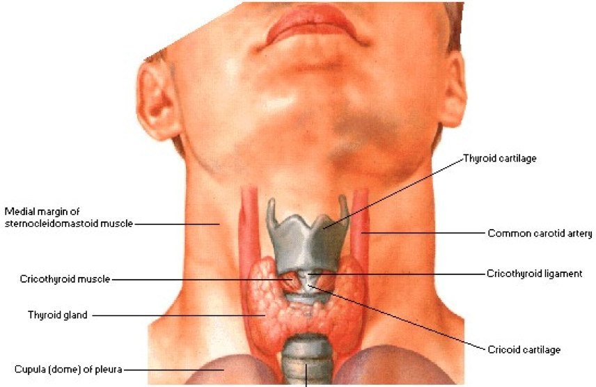
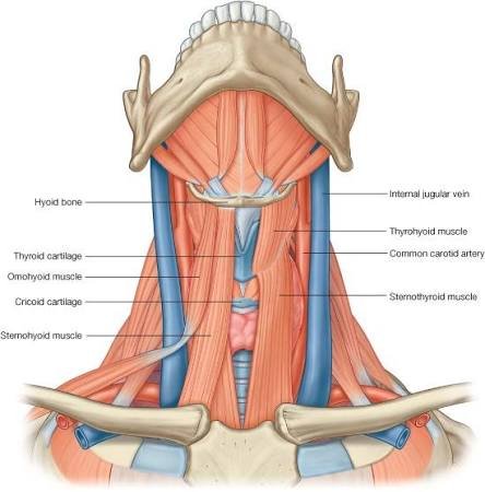
The thyroid gland is the largest dedicated endocrine organ in the human body. Highly vascular and brownish-red, it wraps around the anterior and lateral aspects of the cervical trachea and larynx. It secretes triiodothyronine (T3) and thyroxine (T4), which govern systemic basal metabolic rate, alongside calcitonin, which participates in calcium homeostasis.
The Deep Cervical Fascia & Thyroid Sheaths
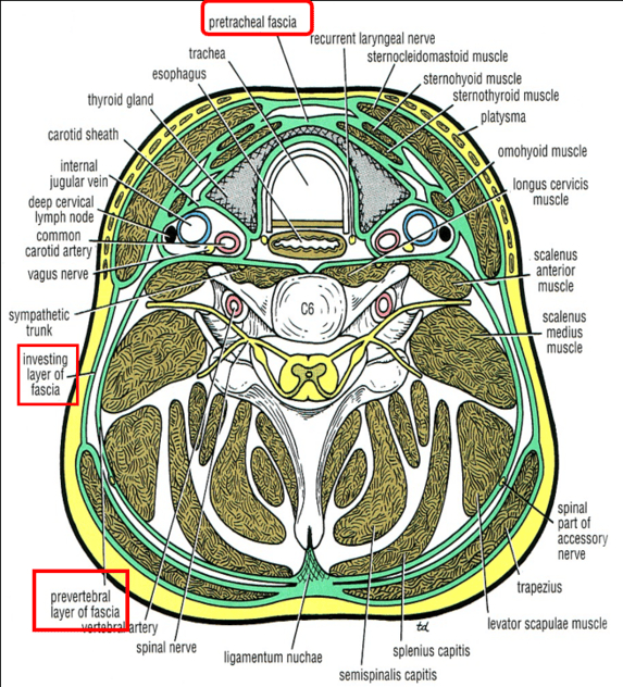
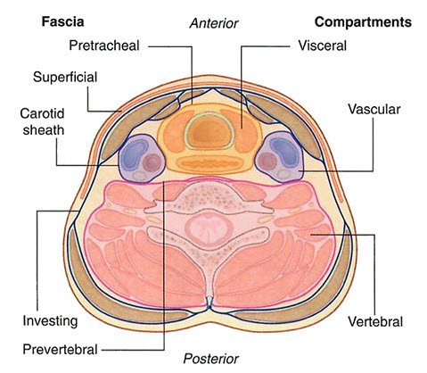
The deep cervical fascia of the neck is divided into three well-defined concentric layers, alongside the specialized paired carotid sheaths:
- 1. Investing Layer: The outermost sleeve that encircles the entire neck, splitting to invest the trapezius and sternocleidomastoid (SCM) muscles, as well as the submandibular and parotid salivary glands.
- 2. Pretracheal Layer: The middle visceral layer limited to the anterior neck. It splits to envelope the thyroid gland, trachea, and esophagus. It extends superiorly to attach to the hyoid bone, thyroid cartilage, and cricoid cartilage, and descends inferiorly into the superior mediastinum to blend with the adventitia of the aortic arch and fibrous pericardium.
- 3. Prevertebral Layer: The deep sleeve that surrounds the cervical vertebral column and its prevertebral and scalene muscles.
- Carotid Sheath: A neurovascular tubular condensation of all three fascial layers enclosing the common and internal carotid arteries medially, the internal jugular vein (IJV) laterally, and the vagus nerve (CN X) posteriorly.
The Two Membranes of the Thyroid Gland
The thyroid gland is surrounded by two distinct membranous envelopes, an arrangement with direct implications for surgical thyroidectomy:
- True Capsule (Fibrous Capsule): A thin, dense connective tissue condensation directly adherent to the thyroid parenchyma. It sends internal septa that divide the gland into functional lobules and individual thyroid follicles.
- False Capsule (Pretracheal Fascial Sheath): A loose outer sheath derived from the visceral split of the pretracheal fascia.
Surgical Plane: The rich capsular venous plexus and the delicate parathyroid glands lie primarily between the true capsule and the false fascial sheath. During thyroidectomy, dissection must remain strictly against or within the true fibrous capsule to avoid avulsing the capsular veins and to avoid devascularizing or inadvertently removing the parathyroid glands.
Morphology & relations
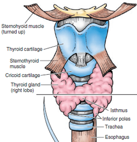
The thyroid gland displays a characteristic butterfly or H-shape, consisting of two lateral lobes (right and left) linked across the midline by a central transverse bridge: the isthmus.
Dimensions & Surface Anatomy of the Lobes
- Pear-Shaped Lobes: Each lateral lobe is conical/pear-shaped, measuring approximately 5 cm in length, 3 cm in width, and 2 cm in depth. The normal adult thyroid weighs between 20 and 25 grams (slightly heavier in females and enlarging during puberty and pregnancy).
- Apex: Directs superiorly and slightly laterally, extending up to the oblique line of the thyroid cartilage. The superior extent is strictly limited by the insertion of the sternothyroid muscle onto this oblique line.
- Base: Directs inferiorly, resting at the level of the fourth or fifth tracheal ring.
- The Isthmus: Measures roughly 1.25 cm vertically and horizontally. It extends across the midline directly in front of the second, third, and fourth rings of the trachea.

The Pyramidal Lobe & Levator Glandulae Thyroideae
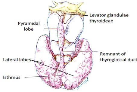
In approximately 50% of individuals, an accessory third lobe—the pyramidal lobe (of Lalouette)—is present. It arises as a conical or finger-like projection from the superior border of the isthmus, typically positioned slightly to the left of the midline.
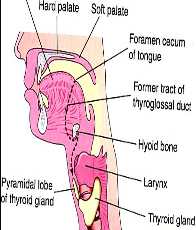
- Embryological Basis: Represents persistent tissue derived from the distal end of the embryonic thyroglossal duct.
- Levator Glandulae Thyroideae: A fibrous or fibromuscular slip connecting the apex of the pyramidal lobe (or the upper border of the isthmus) to the body of the hyoid bone. When muscular, it is derived from infrahyoid muscle mesenchyme.
Anatomical Relations of the Thyroid Lobes
Scroll the table sideways on a smaller screen.
| Boundary / Aspect | Relating Anatomical Structures | Surgical & Clinical Relevance |
|---|---|---|
| Anterolateral Surface |
Overlain by the “4 S’s”: 1. Sternohyoid muscle 2. Sternothyroid muscle 3. Superior belly of omohyoid muscle 4. Sternocleidomastoid (anterior border overlapping inferiorly) |
Infrahyoid strap muscles must be mobilized or retracted laterally along the median avascular white line during anterior surgical neck exposure. |
| Posterior / Posterolateral Surface |
• Carotid sheath and contents: - Common carotid artery (medial) - Internal jugular vein (lateral) - Vagus nerve (CN X, posterior) • Cervical sympathetic trunk (posterior to sheath on prevertebral fascia) |
Enlarged thyroid lobes displace the carotid sheath posterolaterally. Pressure on the cervical sympathetic chain can cause Horner’s syndrome (ptosis, miosis, anhidrosis). |
| Medial Surface |
• Superiorly: Larynx (thyroid and cricoid cartilages) and pharynx (inferior constrictor muscle) • Inferiorly: Trachea and esophagus • Nerves: - External laryngeal nerve (supplying cricothyroid muscle) - Recurrent laryngeal nerve (ascending in tracheoesophageal groove) |
The recurrent laryngeal nerve courses in the tracheoesophageal groove immediately adjacent to the medial surface of the lobe before piercing the larynx under the inferior constrictor. |
| Posterior Border |
• Superior and inferior parathyroid glands • Longitudinal anastomosis between superior and inferior thyroid arteries |
The parathyroid glands lie along this rounded border; careful dissection along this interface is necessary to preserve their blood supply. |
Blood supply & lymphatics
The thyroid gland is an extensively vascularized organ, receiving an estimated blood flow of approximately 4 to 6 mL/min/g (which may increase markedly in hyperthyroid states such as Graves’ disease, producing an audible cervical bruit or palpable thrill).
1. Arterial Supply
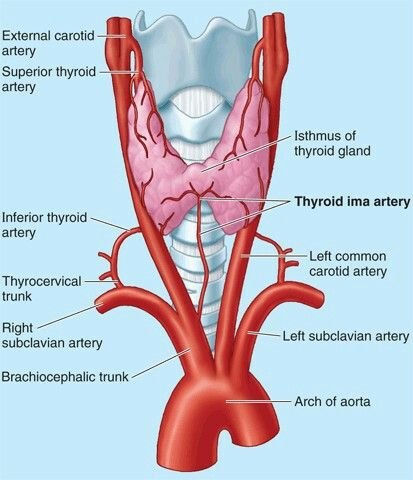
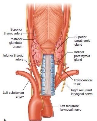
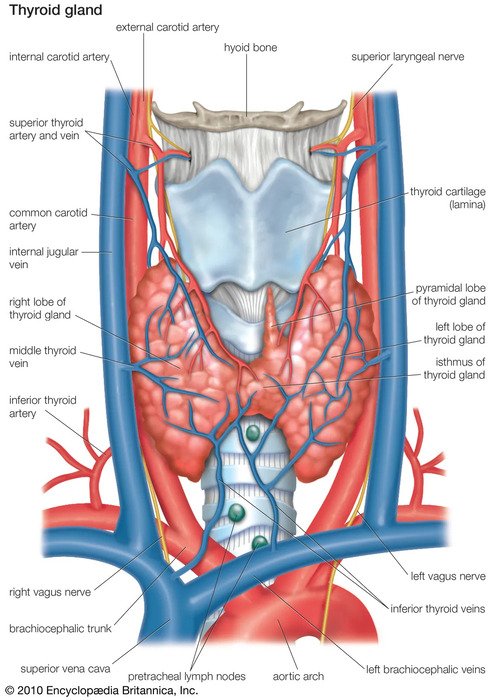
- Superior Thyroid Artery: The first anterior branch of the external carotid artery (arising just above the carotid bifurcation). It descends along the lateral border of the thyrohyoid muscle to reach the superior pole of the thyroid lobe in close anatomical companionship with the external laryngeal nerve.
At the superior pole, it divides into:- Anterior branch: Runs along the superior border of the lobe and isthmus to anastomose with its fellow from the opposite side.
- Posterior branch: Descends down the posterior border of the lobe to anastomose with the ascending branch of the inferior thyroid artery.
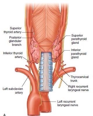
- Inferior Thyroid Artery: Arises from the thyrocervical trunk, a primary branch of the first part of the subclavian artery. It ascends vertically behind the prevertebral fascia, arches medially at the level of the cricoid cartilage (C6) behind the carotid sheath, and pierces the pretracheal fascia to reach the posterior aspect of the lower thyroid pole.
Terminal Branches: Supplies the inferior pole, the posterior parenchyma, and sends crucial end-arterial nutritional branches to both the superior and inferior parathyroid glands. - Thyroidea Ima Artery (Arteria Thyroidea Ima of Neubauer): An inconstant arterial branch present in approximately 3% to 10% of individuals. When present, it arises from the brachiocephalic trunk (innominate artery), the arch of the aorta, or rarely the right common carotid artery. It ascends directly over the anterior surface of the trachea in the midline to enter the inferior border of the isthmus.
Surgical Danger: Represents a recognized cause of sudden, profuse arterial hemorrhage during emergency midline tracheostomy or low anterior neck exploration.
2. Venous Drainage
A dense capsular venous plexus on the surface of the gland is drained by three pairs of primary veins:
- Superior Thyroid Vein: Accompanies the superior thyroid artery; emerges from the upper pole, crosses the common carotid artery, and drains into the internal jugular vein (or occasionally into the facial vein).
- Middle Thyroid Vein: A short, wide, fragile vein emerging from the lateral margin of the lobe. It crosses the common carotid artery almost horizontally to drain directly into the internal jugular vein.
Surgical Hazard: During thyroidectomy, this vessel is placed under direct tension as the thyroid lobe is mobilized medially. If not identified and ligated early, it can tear flush with the IJV wall, resulting in profuse bleeding and air embolism. - Inferior Thyroid Vein: Arises from the lower border of the isthmus and inferior poles as a variable venous plexus. It descends anterior to the trachea and drains into the left brachiocephalic vein (or occasionally into both right and left brachiocephalic veins).
- Vein of Kocher: An occasional accessory middle thyroid vein draining into the IJV.
3. Lymphatic Drainage
The thyroid gland possesses an extensive intrathyroidal lymphatic network that drains in multiple directions, mirroring its vascular pedicles:
- Central Compartment (Level VI): Drains primarily into the pretracheal, paratracheal, and prelaryngeal (Delphian) lymph nodes. The Delphian node sits anterior to the cricothyroid membrane; palpable enlargement is an important clinical indicator of thyroid carcinoma or advanced laryngeal malignancy.
- Lateral Deep Cervical Chain (Levels II, III, and IV): Channels from the upper poles follow the superior thyroid vessels to empty into the upper deep cervical nodes (including the jugulodigastric node). Channels from the lower poles drain into lower deep cervical nodes (jugulo-omohyoid node) and supraclavicular nodes.
- Mediastinal Drainage (Level VII): Lymphatics from the inferior isthmus descend behind the sternum into the superior mediastinal lymph nodes.
Innervation & laryngeal nerves
Autonomic Nerve Supply
The parenchymal autonomic innervation of the thyroid gland is exclusively vasomotor in function; thyroid hormonal synthesis and follicular secretion are controlled endocrinologically by pituitary Thyroid-Stimulating Hormone (TSH):
- Sympathetic Innervation: Derived from the superior, middle, and inferior cervical sympathetic ganglia. Postganglionic fibers travel along the periarterial plexuses of the superior and inferior thyroid arteries, producing vasoconstriction.
- Parasympathetic Innervation: Derived from the superior laryngeal nerve and recurrent laryngeal nerve (branches of the vagus nerve, CN X), conveying vasodilator impulses to the parenchymal microvasculature.

The Recurrent Laryngeal Nerves (RLN): Course & Asymmetry
The recurrent laryngeal nerves supply motor innervation to all intrinsic muscles of the larynx except the cricothyroid muscle (which is supplied by the external laryngeal nerve). They also carry sensory fibers to the laryngeal mucosa below the vocal folds.
- Right Recurrent Laryngeal Nerve: Arises from the right vagus nerve as it crosses anterior to the right subclavian artery. It loops underneath and behind the right subclavian artery (derived embryologically from the right 4th aortic arch), then ascends obliquely medially through the base of the neck to enter the tracheoesophageal groove.
- Left Recurrent Laryngeal Nerve: Arises from the left vagus nerve within the superior mediastinum as it crosses anterior to the aortic arch. It loops underneath the arch of the aorta immediately posterior to the ligamentum arteriosum (derived from the left 6th aortic arch), then ascends vertically through the superior mediastinum and neck within the tracheoesophageal groove. Because of its longer intrathoracic course, the left RLN is more susceptible to compression by mediastinal tumors, aortic aneurysms, or enlarged subcarinal lymph nodes.
Anatomical Variations in RLN Trajectory
As the recurrent laryngeal nerve approaches the lower pole and posterior surface of the thyroid lobe, its spatial relationship with the trachea, esophagus, and inferior thyroid artery varies:
- Within the Tracheoesophageal Groove: Observed in 48.5% of dissections (the most common classical anatomical course).
- Paratracheal Course: Observed in 42.2% of cases (nerve ascends slightly lateral to the groove along the anterolateral tracheal wall).
- Paraesophageal Course: Observed in 5.4% of cases (nerve ascends along the anterior or lateral surface of the esophagus).
- Intraparenchymal Course: Observed in 3.9% of individuals (the nerve traverses directly through the fibrous thyroid capsule and superficial thyroid parenchymal tissue, putting it at high risk during subtotal or lobar resection).

The Non-Recurrent Laryngeal Nerve
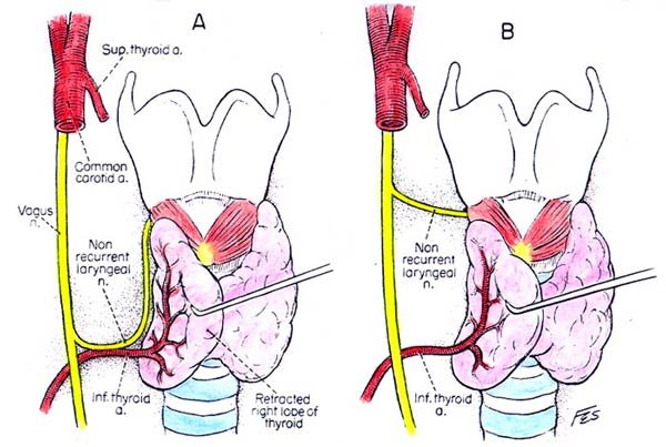
In less than 1% of individuals, an anatomical variant known as the non-recurrent laryngeal nerve occurs, almost exclusively on the right side.
- Developmental Mechanism: Caused by abnormal embryonic involution of the right 4th aortic arch, resulting in an aberrant retroesophageal right subclavian artery (arteria lusoria). The right subclavian artery arises directly from the descending aorta and courses behind the esophagus to reach the right arm.
- Surgical Course: Because there is no vascular arch to snare the nerve caudally, the right laryngeal nerve arises directly from the cervical vagus at the level of the cricoid cartilage and courses transversely directly into the larynx. It is vulnerable to accidental transection during right hemithyroidectomy if the surgeon expects a normal ascending recurrent nerve.
The External Laryngeal Nerve
The external laryngeal nerve is a terminal branch of the superior laryngeal nerve (from CN X). It descends along the inferior constrictor muscle, running in close anatomical proximity to the superior thyroid artery before turning medially to pierce and supply the cricothyroid muscle.
- Action of Cricothyroid: Tenses and elongates the vocal folds, facilitating high-pitched vocal projection.
- Surgical Vulnerability: High ligation of the superior thyroid artery pedicle well above the gland places this nerve at risk. Injury causes a weak, monotonous voice with loss of high pitch and vocal fatigue, particularly disabling for vocalists, teachers, and public speakers.
Parathyroid glands
The parathyroid glands are small, yellow-brown, flattened ovoid endocrine bodies. Typically four in number (two superior and two inferior), they measure approximately 6 mm in length, 3 to 4 mm in width, and 1 to 2 mm in thickness, each weighing roughly 30 to 50 milligrams.

Topography: Superior vs. Inferior Glands
- Superior Parathyroid Glands (Parathyroid IV): Exhibit a remarkably constant anatomical position. They sit along the posterior border of the lateral thyroid lobe at the level of the middle third of the gland, approximately 1 cm superior to the intersection of the inferior thyroid artery and recurrent laryngeal nerve (adjacent to the lower border of the cricoid cartilage). They lie dorsal to the recurrent laryngeal nerve.
- Inferior Parathyroid Glands (Parathyroid III): Exhibit a highly variable anatomical distribution due to their extended embryological migration path alongside the thymus. Typically, they lie along the posterior border of the inferior thyroid pole. They lie ventral to the recurrent laryngeal nerve.
Ectopic Locations: In 1% to 5% of individuals, the inferior parathyroid glands may be found within the thyrothymic ligament, embedded in the cervical or anterior mediastinal thymus, or situated within the carotid sheath. - Intracapsular vs. Extracapsular Position: Parathyroid glands generally reside within the pretracheal fascial sheath (false capsule), but remain outside the true fibrous capsule of the thyroid gland. Occasionally (in up to 3% to 5% of cases), an inferior parathyroid gland may be completely embedded within the thyroid parenchyma (intrathyroidal parathyroid), complicating subtotal resections.

Neurovascular Supply & Lymphatic Drainage
- Arterial Supply: Derived primarily from the inferior thyroid artery (supplying over 80% of both superior and inferior glands via dedicated, minute end-arteries). The superior parathyroid glands may receive branches from the superior thyroid artery or from the longitudinal anastomotic arcade connecting the superior and inferior thyroid arteries.
Surgical Note: Because parathyroid vessels are true end-arteries with minimal collateral circulation, ligating the main trunk of the inferior thyroid artery medial to the parathyroid branches can cause post-thyroidectomy ischemic hypoparathyroidism. - Venous Drainage: Drains into the capsular venous network and subsequently into the superior, middle, and inferior thyroid veins.
- Lymphatic Drainage: Parallels that of the thyroid gland, draining to deep cervical and paratracheal lymph nodes.
- Nerve Supply: Vasomotor autonomic fibers derived from the superior and middle cervical sympathetic ganglia accompany the arterial supply. Parathyroid hormone (PTH) secretion is governed homeostatically by serum ionized calcium levels acting upon calcium-sensing G-protein coupled receptors (CaSR), rather than direct neural control.
Embryological development
The development of the thyroid and parathyroid glands involves the coordinated interaction of the embryonic pharyngeal apparatus and neural crest mesenchyme.
The Pharyngeal Apparatus Architecture
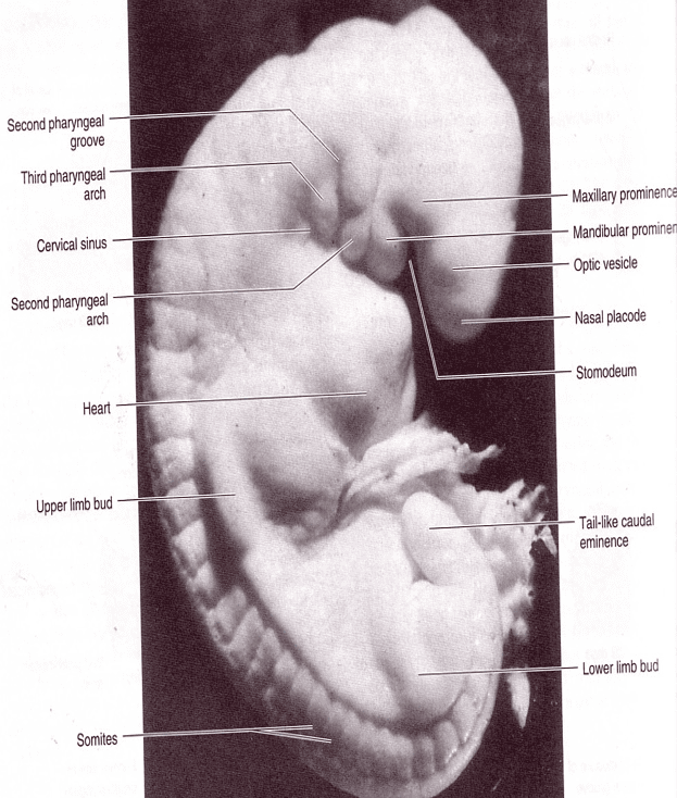
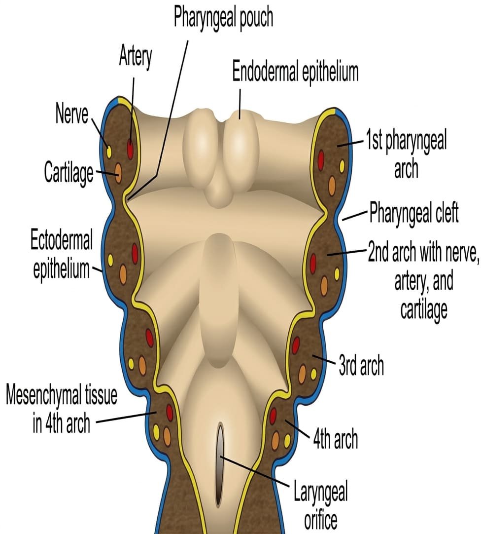
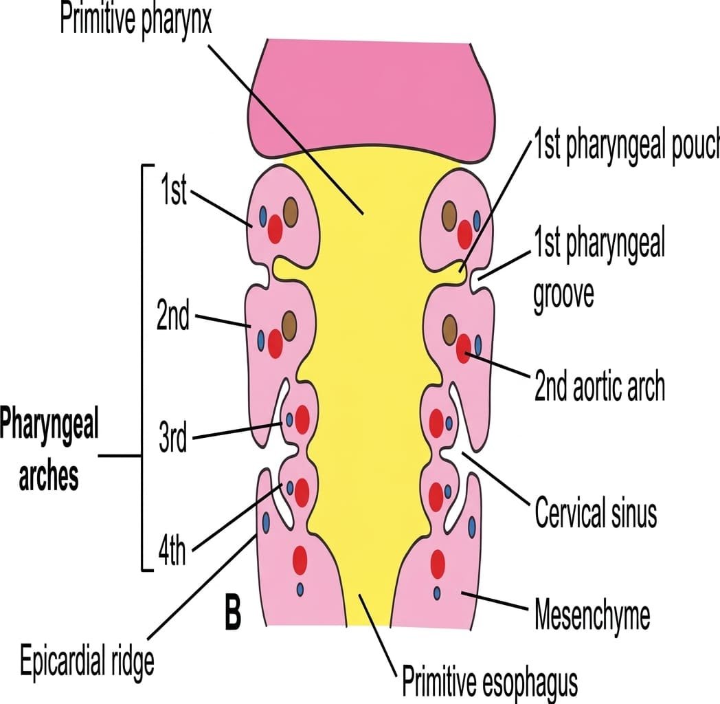
Beginning in the fourth embryonic week, the head and neck region develops from the pharyngeal (branchial) apparatus, comprising four fundamental components:
- 1. Pharyngeal Arches: Six pairs of cylindrical mesodermal masses (the fifth arch involutes rapidly, leaving arches 1, 2, 3, 4, and 6). Each arch contains a core of paraxial and lateral plate mesoderm plus neural crest mesenchyme, a cranial nerve branch, an aortic arch artery, and a cartilaginous bar.
- 2. Pharyngeal Pouches: Four pairs of lateral outpocketings of endodermal epithelium protruding outward between adjacent arches from the lateral walls of the primitive foregut (pharynx). (The fifth pouch is rudimentary or absent).
- 3. Pharyngeal Clefts (Grooves): Four pairs of corresponding external indentations of surface ectoderm separating the arches from the exterior.
- 4. Pharyngeal Membranes: Thin cellular interfaces where endoderm of the pouches contacts ectoderm of the clefts.
Development & Migration of the Thyroid Gland
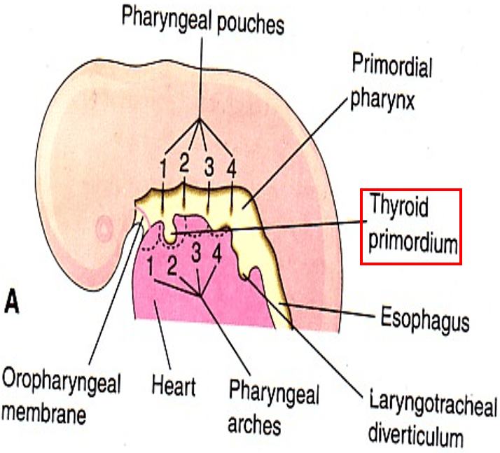
- Earliest Endocrine Gland: The thyroid gland is the first endocrine organ to appear in embryonic life, initiating development on approximately the 24th day after fertilization.
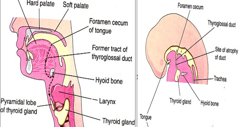
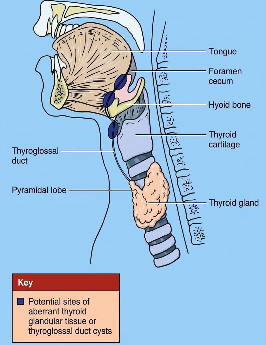
- The Thyroid Diverticulum: Originates as an endodermal epithelial thickening in the midline floor of the primitive pharynx, located between the tuberculum impar and copula at the boundary of the anterior two-thirds and posterior one-third of the developing tongue. This site is marked in adults by the foramen cecum.
- Descent Through the Neck: The endodermal diverticulum invaginates into the underlying mesenchyme, forming a bi-lobed diverticulum that descends anterior to the developing hyoid bone and laryngeal cartilages. During this caudal migration, the gland remains connected to the tongue via a narrow, epithelial-lined cord called the thyroglossal duct.
- Definitive Position & Obliteration: By the 7th embryonic week (day 50), the developing thyroid reaches its definitive adult anatomical position anterior to the 2nd to 4th tracheal rings. The hollow primordium solidifies into follicular cords, and the thyroglossal duct normally undergoes fragmentation, fibrosis, and complete involution.
- Ultimopharyngeal Bodies & C-Cells: The ventral diverticulum of each fourth pharyngeal pouch forms the ultimopharyngeal body. Cells derived from the neural crest migrate into the ultimopharyngeal body, which subsequently fuses with the descending thyroid lobes. These neuroendocrine cells disperse throughout the thyroid parenchyma as parafollicular cells (C-cells), which synthesize and secrete calcitonin.

Development of the Parathyroid Glands & The Descent Paradox
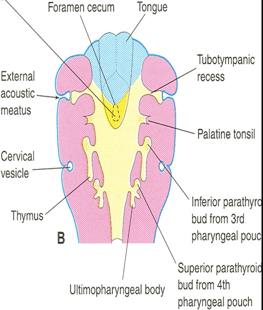
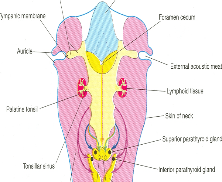
The parathyroid glands develop from the endodermal lining of the third and fourth pharyngeal pouches during the sixth week of embryonic gestation:
- Third Pharyngeal Pouch:
- Dorsal wing: Differentiates into the inferior parathyroid gland (Parathyroid III).
- Ventral wing: Proliferates and fuses across the midline to form the primordium of the thymus gland.
- Fourth Pharyngeal Pouch:
- Dorsal wing: Differentiates into the superior parathyroid gland (Parathyroid IV).
- Ventral wing: Forms the ultimopharyngeal body.
The Parathyroid Descent Paradox Explained
A classic embryological question is: Why do the inferior parathyroid glands arise from the third pouch, while the superior glands arise from the lower, fourth pouch?
As development proceeds, the paired ventral primordia of the thymus lose their connection to the pharyngeal wall and descend caudally toward their definitive home behind the manubrium sterni in the superior mediastinum. Because the inferior parathyroid buds (pouch 3) share a common origin and fascial sheath with the migrating thymus, they are dragged inferiorly down the neck. Eventually, the inferior parathyroids disconnect from the thymus and settle near the inferior pole of the thyroid gland.
In contrast, the superior parathyroids (pouch 4) remain closely associated with the ultimopharyngeal body and the lateral thyroid lobes, undergoing minimal caudal migration. Consequently, they remain near the middle third of the posterior border, higher in the neck than the derivatives of the third pouch.
Congenital anomalies
Disruptions in the migration, canalization, or involution of the thyroid diverticulum and pharyngeal pouches can produce several congenital clinical entities.
Overview of Congenital Thyroid Anomalies
- Agenesis of the Thyroid Gland: Complete failure of the thyroid primordium to form (athyreosis) or failure of one lobe to develop (hemiagenesis, typically the left lobe).
- Congenital Hypothyroidism (Cretinism): Results from thyroid dysgenesis (agenesis, hypoplasia, or ectopia) or inherited defects in thyroid hormone biosynthesis (dyshormonogenesis). Manifests as neonatal jaundice, poor feeding, macroglossia, umbilical hernia, hypotonia, and, if untreated, irreversible cognitive disability and stunted skeletal growth.
- Persistent Thyroglossal Duct: Incomplete obliteration of the epithelial duct leaving a patent fibrous or tubular tract connecting the tongue to the thyroid.
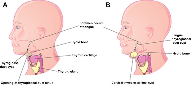
- Thyroglossal Duct Cyst: Fluid-filled cystic degeneration of epithelial remnants along the thyroglossal tract.
- Ectopic Thyroid Gland: Arrest of descent or aberrant migration of thyroid tissue along the thyroglossal pathway.
- Accessory Thyroid Tissue: Detached, functional islands of thyroid tissue situated anywhere along the developmental descent pathway (e.g., within the thymus, attached to the thyrohyoid muscle, or in the submandibular triangle).

Thyroglossal Duct Cysts & Sinuses
- Clinical Features: The most common congenital neck mass, accounting for over 70% of pediatric midline cervical anomalies. Most commonly located immediately inferior to the hyoid bone (65%), followed by suprahyoid and infrahyoid positions, and rarely intralingual at the foramen cecum.
- Diagnostic Hallmark: Because the cyst remains tethered to the foramen cecum by persistent epithelial remnants that traverse or wrap around the hyoid bone, the cyst moves upward on swallowing and upon active tongue protrusion. This distinguishes it from an infected dermoid cyst or enlarged lymph node (which moves only with swallowing).
- Thyroglossal Fistula / Sinus: A sinus opening onto the skin of the anterior neck. Almost always secondary to infection and spontaneous rupture or inadequate surgical incision of a pre-existing thyroglossal cyst; true primary congenital fistulae are rare.
- Surgical Treatment (The Sistrunk Procedure): Simple excision carries a high recurrence rate (>50%) because microscopic tracts arborize through the body of the hyoid bone. The definitive operation is the Sistrunk procedure, which resects the cyst, the central core of the hyoid bone, and a surrounding cuff of base-of-tongue musculature up to the foramen cecum.
Ectopic Thyroid Tissue & Lingual Thyroid
- Lingual Thyroid: The most common form of thyroid ectopia (accounting for 90% of cases). Occurs when the thyroid primordium completely fails to descend, developing entirely at the base of the tongue posterior to the circumvallate papillae.
Clinical Presentation: Symptoms include dysphagia, dysphonia (“hot potato voice”), sensation of a foreign body in the throat, and upper airway obstruction.
Diagnostic Imperative: In approximately 70% to 75% of patients with a lingual thyroid, it is the patient’s only functioning thyroid tissue. Surgical excision without prior radioiodine scanning (or ultrasound confirmation of a normal orthotopic thyroid) will cause iatrogenic hypothyroidism. - Substernal / Retrosternal Ectopic Thyroid: Thyroid descent may proceed into the superior mediastinum or thoracic cavity, situated anterior to the great vessels or behind the esophagus.
Surgical anatomy & thyroidectomy
Thyroid surgery requires precise dissection due to the complex vascular relationships and adjacent neural structures within the anterior visceral compartment of the neck.
Surgical Danger Zones & Laryngeal Nerve Preservation
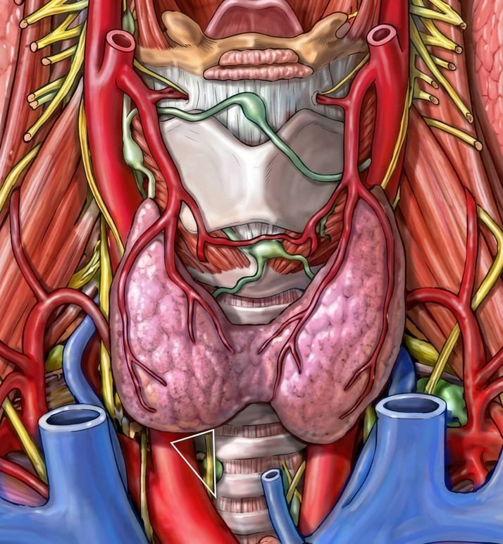
- Superior Pole Dissection (External Laryngeal Nerve): The external laryngeal nerve travels near the superior thyroid artery before turning medially to supply the cricothyroid muscle.
Surgical Rule: To avoid nerve injury, ligate the superior thyroid vessels individually and as close as possible to the upper pole of the thyroid gland. High ligation of the vascular pedicle risks including the nerve in the clamp or suture ligature. - Inferior Pole Dissection (Recurrent Laryngeal Nerve): The inferior thyroid artery approaches the gland from a posterolateral direction, crossing the recurrent laryngeal nerve.
Surgical Triangle: The nerve is identified in a surgical triangle bounded laterally by the common carotid artery, medially by the trachea, and superiorly by the lower border of the thyroid lobe.
Surgical Rule: The relationship between the artery and nerve is variable—the nerve may pass deep to, superficial to, or directly between the terminal branches of the inferior thyroid artery. Therefore, branches of the inferior thyroid artery should be ligated individually on the thyroid capsule rather than ligating the main arterial trunk laterally. This preserves the branch supplying the parathyroid glands and protects the nerve from traction or thermal injury.
Patterns of Laryngeal Nerve Injury
Recurrent Laryngeal Nerve (RLN) Palsy
Because the RLN supplies all intrinsic laryngeal muscles except the cricothyroid, its injury produces characteristic functional deficits depending on laterality:
- Unilateral RLN Injury: The paralyzed vocal cord adopts a paramedian position (between abduction and adduction). The normal contralateral vocal cord compensates by crossing the midline to meet the paralyzed cord during phonation.
Signs: Marked hoarseness of voice, weak/breathy speech, and decreased cough effectiveness, while airway patency and ventilation are generally maintained. - Bilateral RLN Injury: A life-threatening surgical emergency. Both vocal cords adopt a paramedian position, causing severe narrowing of the rima glottidis.
Signs: Marked inspiratory stridor, dyspnea, and acute upper airway obstruction, often requiring immediate endotracheal re-intubation or an emergency bedside tracheostomy. Phonation may paradoxically sound clearer because both cords are close to the midline, but effective ventilation is critically compromised.
External Laryngeal Nerve (ELN) Palsy
Results from trauma or traction to the external branch of the superior laryngeal nerve during superior pole dissection. Denervates the cricothyroid muscle, preventing vocal fold tension.
- Clinical Features: The patient cannot hit high-pitched vocal frequencies. Phonation becomes weak and monotonous, accompanied by early vocal fatigue. Often noticed first by individuals who rely on their voice professionally.
Pathophysiology of Goiter & Compressive Phenomena
Any abnormal enlargement of the thyroid gland is clinically classified as a goiter. Goiters may be diffuse or nodular, euthyroid, hypothyroid, or toxic (hyperthyroid):
- Barriers to Superior Expansion: The superior attachment of the sternothyroid muscle to the oblique line of the thyroid cartilage prevents an enlarging thyroid lobe from extending superiorly into the neck.
Consequently, an expanding goiter grows anteriorly (stretching the strap muscles and creating a visible neck mass) or extends inferiorly through the thoracic inlet into the superior mediastinum, producing a retrosternal (substernal/intrathoracic) goiter. - Tracheal Compression: Compressing the cervical or mediastinal trachea causes progressive exertional dyspnea, positional orthopnea, and audible biphasic inspiratory stridor. Longstanding compression can cause tracheomalacia (softening and atrophy of the tracheal cartilaginous rings), risking complete dynamic airway collapse when the supportive thyroid mass is resected during thyroidectomy.
- Esophageal Compression: Pressure directed posteromedially against the cervical esophagus produces progressive dysphagia, typically more noticeable for solid foods.
- Carotid Sheath & Venous Compression: An expanding mass compresses the low-pressure internal jugular veins against the sternocleidomastoid muscles, causing facial congestion, plethoric cyanosis, and prominent anterior neck venous collaterals.
Pemberton’s Sign: Raising both arms above the head until they touch the ears draws the thyroid mass into the rigid thoracic inlet, worsening venous compression. A positive sign is indicated by marked facial flushing, cyanosis, and jugular venous distention within one minute, signaling significant retrosternal compressive goiter. - Surgical Challenges of the Isthmus: An enlarged isthmus obscures the 2nd, 3rd, and 4th tracheal rings, complicating emergency surgical tracheostomy. In such cases, the isthmus must be divided and retracted to gain access to the underlying trachea.
Post-Thyroidectomy Hypocalcemia & Tetany
Iatrogenic devascularization or inadvertent resection of the parathyroid glands during total or subtotal thyroidectomy causes acute hypoparathyroidism, leading to hypocalcemia (serum ionized Ca2+ < 1.1 mmol/L or total serum Ca2+ < 2.1 mmol/L):
- Neuromuscular Hyperexcitability: Extracellular calcium ions stabilize neuronal and muscle cell membranes by regulating voltage-gated sodium channels. In hypocalcemia, the threshold for sodium channel activation shifts closer to the resting membrane potential, leading to spontaneous depolarization and involuntary muscle contractions (tetany).
- Clinical Manifestations: Early symptoms include circumoral numbness, tingling in the fingertips and toes (paresthesias), muscle cramping, and hyperreflexia. Left untreated, it progresses to painful carpopedal spasms, laryngeal stridor, generalized convulsions, and prolonged QT intervals on ECG.
- Bedside Clinical Signs:
- Chvostek’s Sign: Tapping the facial nerve (CN VII) anterior to the external auditory meatus over the parotid gland triggers involuntary ipsilateral twitching of the facial muscles (especially the upper lip and angle of the mouth).
- Trousseau’s Sign: Inflating a blood pressure cuff on the upper arm to 20 mmHg above systolic pressure for 3 minutes occludes brachial artery flow. The resulting local ischemia enhances hypocalcemic hyperexcitability, precipitating carpopedal spasm (flexion of the wrist and metacarpophalangeal joints, extension of the interphalangeal joints, and adduction of the thumb—“main d’accoucheur”). Trousseau’s sign is more specific and sensitive than Chvostek’s sign.
- Acute Management: Emergency intravenous administration of 10 mL of 10% calcium gluconate infused slowly over 10 to 15 minutes under cardiac monitoring, followed by oral calcium supplementation and active vitamin D (calcitriol).
Quick recap
References
The anatomical and surgical concepts presented in this study note reflect established head and neck surgical and embryological principles, verified against the following core reference texts:
- Gray’s Anatomy: Standring, S. (ed.) (2021) Gray’s Anatomy: The Anatomical Basis of Clinical Practice. 42nd edn. London: Elsevier. [Section 4: Head and Neck; Chapter 30: Neck and Thyroid Gland].
- Last’s Anatomy: Sinnatamby, C.S. (2011) Last’s Anatomy: Regional and Applied. 12th edn. Edinburgh: Churchill Livingstone. [Chapter 6: Head, Neck and Spine – The Thyroid Gland].
- Langman’s Medical Embryology: Sadler, T.W. (2019) Langman’s Medical Embryology. 14th edn. Philadelphia: Wolters Kluwer. [Chapter 17: Head and Neck – Pharyngeal Pouches and Thyroid Gland Development].
- Bailey & Love’s Short Practice of Surgery: Williams, N.S., O’Connell, P.R. and McCaskie, A.W. (eds.) (2018) Bailey & Love’s Short Practice of Surgery. 27th edn. Boca Raton: CRC Press. [Chapter 53: The Thyroid Gland and Thyroglossal Tract; Chapter 54: The Parathyroid Glands].
- Skandalakis’ Surgical Anatomy: Skandalakis, J.E., Colborn, G.L., Weidman, T.A., Foster, R.S., Kingsnorth, A.N. and Skandalakis, L.J. (2004) Skandalakis’ Surgical Anatomy: The Embryologic and Anatomic Basis of Modern Surgery. Athens: PMP. [Chapter 2: Neck – Thyroid and Parathyroid Glands].