
Adrenergic agonists & the presynaptic system: neurochemistry, receptor pharmacology & clinical therapeutics
On this page 14 sections
Sympathetic anatomy & neurochemistry
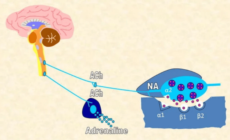
The sympathetic nervous system (SNS) constitutes the thoracolumbar division of the autonomic nervous system, originating from preganglionic neuronal cell bodies located within the intermediolateral cell column of the spinal cord from segments T1 to L2. It coordinates involuntary homeostatic adjustments and drives the classic evolutionary “fight-or-flight” response to physical stress, trauma, hypovolemia, hypoglycemia, and emotional arousal.
Primary Endogenous Adrenergic Transmitters
- Norepinephrine (Noradrenaline, NE): The principal postganglionic sympathetic neurotransmitter synthesized and stored in terminal varicosities of peripheral adrenergic nerves. Directly modulates vascular resistance, visceral motility, and cardiac inotropy.
- Epinephrine (Adrenaline, Epi): Synthesized and stored in the chromaffin cells of the adrenal medulla, which represents a specialized, modified sympathetic ganglion lacking postganglionic axonal processes. Upon splanchnic preganglionic stimulation, the adrenal medulla releases approximately 80% epinephrine and 20% norepinephrine directly into systemic circulation to function as a global endocrine hormone.
- Dopamine (DA): An essential central neurotransmitter within the basal ganglia and limbic system, as well as an endogenous peripheral catecholamine that regulates renal, mesenteric, and coronary vascular beds through specialized dopamine receptors.
Catecholamine synthesis, storage, release & termination
The life cycle of catecholamines within the presynaptic adrenergic nerve terminal involves a tightly regulated sequence: dietary precursor uptake, enzymatic biosynthesis, active vesicular storage, calcium-dependent exocytosis, and rapid signal termination dominated by neuronal reuptake.
1. Biosynthesis of Catecholamines
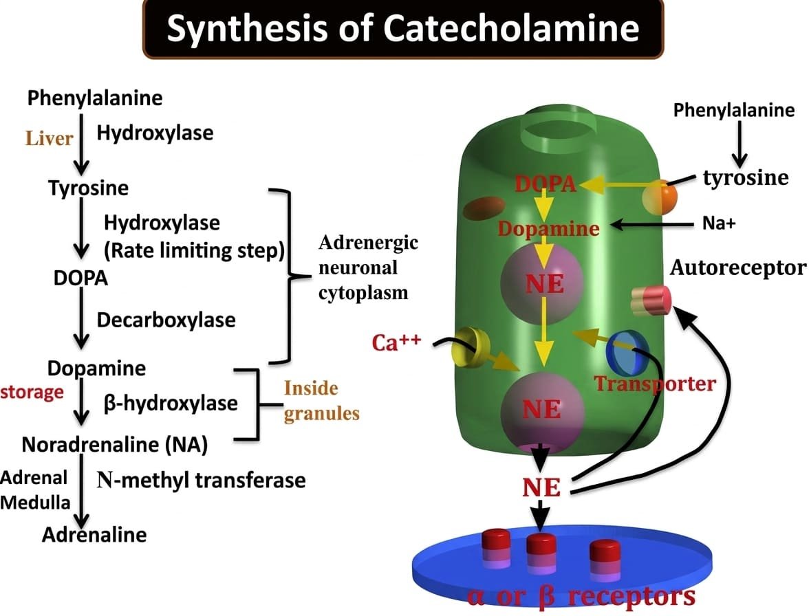
- Precursor Conversion: The essential dietary amino acid phenylalanine is hydroxylated in the liver by phenylalanine hydroxylase to yield L-tyrosine. Tyrosine enters the adrenergic axoplasm via a sodium-dependent active symporter.
- The Rate-Limiting Step: Cytoplasmic tyrosine is hydroxylated to dihydroxyphenylalanine (L-DOPA) by the cytosolic enzyme tyrosine hydroxylase (TH). This step requires molecular oxygen, ferrous iron (Fe2+), and tetrahydrobiopterin (BH4). Tyrosine hydroxylase is subject to end-product feedback inhibition by cytosolic catecholamines and is activated by sympathetic nerve stimulation.
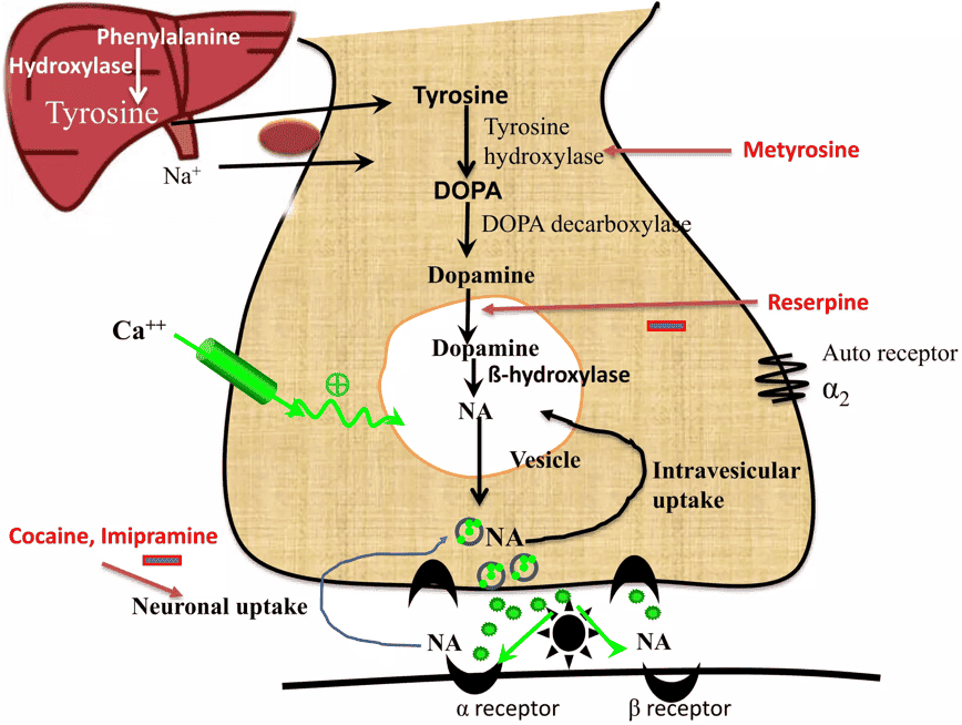
Pharmacological blocker: Metyrosine (α-methyltyrosine) competitively inhibits tyrosine hydroxylase, blocking all downstream catecholamine synthesis (used in managing inoperable pheochromocytoma). - Decarboxylation to Dopamine: L-DOPA is rapidly converted to dopamine by the cytosolic enzyme DOPA decarboxylase (aromatic L-amino acid decarboxylase), utilizing pyridoxal phosphate (vitamin B6) as a cofactor.
- Vesicular Uptake & Norepinephrine Synthesis: Dopamine is actively translocated from the cytoplasm into synaptic storage vesicles by the vesicular monoamine transporter (VMAT-2). Inside the vesicle, dopamine is stereospecifically hydroxylated on its β-carbon to form norepinephrine (NE) by the copper-containing enzyme dopamine β-hydroxylase (DBH), which is bound to the inner vesicular membrane. Ascorbic acid (vitamin C) serves as the necessary electron donor.
- Adrenal Medullary Conversion to Epinephrine: In the adrenal medulla and select phenylethanolamine-containing central neurons, norepinephrine diffuses out of the vesicle back into the cytosol. Here, the enzyme phenylethanolamine N-methyltransferase (PNMT) transfers a methyl group from S-adenosylmethionine (SAMe) to the amino nitrogen, forming epinephrine. Glucocorticoids transported directly from the adjacent adrenal cortex via the intra-adrenal portal vascular system induce high levels of PNMT expression. Newly synthesized epinephrine is pumped back into chromaffin granules via VMAT-2 for definitive storage.
2. Vesicular Storage & Exocytotic Release
- Intravesicular Complexation: Within storage granules, norepinephrine is sequestered in an osmotically inactive, condensed gel complex with adenosine triphosphate (ATP) in a fixed 4:1 molar ratio, bound to the acidic binding protein chromogranin A. This stable complex prevents leakage and protects the catecholamine from degradation by cytosolic enzymes.
- VMAT-2 Inhibition: The plant alkaloid reserpine binds irreversibly to VMAT, permanently abolishing vesicular monoamine transport. Unsequestered dopamine and norepinephrine accumulate unprotected in the cytoplasm, where they undergo rapid deamination by mitochondrial monoamine oxidase (MAO), producing complete, long-lasting depletion of peripheral and central catecholamine stores.
- Exocytosis & Influx of Calcium: Propagation of an action potential down the postganglionic sympathetic axon depolarizes the terminal varicosity, opening voltage-gated N-type calcium (Ca2+) channels. Inward Ca2+ flux activates synaptotagmin and triggers SNARE-complex-mediated fusion of the synaptic vesicle with the neuronal plasma membrane, extruding norepinephrine, ATP, dopamine β-hydroxylase, and chromogranin A into the synaptic cleft.
- Release Inhibition: The historic adrenergic neuron-blocking drugs guanethidine and bretylium accumulate within adrenergic storage vesicles, displace norepinephrine, stabilize vesicular membranes, and prevent calcium-dependent exocytosis.
3. Termination of Action & Metabolism
Unlike the cholinergic system, where acetylcholine is rapidly destroyed in the cleft by acetylcholinesterase, the actions of norepinephrine are terminated primarily by physical removal from the synaptic cleft:
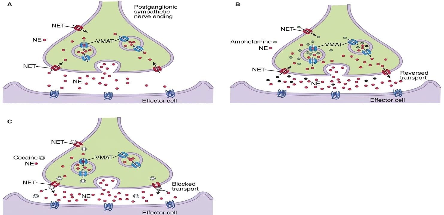
- Neuronal Reuptake (Uptake 1 / NET): Quantitatively the most important termination mechanism, clearing approximately 75% to 80% of released norepinephrine from the synapse back into the presynaptic varicosity via the high-affinity, sodium/chloride-dependent Norepinephrine Transporter (NET). Once internalized, roughly half the catecholamine is pumped back into vesicles by VMAT-2 for re-release, while the remainder is metabolized by mitochondrial enzymes.
NET Inhibitors: Cocaine and tricyclic antidepressants (TCAs: amitriptyline, imipramine) block NET directly. This prevents synaptic clearance, causing massive catecholamine accumulation at postjunctional receptors, driving acute hypertension, tachycardia, and euphoric central actions. - Extraneuronal Uptake (Uptake 2 / EMT): Circulating or non-reabsorbed catecholamines are transported into surrounding vascular smooth muscle, cardiac myocytes, hepatocytes, and endothelial cells by the low-affinity, high-capacity Extraneuronal Monoamine Transporter (EMT / OCT-3).
- Enzymatic Catabolism: Handled by two coordinated enzymes:
- Monoamine Oxidase (MAO): A flavin-containing enzyme anchored to the outer mitochondrial membrane within neuronal axoplasm and intestinal/hepatic tissues. Metabolizes intraneuronal monoamines via oxidative deamination into aldehyde derivatives. Exists in two isoforms: MAO-A (prefers serotonin, norepinephrine, and epinephrine; inhibited by moclobemide) and MAO-B (prefers dopamine and phenylethylamine; selectively inhibited by selegiline).
- Catechol-O-Methyltransferase (COMT): A cytosolic enzyme present primarily in extraneuronal tissues (liver, kidneys, vascular wall). Transfers a methyl group from SAMe to the 3-hydroxyl position of the catechol ring.
- End-Product Excretion (VMA): Sequential or parallel oxidation and methylation of epinephrine and norepinephrine yields metanephrine and normetanephrine, which are converted into vanillylmandelic acid (VMA / 4-hydroxy-3-methoxymandelic acid) and 3-methoxy-4-hydroxyphenylethylene glycol (MOPEG). VMA and metanephrines are excreted in the urine, serving as definitive biochemical diagnostic markers for catecholamine-secreting neuroendocrine tumors (pheochromocytoma and neuroblastoma).
Adrenoceptors & signal transduction
Raymond Ahlquist (1948) demonstrated that sympathomimetic drugs act on two distinct receptor classes—alpha (α) and beta (β)—distinguished by differential agonist potency rank-orders. Modern molecular cloning establishes that all adrenergic and dopaminergic receptors belong to the superfamily of seven-transmembrane, rhodopsin-like G-protein coupled receptors (GPCRs).
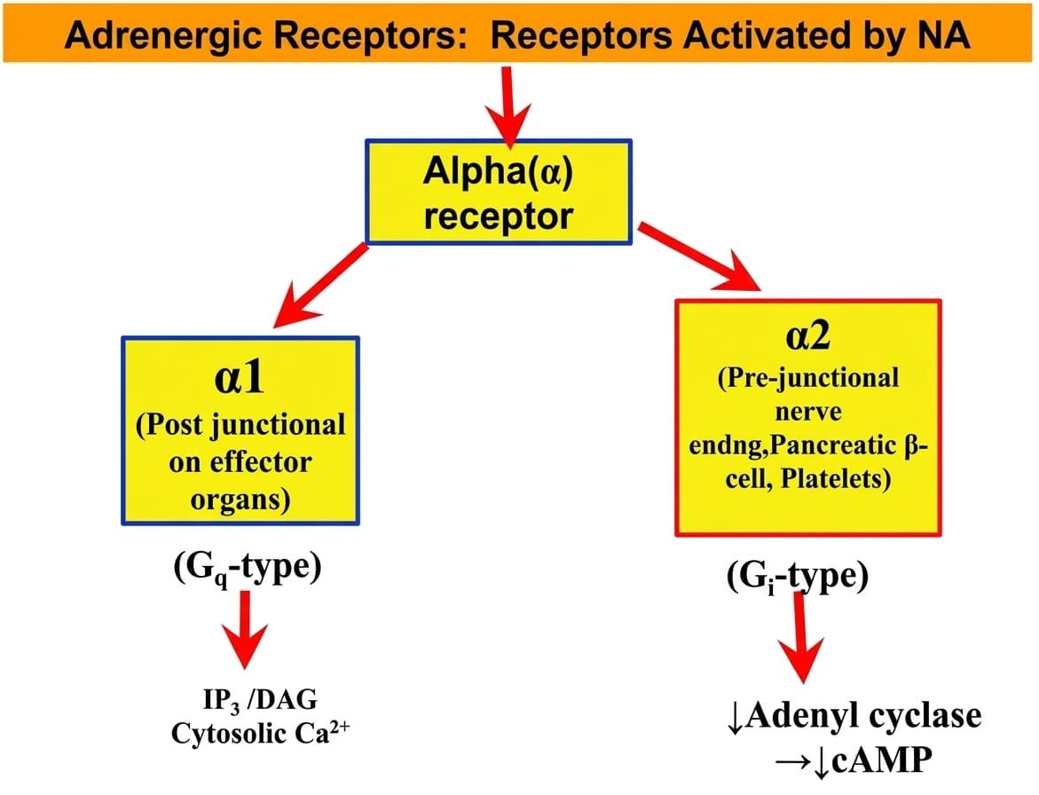
1. The Alpha-Adrenergic Family (α1 and α2)
α1-Adrenoceptors (Postjunctional Excitatory)
- G-Protein Coupling & Effector: Coupled primarily to Gq/11 proteins. Agonist activation stimulates membrane-bound phospholipase C-β (PLC-β), which hydrolyzes phosphatidylinositol 4,5-bisphosphate (PIP2) into two second messengers:
- Inositol 1,4,5-trisphosphate (IP3): Diffuses through the cytoplasm to bind IP3 receptor ligand-gated calcium channels on the sarcoplasmic/endoplasmic reticulum, releasing stored Ca2+ into the cytoplasm. Elevated intracellular Ca2+ binds calmodulin to activate myosin light-chain kinase (MLCK), driving vascular smooth muscle contraction.
- Diacylglycerol (DAG): Remains in the plasma membrane to activate protein kinase C (PKC), which phosphorylates downstream target proteins. Also stimulates phospholipase A2 (PLA2), releasing arachidonic acid and prostaglandin metabolites.
- Agonist Potency Profile: Epinephrine ≥ Norepinephrine >> Isoproterenol.
- Subtype Diversity:
- α1A: Dominant in human prostate stroma, prostatic urethra, and bladder trigone. Mediates smooth muscle contraction. Target of uroselective antagonists (e.g., tamsulosin).
- α1B: Highly expressed in cardiac tissue; facilitates cardiomyocyte protein synthesis and hypertrophy.
- α1D: Predominant in major arterial conductance vessels (aorta and coronary arteries); mediates vasoconstriction.
α2-Adrenoceptors (Autoinhibitory & Central)
- G-Protein Coupling & Effector: Coupled to pertussis toxin-sensitive Gi/o proteins. The αi subunit directly inhibits adenylyl cyclase, lowering intracellular cAMP and suppressing protein kinase A (PKA) phosphorylation. Concurrently, the Gβγ dimer complex directly inhibits presynaptic voltage-gated N-type Ca2+ channels (suppressing transmitter exocytosis) and opens inwardly rectifying G-protein-coupled potassium channels (GIRK), hyperpolarizing neuronal membranes.
- Primary Locations:
- Presynaptic Nerve Terminals: Serve as autoinhibitory feedback sensors; released NE activates α2A autoreceptors to halt further NE release.
- Brainstem Vasomotor Center: Located postsynaptically within the nucleus tractus solitarius (NTS) and ventrolateral medulla; suppresses central sympathetic outflow.
- Pancreatic β-Cells: Directly suppresses insulin exocytosis.
- Blood Platelets: Mediates epinephrine-induced platelet aggregation.
- Subtypes: α2A (mediates central sympatholysis, sedation, and analgesia), α2B (mediates initial peripheral vasoconstriction), and α2C (modulates striatal dopamine and emotional processing).
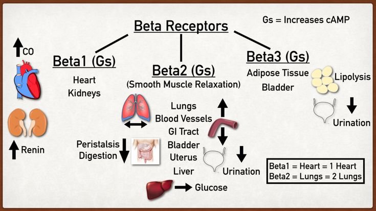
2. The Beta-Adrenergic Family (β1, β2, and β3)
β1-Adrenoceptors (“Cardiac / Renal”)
- Location: Myocardium (sinoatrial node, AV node, conduction system, atrial and ventricular myocytes), salivary glands, and juxtaglomerular (JG) cells of the renal afferent arterioles.
- Effector Mechanism: Gs-mediated adenylyl cyclase activation → elevated cAMP → PKA activation. In the heart, PKA phosphorylates:
- L-type Ca2+ channels (increasing inward Ca2+ current).
- Ryanodine receptors (RyR2) on the sarcoplasmic reticulum (amplifying calcium-induced calcium release).
- Phospholamban (accelerating SERCA-2 calcium reuptake, speeding relaxation [positive lusitropy]).
- Renal Action: Directly stimulates renin transcription and release from JG cells, activating the renin-angiotensin-aldosterone system (RAAS).
- Agonist Potency: Isoproterenol > Epinephrine = Norepinephrine.
β2-Adrenoceptors (“Smooth Muscle Relaxant / Metabolic”)
- Location: Bronchiolar smooth muscle, vascular smooth muscle of skeletal muscle and splanchnic beds, uterine myometrium, gastrointestinal tract, liver, and skeletal muscle motor terminals.
- Smooth Muscle Relaxation Mechanism: PKA phosphorylates and inactivates myosin light-chain kinase (MLCK). Concurrently, PKA enhances Ca2+ extrusion via the sarcolemmal Ca2+-ATPase pump and hyperpolarizes the membrane by opening large-conductance Ca2+-activated K+ channels (BKCa), causing profound relaxation.
- Metabolic & Skeletal Actions: Stimulates hepatic and skeletal glycogenolysis, activates the Na+/K+-ATPase pump (shifting K+ into skeletal muscle cells, causing hypokalemia), and enhances acetylcholine release at lower motor neuron endings, precipitating physiological tremor.
- Agonist Potency: Isoproterenol > Epinephrine >> Norepinephrine (NE exhibits very weak affinity for β2).
β3-Adrenoceptors (“Metabolic / Adipose / Detrusor”)
- Location: White and brown adipose tissue, urinary bladder detrusor muscle, and gallbladder.
- Actions: In adipocytes, PKA phosphorylates hormone-sensitive lipase (HSL) and perilipin, driving lipolysis (hydrolyzing triglycerides into free fatty acids and glycerol) and stimulating thermogenesis via uncoupling protein-1 (UCP-1 / thermogenin) in brown fat. In the urinary bladder, β3 activation relaxes the detrusor muscle, increasing bladder capacity (clinical target of mirabegron for overactive bladder).
- Agonist Potency: Isoproterenol = Norepinephrine > Epinephrine.
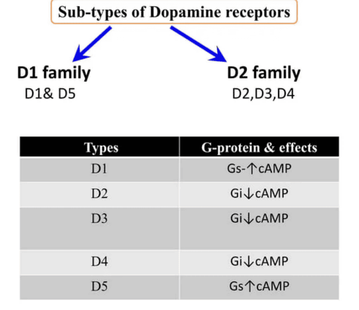
3. The Dopaminergic Family (D1 through D5)
- D1-Like Family (D1 and D5): Coupled via Gs proteins to stimulate adenylyl cyclase, elevating intracellular cAMP. Located in the renal, mesenteric, coronary, and cerebral arterial beds, where activation causes vascular smooth muscle relaxation and profound vasodilation, increasing renal blood flow, glomerular filtration rate (GFR), and natriuresis. Target of the selective hypertensive emergency agent fenoldopam.
- D2-Like Family (D2, D3, and D4): Coupled via Gi/o proteins to inhibit adenylyl cyclase, decrease cAMP, open potassium channels, and close calcium channels. Abundant presynaptically on autonomic nerves and within the central nervous system (striatum, substantia nigra, pituitary lactotrophs, and the area postrema chemoreceptor trigger zone).
Chemical & mechanistic classification
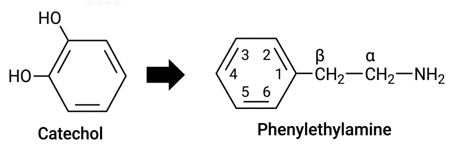
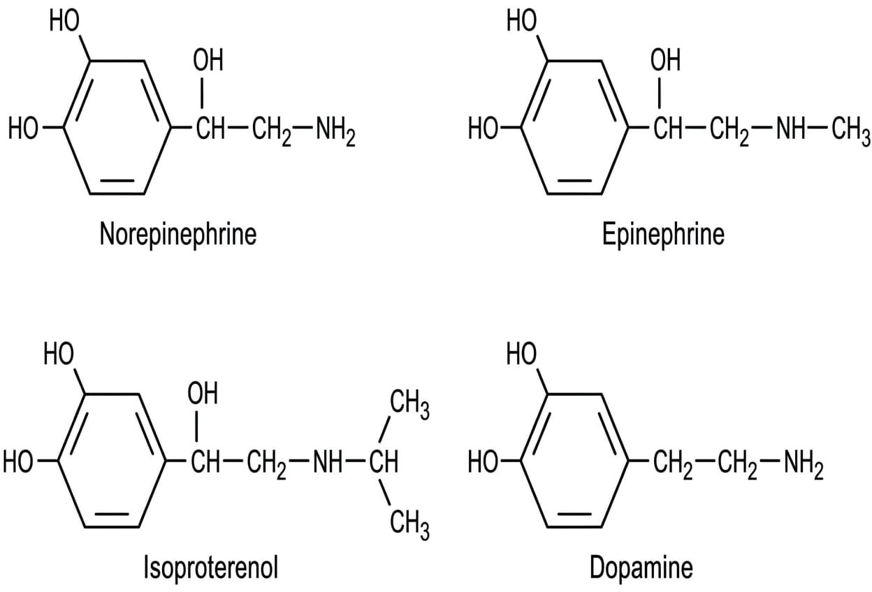
1. Chemical Classification: Catecholamines vs. Non-Catecholamines
Sympathomimetics are derived chemically from β-phenylethylamine. Modifications to the aromatic ring or aliphatic side chain dictate pharmacokinetic and pharmacodynamic profiles:
| Pharmacological Parameter | Catecholamines (Epi, NE, DA, Isoproterenol, Dobutamine) | Non-Catecholamines (Ephedrine, Amphetamine, Phenylephrine) |
|---|---|---|
| Chemical Structure | Possess 3,4-dihydroxy substitutions on the benzene ring (catechol nucleus) | Lack one or both hydroxyl groups on the aromatic ring, or possess modifications |
| Potency | Extremely high affinity and potency at α and β adrenoceptors | Generally lower potency relative to natural catecholamines |
| Oral Bioavailability | Inactive orally; rapidly degraded by intestinal mucosal and hepatic COMT/MAO | Effective orally; completely resistant to COMT degradation |
| Duration of Action | Very short (half-life measured in minutes); require continuous IV infusion | Prolonged duration of action (half-life measured in hours) |
| Blood-Brain Barrier (CNS Entry) | Polar hydroxyl groups prevent penetration across the blood-brain barrier; negligible direct CNS actions | Highly lipophilic; penetrate the blood-brain barrier readily, exerting powerful central stimulant effects |
| Enzymatic Degradation | Rapidly metabolized by both COMT and MAO | Resistant to COMT; agents with an α-methyl group (amphetamine, ephedrine) also resist MAO |
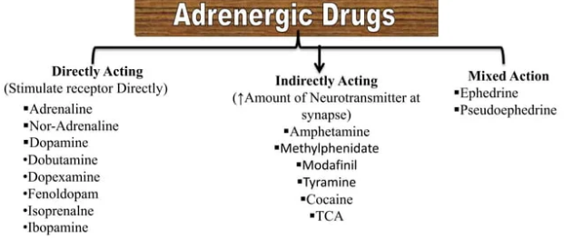
2. Mechanistic Classification: Direct, Indirect & Mixed
- Direct-Acting Agonists: Directly bind and activate postjunctional or prejunctional adrenergic receptors, mimicking endogenous catecholamines.
Examples: Epinephrine, norepinephrine, dopamine, isoproterenol, phenylephrine, methoxamine, clonidine, dobutamine, salbutamol, terbutaline.
Characteristic Response: Display receptor supersensitivity (an exaggerated response) in denervated tissues or following pretreatment with reserpine or cocaine, because the loss of reuptake mechanisms leaves more drug molecules free to stimulate postjunctional receptors. - Indirect-Acting Agonists: Do not activate adrenoceptors directly; instead, they increase the concentration of endogenous norepinephrine within the synaptic cleft:
- Releasing Agents (Displacement Inducers): Taken up into the presynaptic terminal via NET, where they reverse VMAT to displace norepinephrine from storage vesicles into the cytoplasm. Accumulating cytoplasmic NE is then pumped backwards through NET into the synaptic cleft via reverse transport. Examples: amphetamine, methamphetamine, methylphenidate, tyramine.
- Neuronal Uptake Inhibitors: Bind and occlude the NET transporter, preventing synaptic clearance. Examples: cocaine, tricyclic antidepressants.
- Mixed-Acting Agonists: Act through dual mechanisms: they directly bind and activate α and β adrenoceptors, while simultaneously entering the presynaptic varicosity to stimulate the displacement and release of stored norepinephrine.
Examples: Ephedrine, pseudoephedrine, metaraminol, phenylpropanolamine. Display partial resistance to tachyphylaxis due to their direct receptor agonist component.
Cardiovascular actions & hemodynamics
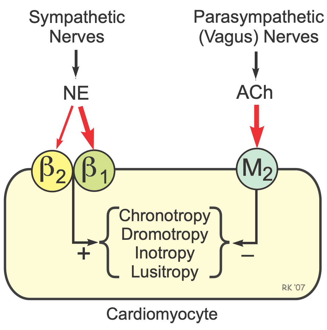
1. Direct Cardiac Effects (β1-Mediated)
Direct stimulation of myocardial β1 (and to a lesser degree β2) receptors by sympathomimetics produces four cardinal cardiac responses:
- Positive Inotropy: Increased myocardial contractility and force of contraction. PKA phosphorylation of sarcolemmal L-type Ca2+ channels and phospholamban increases peak systolic tension and stroke volume.
- Positive Chronotropy: Increased heart rate. PKA phosphorylation of hyperpolarization-activated cyclic nucleotide-gated (HCN) funny current (If) channels in the sinoatrial node accelerates the rate of phase 4 spontaneous diastolic depolarization.
- Positive Dromotropy: Increased impulse conduction velocity through the atrioventricular (AV) node, accompanied by a shortening of the AV nodal refractory period (shortened PR interval on ECG).
- Positive Lusitropy: Accelerated myocardial relaxation rate during diastole via enhanced SERCA-2 calcium reuptake, preserving diastolic filling time despite tachycardia.
- Thermodynamic Cost & Arrhythmogenicity: Enhanced automaticity of latent pacemaker tissue (Purkinje fibers) can induce sinus tachycardia, premature ventricular contractions, and ventricular fibrillation. Concurrently, stroke volume and cardiac output rise at the expense of a marked increase in myocardial oxygen consumption (MVO2). Cardiac mechanical efficiency (external cardiac work divided by oxygen consumed) decreases.
2. Vascular Smooth Muscle Effects
- α1-Mediated Vasoconstriction: Predominates in cutaneous, mucosal, mesenteric, and renal vascular beds. Increases total peripheral vascular resistance (PVR) and elevates systemic diastolic blood pressure.
- β2-Mediated Vasodilation: Predominates in the resistance arterioles supplying skeletal muscle, the splanchnic circulation, and the coronary circulation. Lowers vascular resistance in these specific beds.
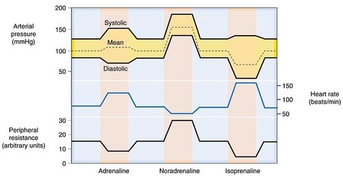
3. Comparative Hemodynamics of the Classical Catecholamines
| Cardiovascular Parameter | Epinephrine (Moderate Dose) | Norepinephrine | Isoproterenol |
|---|---|---|---|
| Receptor Selectivity | α1, α2, β1, β2 | α1, α2, β1 (negligible β2) | β1, β2, β3 (no α) |
| Systolic Blood Pressure (SBP) | ↑↑ (Increased via direct cardiac β1 inotropy and α1) | ↑↑↑ (Markedly increased via α1 constriction + β1) | ↑ or ↔ (Moderate rise via β1 stroke volume) |
| Diastolic Blood Pressure (DBP) | ↓ (Decreased via predominant skeletal β2 vasodilation) | ↑↑ (Markedly increased via generalized α1 vasoconstriction) | ↓↓↓ (Profoundly decreased via pure β2 vasodilation) |
| Mean Arterial Pressure (MAP) | Slight ↑ or ↔ (Pulse pressure widens) | ↑↑↑ (Substantially increased) | ↓↓ (Significantly decreased) |
| Peripheral Vascular Resistance (PVR) | Slight ↓ (Skeletal β2 dilation offsets cutaneous α1) | ↑↑↑ (Intense generalized vasoconstriction) | ↓↓↓ (Marked decrease across all vascular beds) |
| Heart Rate (Chronotropy) | ↑↑ (Direct cardiac β1 tachycardia dominates) | ↓↓ (Reflex bradycardia): Carotid sinus baroreceptors detect severe MAP rise, triggering vagal discharge that overrides direct β1 chronotropy | ↑↑↑ (Marked tachycardia via direct cardiac β1 plus reflex sympathetic response to low DBP) |
| Cardiac Output (CO) | ↑↑↑ (Markedly increased) | ↔ or slight ↓ (Due to intense afterload and bradycardia) | ↑↑↑ (Markedly increased) |
4. Dale’s Vasomotor Reversal
Sir Henry Dale (1913) demonstrated that the blood pressure response to epinephrine can be inverted using receptor-blocking drugs:
- Normal Biphasic Epinephrine Response: An intravenous bolus of moderate-dose epinephrine produces an initial sharp rise in blood pressure (driven by cardiac β1 inotropy and peripheral vascular α1 vasoconstriction). As circulating concentrations decay, high-affinity vascular β2 receptors remain occupied longer than lower-affinity α1 receptors, revealing a late, secondary fall in blood pressure below baseline (the β2 vasodilatory tail).
- The Reversal Phenomenon: When an animal or patient is pretreated with an α-adrenergic antagonist (e.g., phentolamine, phenoxybenzamine), the α1 vasoconstrictor effect of a subsequent dose of epinephrine is completely blocked. The drug can now activate only vascular β2 adrenoceptors, resulting in an unexpected, paradoxical precipitous drop in blood pressure (pure vasodilation). This classic phenomenon is designated Dale’s vasomotor reversal.
- Clinical Implication: Dale’s reversal occurs strictly with mixed agonists possessing both α and β activity (epinephrine). It does not occur with pure α agonists like phenylephrine (which simply has its pressor response blocked to zero) or with norepinephrine (which lacks sufficient β2 activity to drive vasodilation).
Visceral & metabolic pharmacology
1. Respiratory System
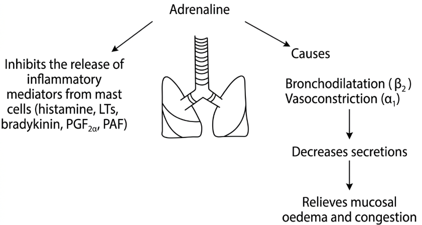
- Bronchodilation (β2): Bronchial smooth muscle lacks direct sympathetic innervation; however, it expresses abundant circulating catecholamine-sensitive β2 receptors. Activation stimulates adenylyl cyclase, lowering intracellular calcium, inactivating MLCK, and producing potent bronchodilation, dramatically reducing airway resistance.
- Mucosal Decongestion (α1): Constricts the capacitance and resistance vessels of the nasal and respiratory submucosa, reducing hydrostatic capillary pressure, promoting fluid reabsorption, and shrinking congested nasal mucous membranes.
- Mast Cell Stabilization (β2): Inhibits IgE-antigen-mediated degranulation of mast cells and basophils, suppressing the release of histamine, leukotrienes (LTC4, LTD4), prostaglandin D2, and platelet-activating factor (PAF).
2. The Eye & Intraocular Pressure Dynamics
- Mydriasis (α1): The pupillary dilator (radial) muscle of the iris contains exclusively α1 receptors. Sympathomimetic stimulation contracts this muscle, dilating the pupil (mydriasis). Because ciliary muscle accommodation is governed purely by muscarinic receptors, adrenergic mydriasis occurs without cycloplegia (near vision remains intact).
- Intraocular Pressure (IOP) Modulation: Adrenergic receptors regulate aqueous humor production and drainage through three mechanisms:
- α1-Adrenoceptors: Cause vasoconstriction of afferent ciliary arterioles, reducing ultrafiltration and transiently lowering aqueous humor formation.
- α2-Adrenoceptors: Located on the ciliary epithelium; activation inhibits adenylyl cyclase, directly decreasing aqueous humor production, while concurrently enhancing uveoscleral outflow. Clinical agents: apraclonidine and brimonidine (used topically in open-angle glaucoma).
- β2-Adrenoceptors: Stimulate ciliary epithelial secretion via cAMP, increasing aqueous humor production. Topical β-blockers (e.g., timolol, betaxolol) decrease aqueous secretion, serving as cornerstones of chronic glaucoma therapy.
3. Gastrointestinal & Genitourinary Systems
- Gastrointestinal Tract: Sympathetic activation reduces gastrointestinal propulsive activity via a coordinated three-receptor mechanism:
- α2-Presynaptic Receptors: Located on postganglionic parasympathetic cholinergic nerve terminals within the myenteric (Auerbach’s) plexus; activation inhibits acetylcholine release.
- β2-Receptors: Located directly on longitudinal and circular visceral smooth muscle; activation increases cAMP-PKA, inactivating MLCK to produce direct relaxation.
- α1-Receptors: Open membrane potassium channels, causing hyperpolarization that contributes to visceral smooth muscle relaxation while simultaneously contracting GI sphincters (ileocecal and internal anal sphincters).
- Urinary Bladder:
- β2 and β3 Receptors: Predominate in the detrusor smooth muscle; activation drives detrusor relaxation, promoting urine storage.
- α1A Receptors: Highly concentrated in the bladder neck, trigone, and prostatic urethra; activation produces sustained contraction, preventing voiding and potentially precipitating urinary retention.
- Uterine Myometrium: Responses depend on endocrine status. Non-pregnant uterus contracts under α1 stimulation. In the third trimester of pregnancy and during parturition, β2 receptors dominate, mediating profound uterine relaxation. Selective β2 agonists (ritodrine, isoxsuprine, terbutaline) are used clinically as tocolytics to arrest premature labor.
- Male Sex Organs: α1 stimulation contracts the smooth muscle of the vas deferens, prostate, and seminal vesicles, driving physiological emission and ejaculation (remember: “Point and Shoot” – Parasympathetic Erection, Sympathetic Ejaculation).
4. Metabolic & Endocrine Effects
- Carbohydrate Metabolism: Epinephrine induces acute hyperglycemia through:
- Hepatic & Skeletal Glycogenolysis (β2): PKA phosphorylates and activates phosphorylase kinase, which activates glycogen phosphorylase to break down glycogen into glucose-6-phosphate. In the liver, glucose-6-phosphatase releases free glucose into the blood. Skeletal muscle lacks this phosphatase; glycogen is metabolized into lactate, which enters the circulation.
- Gluconeogenesis: Stimulates hepatic de novo glucose synthesis from lactate, amino acids, and glycerol.
- Pancreatic Endocrine Control: α2 receptors on pancreatic β-cells directly suppress insulin secretion, while β2 receptors stimulate glucagon release from α-cells. The α2 inhibitory effect predominates, preventing insulin from counteracting stress-induced hyperglycemia.
- Peripheral Insulin Resistance: α1-receptor stimulation reduces insulin receptor signaling by decreasing GLUT-4 glucose transporter translocation in skeletal muscle.
- Lipid Metabolism: β1 and β3 adrenoceptors stimulate hormone-sensitive lipase via cAMP-PKA, accelerating the hydrolysis of triglycerides into free fatty acids and glycerol (lipolysis). Free fatty acids serve as oxidizable substrates for myocardium and skeletal muscle, driving a calorigenic state characterized by increased systemic oxygen consumption.
- Renin Secretion: Renal juxtaglomerular cells express β1 receptors that stimulate renin release, triggering angiotensin II formation and aldosterone secretion.
- Potassium Homeostasis: β2 receptors stimulate the sarcolemmal Na+/K+-ATPase pump in skeletal muscle, driving an inward potassium flux that causes hypokalemia.
Direct-acting catecholamines
1. Epinephrine (Adrenaline)
A non-selective, high-potency agonist stimulating α1, α2, β1, β2, and β3 receptors. Rapidly metabolized by liver and intestinal MAO and COMT; ineffective orally. Injected intramuscularly (IM) into the anterolateral thigh for emergency systemic absorption.
- Drug of Choice in Anaphylactic Shock: Acts as the definitive physiological antagonist to histamine and leukotrienes:
- α1 vasoconstriction reverses severe hypotension and reduces laryngeal and mucosal edema.
- β2 relaxation reverses life-threatening bronchospasm.
- β1 inotropy supports depressed cardiac output and blood pressure.
- β2 activation on mast cell membranes halts further autacoid degranulation.
- Cardiac Arrest: Administered IV or intraosseously (1 mg of 1:10,000 every 3–5 minutes) in ventricular fibrillation, pulseless electrical activity (PEA), and asystole to elevate coronary and cerebral perfusion pressures via α1 vasoconstriction.
- Local Anesthetic Adjuvant: Formulated with local anesthetics (e.g., 1:100,000 or 1:200,000 with lignocaine/lidocaine) to produce local vasoconstriction. This delays systemic anesthetic absorption, prolongs duration of anesthesia up to twofold, minimizes peak systemic anesthetic toxicity, and provides operative hemostasis. (Contraindicated in end-arterial territories: digits, tip of nose, penis, and pinna due to gangrene risk).
- Epistaxis & Superficial Bleeding: Applied topically on gauze packs (1:100,000 dilution) as an immediate hemostatic/styptic agent.
2. Norepinephrine (Noradrenaline)
A potent agonist at α1, α2, and β1 receptors with negligible action on β2 receptors. Not absorbed orally; administered strictly by continuous central IV infusion (4 to 16 μg/min) because extravasation causes ischemic tissue necrosis.
- First-Line Inotrope & Vasopressor in Septic Shock: Intense α1 vasoconstriction restores total peripheral resistance and organ perfusion pressure in vasoplegic distributive shock without inducing excessive β2-mediated vasodilation.
- Hemodynamics: Increases systolic and diastolic blood pressure, increases systemic vascular resistance, and produces a baroreceptor-mediated reflex reduction in heart rate.
3. Dopamine
An endogenous catecholamine that exhibits distinct, dose-dependent receptor selectivity:
- Low “Renal” Dose (0.5 to 2 μg/kg/min): Selectively activates vascular D1 receptors in renal, mesenteric, and coronary beds, stimulating adenylyl cyclase to induce vasodilation, increasing renal blood flow, glomerular filtration rate, and urinary sodium excretion.
- Intermediate “Cardiac” Dose (2 to 10 μg/kg/min): Directly stimulates myocardial β1 receptors and releases endogenous norepinephrine from presynaptic varicosities, producing positive inotropy, modest tachycardia, and increased cardiac output.
- High “Vasopressor” Dose (>10 μg/kg/min): Overwhelms D1 and β receptors to stimulate vascular α1 receptors, producing systemic vasoconstriction and elevating peripheral vascular resistance.
- Clinical Uses: Used in cardiogenic and septic shock accompanied by oliguria. Does not cross the blood-brain barrier.
4. Dobutamine
A synthetic catecholamine analog of dopamine administered as a racemic mixture: the (−)-enantiomer is a potent α1 agonist and weak β agonist; the (+)-enantiomer is a potent β1 and β2 agonist and potent α1 competitive antagonist. The net clinical effect is selective β1 receptor stimulation with balanced, minimal vascular actions.
- Inotropic Selectivity: Produces a prominent positive inotropic effect with minimal chronotropic effect compared to isoproterenol or dopamine. Increases cardiac stroke volume and cardiac output with a modest rise in heart rate and lower oxygen consumption cost.
- Clinical Uses: First-line inotropic agent for short-term management of acute decompensated heart failure and cardiogenic shock. Also used in pharmacological cardiac stress echocardiography.
5. Isoproterenol (Isoprenaline)
A synthetic non-selective β1, β2, and β3 agonist devoid of α-adrenergic activity. Produces powerful cardiac stimulation (β1) alongside profound peripheral vasodilation (β2).
- Clinical Profile: Produces marked tachycardia, increased cardiac output, and a sharp fall in diastolic blood pressure and total peripheral resistance. Extensively cleared by COMT; ineffective orally.
- Clinical Indications: Emergency management of severe refractory bradycardia, torsades de pointes, and partial or complete atrioventricular (AV) nodal heart block awaiting temporary transvenous pacemaker insertion.
6. Fenoldopam
A synthetic benzazepine derivative that acts as a rapid, selective postjunctional D1-receptor agonist with moderate affinity for α2 receptors, lacking action at D2, α1, or β receptors. Administered by continuous IV infusion.
- Clinical Profile: Induces rapid vasodilation across renal, mesenteric, and coronary arterial beds, lowering systemic vascular resistance and blood pressure while maintaining or increasing renal blood flow, GFR, and sodium excretion.
- Clinical Indication: Hypertensive emergencies (especially in patients with renal impairment). Eliminates the risk of cyanide or thiocyanate toxicity seen with sodium nitroprusside.
Selective alpha agonists & central agents
1. Selective Postjunctional α1-Agonists
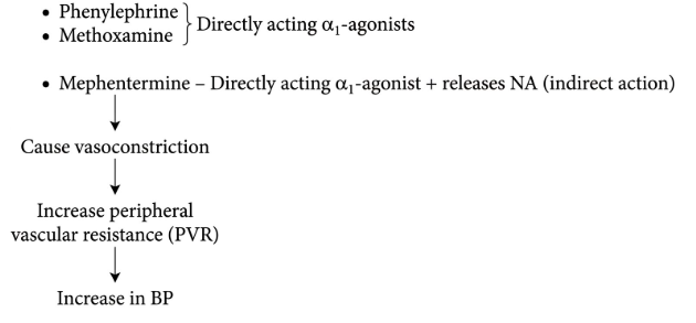
- Phenylephrine & Methoxamine: Synthetic non-catecholamines lacking catechol hydroxyl groups; resistant to COMT degradation and longer-acting than norepinephrine.
- Hemodynamics: Induce direct vasoconstriction, elevating systolic and diastolic blood pressure and total peripheral resistance, triggering pronounced baroreceptor-mediated reflex bradycardia.
- Hypotensive States: Administered parenterally to correct acute hypotension induced by spinal anesthesia or general anesthetics. (Contraindicated in hypovolemic shock, where vasoconstriction further compromises microcirculatory perfusion).
- Paroxysmal Supraventricular Tachycardia (PSVT): Historically used IV to terminate re-entrant AV nodal arrhythmias by driving a sudden rise in blood pressure, triggering intense reflex vagal discharge to slow AV nodal conduction.
- Topical Ophthalmic Use: Instilled as eye drops (2.5% or 10%) to induce rapid mydriasis for retinal examination without paralyzing accommodation (cycloplegia).
- Topical Nasal Decongestant: Applied locally as drops or sprays to relieve nasal congestion.
- Midodrine: An orally active prodrug enzymatically hydrolyzed to the selective α1 agonist desglymidodrine (peak effect at 1 hour). Produces arteriolar and venous constriction, elevating blood pressure. Used to treat disabling orthostatic (postural) hypotension. Adverse effect: supine hypertension, piloerection (goosebumps), and urinary retention.
- Topical Imidazoline Decongestants: Oxymetazoline, Xylometazoline, and Naphazoline are partial α1 and α2 agonists that produce rapid, prolonged constriction of congested nasal mucosal vasculature.
Adverse Effect – Rhinitis Medicamentosa: Use exceeding 3 to 5 consecutive days induces receptor desensitization and local ischemia, causing severe rebound nasal congestion upon drug cessation.
2. Centrally Acting α2-Agonists (Sympatholytics)
Clonidine
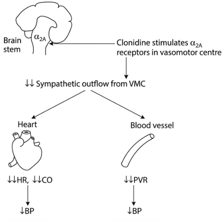
- Mechanism: An imidazoline derivative that crosses the blood-brain barrier to stimulate postjunctional α2A receptors in the nucleus tractus solitarius (NTS) of the lower brainstem. Suppresses central sympathetic outflow, reducing plasma norepinephrine, resting heart rate, and total peripheral resistance. Postural baroreflex pathways are preserved; therefore, clonidine rarely causes orthostatic hypotension. (Acute rapid IV injection causes a transient, paradoxically brief rise in blood pressure due to initial stimulation of peripheral vascular α2B receptors before central sympatholysis supervenes).
- Clinical Uses: Essential hypertension, hypertensive urgency, opioid and alcohol withdrawal management (blunts sympathetic hyperarousal), menopausal hot flushes, prophylaxis of migraine, and autonomic diabetic diarrhea (enhances intestinal fluid and electrolyte absorption).
- Rebound Hypertension (Withdrawal Crisis): Abrupt discontinuation after chronic use triggers life-threatening rebound hypertension, tachycardia, headache, and tremors caused by the sudden release of uninhibited catecholamines acting on upregulated postjunctional receptors. Managed by reinstituting clonidine or administering combined α- and β-blockers (e.g., phentolamine alongside labetalol).
α-Methyldopa
- Mechanism: An aromatic amino acid prodrug transported across the blood-brain barrier via L-type amino acid transporters. In central adrenergic neurons, it is decarboxylated by DOPA decarboxylase to α-methyldopamine, then hydroxylated by DBH into α-methylnorepinephrine (a “false transmitter”). α-Methylnorepinephrine is stored in vesicles, released into the synapse, and selectively stimulates central medullary α2 receptors, suppressing central sympathetic outflow and lowering peripheral vascular resistance.
- Drug of Choice in Pregnancy: First-line agent for managing chronic hypertension and preeclampsia in pregnant women due to decades of proven fetal hemodynamic safety.
- Adverse Effects: Sedation, mental lassitude, dry mouth, nasal stuffiness, hyperprolactinemia, drug-induced hepatitis, and development of a positive direct Coombs test (autoimmune hemolytic anemia in 1–5% of chronic users).
Other α2 Agonists
- Dexmedetomidine: A highly selective α2 agonist (α2:α1 ratio 1600:1) administered IV for sedation of intubated patients in intensive care units (ICU) and procedural anesthesia. Produces conscious sedation and analgesia without depressing respiratory drive.
- Apraclonidine & Brimonidine: Topically applied α2 agonists that decrease intraocular pressure in open-angle glaucoma by suppressing aqueous humor secretion and increasing uveoscleral outflow. Brimonidine crosses the blood-brain barrier less than clonidine, minimizing systemic hypotension.
- Tizanidine: A centrally acting α2 agonist in the spinal cord used as a skeletal muscle relaxant (spasmolytic) to treat spasticity in multiple sclerosis, stroke, and spinal trauma.
Selective beta agonists & tocolytics
1. Short-Acting β2 Agonists (SABAs)
Salbutamol (Albuterol) and Terbutaline are non-catecholamine agonists engineered with bulky tertiary butyl amino substitutions and altered ring hydroxyl positions (saligenin or resorcinol rings), conferring high selectivity for β2 receptors over β1 receptors, resistance to COMT degradation, and oral/inhalational bioavailability.
- Acute Bronchospasm: Administered via metered-dose inhalers (MDI) or nebulization as first-line rescue therapy for acute asthma attacks and COPD exacerbations. Rapid onset (<5 minutes); duration 4 to 6 hours.
- Treatment of Acute Hyperkalemia: Inhaled or IV salbutamol stimulates β2 receptors on skeletal muscle sarcolemma, activating the Na+/K+-ATPase pump to shift potassium from the extracellular fluid into the intracellular compartment, rapidly lowering serum K+.
2. Long-Acting β2 Agonists (LABAs)
- Salmeterol & Formoterol: Highly lipophilic molecules with extended aliphatic side chains that anchor into the cell membrane adjacent to the β2 receptor. Provide sustained bronchodilation lasting ≥12 hours. Used for chronic maintenance therapy in moderate-to-severe asthma and COPD (always co-prescribed with inhaled corticosteroids [ICS] in asthma to prevent fatal asthma exacerbations).
- Ultra-LABAs: Indacaterol, vilanterol, and olodaterol provide 24-hour bronchodilation, permitting once-daily maintenance dosing in COPD.
3. Uterine Relaxants (Tocolytics)
Ritodrine and Isoxsuprine (alongside salbutamol and terbutaline) are selective β2 agonists administered orally or by continuous IV infusion to arrest premature labor. Activation of myometrial β2 receptors stimulates cAMP-PKA, phosphorylating MLCK, lowering free cytosolic Ca2+, and arresting active uterine contractions to prolong pregnancy for 48 to 72 hours. This therapeutic window permits maternal administration of antenatal corticosteroids (dexamethasone or betamethasone) to accelerate fetal lung surfactant production.
Adverse Effects of β2 Agonists
Clinical connection
Systemic Complications of β2 Agonist Therapy
- Skeletal Muscle Tremor: Direct β2 stimulation on neuromuscular terminals facilitates excessive acetylcholine exocytosis, producing dose-limiting fine muscle tremors (propranolol-sensitive).
- Tachycardia & Palpitations: Results from two additive mechanisms: (1) partial loss of selectivity at higher doses directly activating cardiac β1 receptors; and (2) reflex sympathetic activation secondary to β2-mediated peripheral vasodilation.
- Metabolic Hyperglycemia: Accelerated glycogenolysis and gluconeogenesis can destabilize glycemic control in diabetic patients.
- Hypokalemia: Excessive stimulation of the Na+/K+-ATPase pump can trigger cardiac arrhythmias in predisposed patients.
Indirect- & mixed-acting sympathomimetics
1. Ephedrine & Pseudoephedrine (Mixed-Acting)
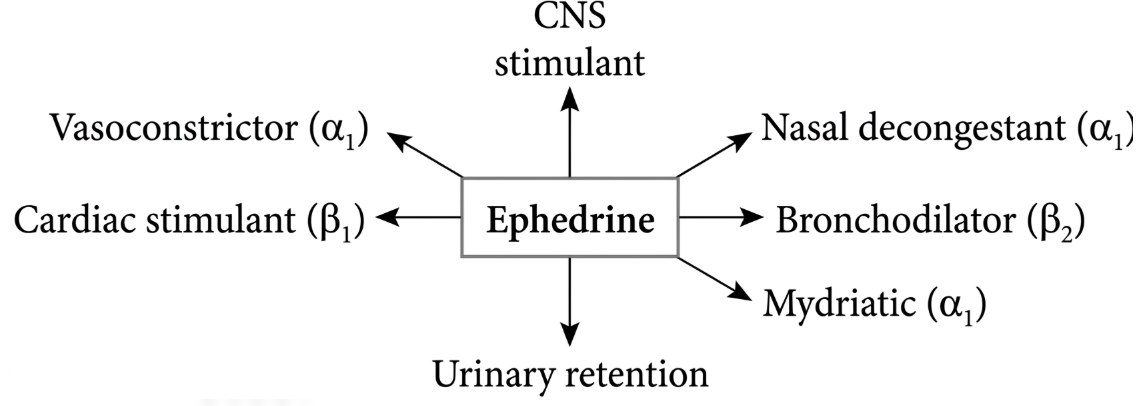
- Ephedrine: A naturally occurring non-catecholamine alkaloid extracted from plants of the genus Ephedra (Ma Huang). Acts by directly stimulating α and β adrenoceptors while entering presynaptic terminals to displace stored norepinephrine. Because it possesses an α-methyl group and lacks catechol hydroxyls, it is completely resistant to MAO and COMT, displaying high oral bioavailability, prolonged duration of action, and blood-brain barrier permeability. Used to prevent or treat hypotension during spinal anesthesia. Displays tachyphylaxis upon repeated administration.
- Pseudoephedrine: A stereoisomer of ephedrine with fewer central stimulant actions and lower β2 affinity. Widely formulated as an oral over-the-counter (OTC) decongestant for symptomatic management of allergic rhinitis and upper respiratory tract viral infections.
- Phenylpropanolamine (PPA): Chemically similar mixed agonist used historically as an oral decongestant and appetite suppressant, largely removed from clinical markets due to an increased risk of hemorrhagic stroke in young women.
2. Amphetamines & Psychostimulants (Indirect-Acting Releasers)
- Amphetamine, Dextroamphetamine & Methamphetamine: Basic non-catecholamine compounds that readily cross the blood-brain barrier. Enter presynaptic monoaminergic nerve terminals via NET and DAT, competitively displace norepinephrine and dopamine from storage vesicles via VMAT-2, and drive reverse transport through NET and DAT into the synaptic cleft.
- Central Actions: Marked elevation in cortical and reticular alertness, decreased fatigue, insomnia, euphoria, heightened concentration, generalized psychomotor stimulation, and suppression of the lateral hypothalamic feeding center (anorexia).
- Therapeutic Indications: Narcolepsy (uncontrollable daytime sleep attacks) and Attention-Deficit/Hyperactivity Disorder (ADHD) in children, where paradoxical calming and improved focus result from enhanced dopamine and norepinephrine signaling in the prefrontal cortex. Methylphenidate is preferred over amphetamine in ADHD due to lower abuse potential.
- Adverse Effects & Toxicity: Severe insomnia, tremor, anxiety, paranoid psychosis, hypertension, tachycardia, palpitations, and cardiac arrhythmias. Banned as an illicit performance-enhancing agent in athletes.
- Management of Acute Overdose: Amphetamine is a basic amine (pKa ≈ 9.9). Acidification of urine with ascorbic acid (vitamin C) or ammonium chloride converts the drug into its charged, non-absorbable ammonium form, preventing renal tubular reabsorption and accelerating clearance. Central agitation is managed with benzodiazepines, and severe hypertension is treated with sodium nitroprusside or phentolamine.
- Modafinil: A wakefulness-promoting psychostimulant that inhibits dopamine and norepinephrine reuptake while modulating GABA and glutamate transmission. First-line agent for narcolepsy, shift work sleep disorder, and obstructive sleep apnea. Produces less psychomotor euphoria and lower abuse liability than classic amphetamines.
3. Anorectic Agents (Appetite Suppressants)
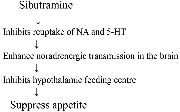
- Sibutramine: A combined norepinephrine and serotonin (5-HT) reuptake inhibitor that enhances central satiety signaling while stimulating peripheral β3-mediated thermogenesis in brown adipose tissue. Withdrawn from clinical use due to increased cardiovascular morbidity (myocardial infarction, stroke).
- Fenfluramine & Dexfenfluramine: Indirect agents that selectively trigger central serotonin release, suppressing appetite without amphetamine-like dopamine-mediated psychomotor agitation or addiction liability. Withdrawn internationally due to association with pulmonary arterial hypertension and cardiac valvular fibroplasia.
4. The Tyramine “Cheese Reaction”
Tyramine is a dietary monoamine byproduct of bacterial tyrosine fermentation present in high concentrations in aged cheeses, red wines, draft beers, smoked or pickled meats, and yeast supplements. Normally, ingested dietary tyramine is completely catabolized in the intestinal mucosa and liver by MAO-A, preventing systemic absorption.
| Food Commodity | Tyramine Content (mg per Average Serving) | Clinical Significance in Patients on MAO Inhibitors |
|---|---|---|
| Natural or Aged Cheeses (Cheddar, Gruyère, Stilton) | Nil to 130 mg | Consistently dangerous; a single serving can exceed the threshold for hypertensive crisis |
| Smoked or Pickled Fish (Herring) | Nil to 198 mg | Extremely high risk of severe hypertensive reaction |
| Fermented Sausages (Salami, Pepperoni, Summer Sausage) | Nil to 74 mg | Dangerous; sufficient to precipitate severe cardiovascular crisis |
| Yeast Supplements (Brewer’s Yeast Extracts) | 2 to 68 mg | High risk; consistently dangerous in concentrated form |
| Beer (Unpasteurized or Tap) | 4 to 45 mg | Moderate to high risk upon multi-unit consumption |
| Red Wine (Chianti) | Nil to 3 mg | Variable risk; generally low to moderate unless consumed in high volume |
| Broad Beans (Fava Beans) | Negligible tyramine, but contains free L-DOPA | Dangerous; direct DOPA precursor bypasses rate-limiting tyrosine hydroxylase |
| Chocolate | Negligible tyramine, but contains phenylethylamine | Can trigger mild central and cardiovascular stimulation in high amounts |
Clinical Note: In a patient treated with an irreversible, non-selective monoamine oxidase inhibitor (e.g., phenelzine, tranylcypromine), ingestion of as little as 20 to 50 mg of tyramine allows intact dietary tyramine to enter the systemic circulation. Tyramine is transported into presynaptic adrenergic terminals, displacing massive quantities of norepinephrine into the synaptic cleft, precipitating an acute hypertensive crisis, intracranial hemorrhage, and cardiac arrhythmias. Emergency treatment: IV α-adrenergic blockers (e.g., phentolamine).
Emergency protocols & clinical vignettes
1. Emergency Protocol: Anaphylactic Shock
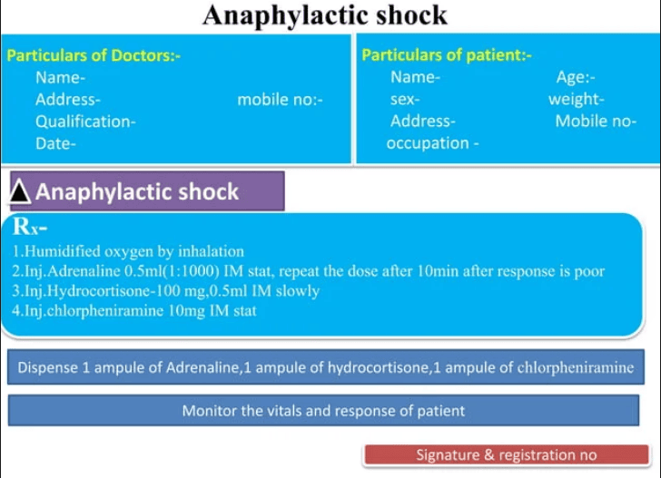
Anaphylaxis is a severe, life-threatening systemic type I hypersensitivity reaction characterized by rapid airway compromise, bronchospasm, and circulatory collapse. Epinephrine is the primary, life-saving therapeutic intervention:
- Route of Choice: Administered Intramuscularly (IM) into the anterolateral mid-thigh (vastus lateralis muscle). The vastus lateralis has extensive vascularity, achieving peak plasma drug concentrations significantly faster and more reliably than subcutaneous (SC) or deltoid injection. (Subcutaneous absorption is impaired by local α1 vasoconstriction, delaying therapeutic delivery).
- Dosing Protocol:
- Adult dose: 0.3 to 0.5 mg (0.3 to 0.5 mL of 1:1000 [1 mg/mL] solution) IM stat.
- Pediatric dose: 0.01 mg/kg (0.01 mL/kg of 1:1000 solution) IM up to a maximum single dose of 0.3 mg.
- Repeat every 5 to 15 minutes as needed based on clinical response.
- Secondary Adjuvants: Once epinephrine is administered, secondary medications are initiated: high-flow oxygen, aggressive crystalloid fluid boluses, IV antihistamines (H1 blocker: chlorpheniramine 10 mg IM/IV; H2 blocker: ranitidine), and IV corticosteroids (hydrocortisone 100 to 200 mg IM/IV) to prevent biphasic late-phase inflammatory reactions.
2. Diagnostic Localization of Horner’s Syndrome
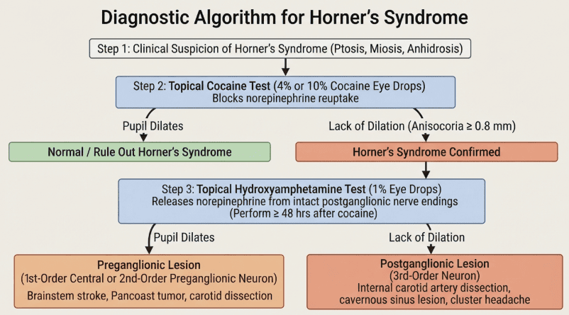
Horner’s syndrome results from interruption of the three-neuron oculosympathetic pathway, characterized by ipsilateral miosis, partial ptosis, and facial anhidrosis. Autonomic pharmacology provides the definitive tools to diagnose and localize the anatomical lesion:
- Step 1 – Confirmation of Diagnosis (The Cocaine Test): Cocaine blocks the NET transporter, preventing reuptake of physiologically released norepinephrine in the synaptic cleft of the iris dilator muscle:
- Normal Eye: Accumulating synaptic norepinephrine stimulates α1 receptors, causing robust pupillary dilation.
- Horner’s Eye: Because sympathetic drive is interrupted, no basal norepinephrine is released; cocaine has no transmitter to trap, and the pupil fails to dilate. An increase in anisocoria confirms the diagnosis of Horner’s syndrome. (Alternatively, topical apraclonidine 0.5% dilates the Horner pupil due to denervation supersensitivity while having minimal effect on a normal pupil, reversing the anisocoria).
- Step 2 – Anatomical Localization (The Hydroxyamphetamine Test): Hydroxyamphetamine stimulates the release of preformed norepinephrine from intact postganglionic (third-order) adrenergic nerve endings:
- Preganglionic Lesion (First- or Second-Order Neuron, e.g., Pancoast Tumor): The postganglionic third-order neuron remains anatomically intact and maintains its normal vesicular stores of norepinephrine. Hydroxyamphetamine releases this stored norepinephrine, producing normal pupillary dilation.
- Postganglionic Lesion (Third-Order Neuron, e.g., Internal Carotid Artery Dissection): The distal axonal terminals of the postganglionic neuron have degenerated, losing their catecholamine stores. Hydroxyamphetamine fails to dilate the affected pupil.
- Phenylephrine Confirmation: A dilute direct α1 agonist (e.g., 0.1% phenylephrine) has minimal effect on a normal pupil, but induces marked mydriasis in a postganglionic lesion due to pronounced postjunctional denervation supersensitivity.
3. Clinical “Explain Why” Master Vignettes
Vignette 1: Why does Clonidine improve diarrhea in diabetics with autonomic neuropathy?
Long-standing diabetes mellitus often damages enteric autonomic nerves, impairing normal sympathetic inhibitory modulation of the gut. This leads to unopposed parasympathetic secretomotor tone, hypermotility, and secretory diarrhea. Clonidine stimulates α2 receptors within the enteric nervous system and enterocytes. This exerts a dual therapeutic effect: (1) it directly stimulates mucosal α2 receptors to enhance sodium, chloride, and water absorption from the intestinal lumen; and (2) it activates presynaptic α2 autoreceptors on cholinergic terminals, suppressing acetylcholine release and reducing propulsive peristalsis.
Vignette 2: Why is Clonidine an effective antihypertensive that avoids orthostatic hypotension?
Unlike peripheral α-blockers (e.g., prazosin) or ganglionic blockers that disable postural vasoconstrictor reflexes, clonidine acts centrally within the nucleus tractus solitarius (NTS) of the medulla. By stimulating central α2A receptors, it resets central baroreflex setpoints, reducing basal sympathetic outflow to the heart and vasculature while leaving peripheral postjunctional α1 receptors fully responsive. When the patient stands upright, acute pooling of blood in the lower limbs triggers intact peripheral compensatory baroreceptor reflex arc signals, eliciting normal venous and arterial constriction and preventing orthostatic hypotension.
Vignette 3: Why is Adrenaline the drug of choice for Anaphylaxis but avoided in Cardiogenic Shock?
Anaphylaxis is a multi-organ systemic allergic crisis requiring simultaneous reversal of histamine across multiple targets: α1 vasoconstriction restores systemic pressure and resolves upper airway edema, β2 bronchodilation arrests bronchospasm, and β2 mast cell stabilization halts further autacoid degranulation. In contrast, cardiogenic shock stems from primary failure of the cardiac pump (e.g., massive myocardial infarction). Administering adrenaline in cardiogenic shock is hazardous: its intense β1 chronotropic and inotropic actions massively increase myocardial oxygen demand (MVO2) and promote lethal ventricular arrhythmias in an already ischemic myocardium, while its α1 vasoconstriction drastically increases cardiac afterload. For cardiogenic shock, dobutamine (selective inotrope without excessive chronotropy or vasoconstriction) or norepinephrine is preferred.
Vignette 4: Why was Fenfluramine preferred over Amphetamine as an anorectic agent?
Amphetamine suppresses appetite by stimulating the release of dopamine and norepinephrine within the central nervous system. However, this same dopaminergic stimulation in the nucleus accumbens drives intense psychomotor stimulation, insomnia, physical dependence, and profound abuse and addiction liability. Fenfluramine was developed to bypass catecholaminergic reward circuits; it selectively promotes the release and inhibits the reuptake of serotonin (5-HT) in the hypothalamic feeding center. This induces satiety without producing amphetamine-like central excitation, euphoria, or addictive potential (though later withdrawn due to secondary pulmonary hypertension and cardiac valvulopathy).
Drug summary tables
| Therapeutic Category | Representative Agents | Receptor Target(s) | Clinical Indications & Practical Notes |
|---|---|---|---|
| Pressor Agents | Norepinephrine, Phenylephrine, Methoxamine, Mephentermine, Ephedrine | α1 >> β | Septic shock (NE), spinal anesthesia hypotension (Phenylephrine), hypotensive emergencies; elevate PVR and MAP |
| Cardiac Stimulants | Epinephrine, Dobutamine, Isoproterenol, Dopamine | β1, β2, D1 | Cardiac arrest (Epi), acute cardiogenic shock (Dobutamine, DA), severe AV block (Isoproterenol); increase inotropy and CO |
| Bronchodilators | Salbutamol, Terbutaline, Salmeterol, Formoterol, Bambuterol, Epinephrine | β2 selective (> β1) | Acute asthma and COPD rescue (Salbutamol), nocturnal/maintenance asthma (Salmeterol); relax bronchiolar smooth muscle |
| Nasal Decongestants | Oxymetazoline, Xylometazoline, Naphazoline, Pseudoephedrine, Phenylephrine | α1, α2 | Allergic rhinitis, acute coryza, sinusitis; topical imidazoline use limited to <3–5 days to avoid rhinitis medicamentosa |
| CNS Stimulants | Amphetamine, Dextroamphetamine, Methamphetamine, Methylphenidate, Modafinil | Indirect (DAT, NET, VMAT release) | Narcolepsy, Attention-Deficit/Hyperactivity Disorder (ADHD); elevate cortical arousal and attention |
| Anorectics (Appetite Suppressants) | Sibutramine, Fenfluramine, Dexfenfluramine | 5-HT / NET reuptake & release; β3 | Obesity management (historically); suppress lateral hypothalamic feeding center; largely withdrawn for cardiovascular toxicity |
| Uterine Relaxants (Tocolytics) | Ritodrine, Isoxsuprine, Terbutaline, Salbutamol | β2 selective | Arrest premature labor (delay delivery 48–72 hours to administer maternal antenatal steroids for fetal lung maturity) |
| Centrally Acting Antihypertensives | Clonidine, α-Methyldopa, Guanfacine, Guanabenz | Central α2A | Chronic hypertension, preeclampsia in pregnancy (α-Methyldopa), opioid withdrawal; suppress central sympathetic outflow |
| Ophthalmic Agents | Phenylephrine, Apraclonidine, Brimonidine | α1 (mydriasis), α2 (glaucoma) | Mydriasis without cycloplegia for fundoscopy (Phenylephrine); open-angle glaucoma (Brimonidine decreases aqueous humor) |
| Local Anesthetic Adjuvants & Hemostatics | Epinephrine (1:100,000 to 1:200,000) | α1 | Prolong local anesthesia duration, reduce surgical field bleeding, manage epistaxis; avoid in end-artery terminal digits |
Quick recap
References
The following authoritative medical pharmacology references and clinical guidelines were verified in the synthesis of this curriculum study note:
- Katzung’s Basic & Clinical Pharmacology: Katzung, B.G. and Vanderah, T.W. (2021) Basic & Clinical Pharmacology. 15th edn. New York: McGraw-Hill Education. [Chapter 9: Adrenoceptor Agonists & Sympathomimetic Drugs; Chapter 6: Introduction to Autonomic Pharmacology].
- Goodman & Gilman’s Pharmacological Basis of Therapeutics: Brunton, L.L., Hilal-Dandan, R. and Knollmann, B.C. (2018) Goodman & Gilman’s The Pharmacological Basis of Therapeutics. 13th edn. New York: McGraw-Hill Education. [Chapter 8: Neurotransmission: The Autonomic and Somatic Motor Nervous Systems; Chapter 12: Adrenergic Agonists and Antagonists].
- Lippincott Illustrated Reviews: Pharmacology: Whalen, K. (2019) Lippincott Illustrated Reviews: Pharmacology. 7th edn. Philadelphia: Wolters Kluwer. [Chapter 6: Adrenergic Agonists].
- Rang & Dale’s Pharmacology: Ritter, J.M., Flower, R., Henderson, G., Loke, Y.K., MacEwan, D. and Rang, H.P. (2020) Rang & Dale’s Pharmacology. 9th edn. Edinburgh: Elsevier. [Chapter 15: Noradrenergic Transmission].
- Uganda Clinical Guidelines: Ministry of Health Uganda (2023) Uganda Clinical Guidelines: National Guidelines for the Management of Common Conditions. Kampala: Ministry of Health. [Section 1.1: Anaphylactic Shock Management; Section 7.2: Hypertensive Disorders in Pregnancy].