
Cholinoceptor blocking drugs: antimuscarinic, ganglionic & neuromuscular blockers
Overview & receptor classes
Cholinoceptor blocking drugs (also known as cholinergic antagonists, cholinergic blockers, or anticholinergic agents) are pharmacological compounds that bind selectively to acetylcholine receptors without initiating receptor-mediated intracellular signaling. By occupying the orthosteric binding site or allosteric channels, these agents prevent endogenous acetylcholine (ACh) or exogenously administered cholinomimetic agonists from exerting their physiological actions.
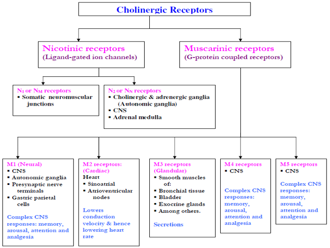
Primary Cholinergic Receptor Subtypes
1. Nicotinic Receptors (Ligand-Gated Ion Channels)
Pentameric ion channels that mediate rapid depolarization through direct influx of Na+ and Ca2+ ions upon binding two molecules of ACh:
- N1 or NM Subtype: Localized exclusively at the postjunctional motor endplate of the somatic neuromuscular junction. Triggers skeletal muscle contraction.
- N2 or NN Subtype: Localized on postganglionic neuronal cell bodies across all autonomic ganglia (both sympathetic and parasympathetic), within the central nervous system, and on neuroendocrine chromaffin cells of the adrenal medulla (mediating epinephrine and norepinephrine release into the bloodstream).
2. Muscarinic Receptors (G-Protein Coupled Receptors)
Seven-transmembrane metabotropic receptors that regulate intracellular second-messenger enzymes:
- M1 (“Neural”): Coupled via Gq/11 proteins to stimulate phospholipase C (PLC), producing inositol trisphosphate (IP3) and diacylglycerol (DAG). Distributed in the central nervous system (mediating memory, arousal, attention, and analgesia), autonomic ganglia, presynaptic nerve terminals, and gastric parietal cells.
- M2 (“Cardiac”): Coupled via Gi/o proteins to inhibit adenylyl cyclase (lowering cyclic AMP [cAMP]) and open inwardly rectifying potassium channels (KAch). Concentrated in the heart (sinoatrial [SA] node and atrioventricular [AV] node), where it decreases conduction velocity, lowers automaticity, and decreases heart rate. Also acts as an inhibitory autoreceptor on postganglionic parasympathetic and sympathetic nerve terminals.
- M3 (“Glandular / Smooth Muscle”): Coupled via Gq/11 to the IP3/DAG pathway, mobilizing intracellular Ca2+. Located on visceral smooth muscle (bronchial tree, gastrointestinal tract, urinary bladder detrusor), exocrine glands (salivary, lacrimal, bronchial, eccrine sweat glands), and vascular endothelium (mediating nitric oxide-dependent vasodilation).
- M4 & M5 Receptors: Located primarily within the central nervous system; participate in complex striatal dopaminergic modulation, memory, extrapyramidal motor processing, and pain transmission.
Classification of cholinergic antagonists
Cholinergic antagonists fall into three major clinical classes based on the specific receptor populations and anatomical divisions they block: antimuscarinic agents (parasympathoplegics), ganglionic blockers (sympatho- and parasympathoplegics), and neuromuscular junction blockers.
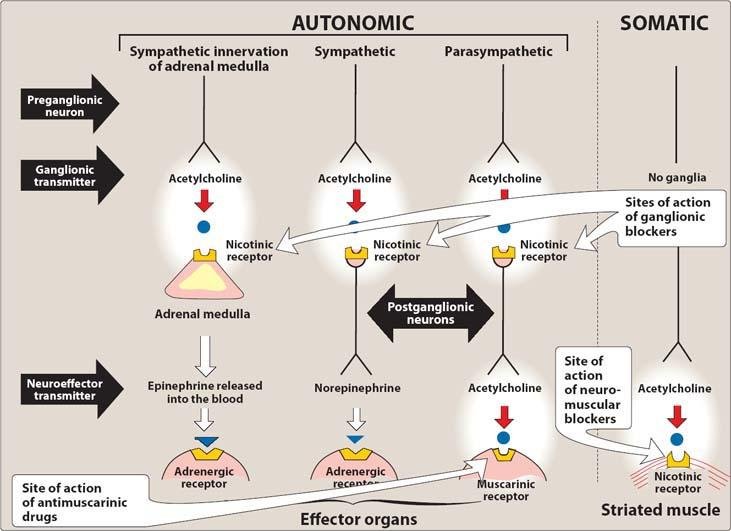

Clinical Significance of the Three Classes
- 1. Muscarinic Antagonists: The most clinically versatile group. By selectively or non-selectively preventing ACh from binding muscarinic receptors at parasympathetic neuroeffector junctions, parasympathetic innervation is interrupted, leaving sympathetic tone unopposed. In addition, they block the exceptional sympathetic cholinergic nerves supplying eccrine sweat glands. Because they do not block nicotinic receptors, they exert negligible effects on skeletal neuromuscular junctions or autonomic ganglia at therapeutic doses.
- 2. Neuromuscular Blocking Agents (NMBDs): Interfere with transmission between somatic motor nerve terminals and nicotinic (NM) receptors on the skeletal muscle endplate. Used as essential adjuvant relaxants in surgical anesthesia, endotracheal intubation, and orthopedic procedures.
- 3. Ganglionic Blockers: Inhibit nicotinic (NN) receptors within both sympathetic and parasympathetic ganglia. Because they indiscriminately eliminate all autonomic outflow, their wide-ranging, poorly tolerated side effects render them obsolete for chronic clinical therapy, though they remain important physiological tools.
Antimuscarinics: chemistry & pharmacokinetics
Natural Alkaloids & Chemical Architecture
Atropine and its naturally occurring congener scopolamine (hyoscine) are tertiary amine alkaloid esters of tropic acid combined with complex organic bases (tropine and scopine, respectively):
- Botanical Sources: Atropine is derived from Atropa belladonna (deadly nightshade) and Datura stramonium (sacred Datura, jimsonweed, or thorn apple). Scopolamine is isolated from Hyoscyamus niger (henbane).
- Stereochemistry: Naturally occurring plant-derived atropine is optically active l-hyoscyamine. During industrial extraction and chemical purification, it readily racemizes into an equimolar racemic mixture of d- and l-hyoscyamine, known pharmacologically as atropine. Nearly all antimuscarinic activity resides in the l-enantiomer.

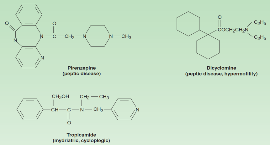
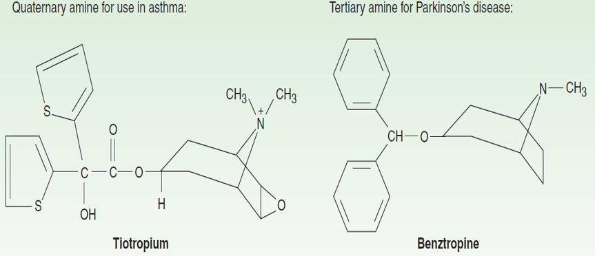
Pharmacokinetic Principles: Absorption, Distribution & Elimination
- Absorption: Natural tertiary alkaloids and synthetic tertiary amines (atropine, scopolamine, tropicamide, oxybutynin) are lipophilic and rapidly absorbed across the gastrointestinal mucosa, conjunctival membranes, and intact skin. Scopolamine is effectively delivered via transdermal patches. Conversely, quaternary amines (ipratropium, tiotropium, glycopyrrolate, propantheline) carry a permanent positive charge, limiting oral bioavailability to only 10% to 30% due to poor lipid membrane partition.
- Distribution: Tertiary agents distribute extensively throughout whole-body tissues. Significant concentrations cross the blood-brain barrier within 30 to 60 minutes. Scopolamine achieves rapid, complete CNS equilibrium, yielding greater central sedative and amnestic actions than atropine. Quaternary amine derivatives are largely excluded from the CNS at therapeutic doses, restricting their pharmacological profile to peripheral tissues.
- Metabolism & Excretion: Elimination of atropine from plasma is biphasic:
- Rapid distribution phase half-life: t1/2 ≈ 2 hours.
- Slow elimination phase half-life: t1/2 ≈ 13 hours.
- Organ-Specific Dissociation Kinetics: While systemic antimuscarinic effects decline rapidly across visceral tissues, ocular blockade persists markedly longer: effects on the iris and ciliary muscle persist for ≥ 72 hours and up to 7 to 10 days due to drug trapping in pigmented ocular structures.
| Drug Name | Chemical Class | Absorption & Tissue Distribution | CNS Penetration & Clinical Impact |
|---|---|---|---|
| Atropine (dl-Hyoscyamine) | Tertiary amine (Natural) | Rapid oral and mucosal absorption; widely distributed | Good CNS entry; mild initial stimulation, delayed central sedation |
| Scopolamine (Hyoscine) | Tertiary amine (Natural) | Excellent oral, mucosal, and transdermal absorption | Marked, rapid CNS penetration; causes profound drowsiness, amnesia, and antiemesis |
| Benztropine / Trihexyphenidyl | Tertiary amine (Synthetic) | Well absorbed orally; crosses blood-brain barrier readily | Centrally targeted; re-establishes striatal dopamine/ACh balance in Parkinsonism |
| Tropicamide / Cyclopentolate | Tertiary amine (Synthetic) | Rapid transcorneal absorption after ocular instillation | Short-acting mydriasis and cycloplegia; limited systemic central toxicity |
| Pirenzepine / Telenzepine | Tertiary amine (Synthetic) | Moderate oral absorption; selective for M1 receptors | Low to moderate CNS penetration at therapeutic anti-ulcer doses |
| Oxybutynin | Tertiary amine (Synthetic) | High oral, transdermal, and intravesical absorption | Moderate CNS penetration; can induce cognitive blunting and sedation in the elderly |
| Ipratropium / Tiotropium | Quaternary amine (Synthetic) | Inhaled; negligible gastrointestinal or mucosal absorption | Completely excluded from CNS; minimal systemic anticholinergic adverse effects |
| Glycopyrrolate / Propantheline | Quaternary amine (Synthetic) | Poor oral bioavailability (10–30%); peripheral distribution | Negligible CNS penetration; useful for peripheral antispasmodic and antisecretory therapy |
Pharmacodynamics & receptor interactions
Mechanism of Competitive & Inverse Agonism
Atropine causes reversible, competitive (surmountable) blockade of muscarinic cholinoceptors. The parallel rightward shift in the agonist log concentration-response curve can be completely surmounted by increasing concentrations of ACh or cholinomimetic agonists.
- Molecular Signaling Interruption: Atropine prevents agonist-induced G-protein activation, halting Gq-mediated stimulation of phospholipase C (blocking IP3/DAG production) and preventing Gi-mediated inhibition of adenylyl cyclase.
- The Inverse Agonist Concept: While classical pharmacology considered antimuscarinics to be neutral competitive antagonists, molecular cloning shows that muscarinic receptors display baseline constitutive activity (spontaneous G-protein coupling in the absence of agonist). Most clinical antimuscarinics—including atropine, pirenzepine, trihexyphenidyl, ipratropium, and glycopyrrolate—are actually inverse agonists that bind and stabilize the inactive (R) receptor conformation, suppressing constitutive basal signaling.
Differential Tissue Sensitivity to Antimuscarinic Blockade
Target organs display markedly different sensitivity thresholds to muscarinic blockade. Doses must be titrated carefully to achieve therapeutic effects without precipitating broad anticholinergic toxicity:
- Highest Sensitivity: Salivary, bronchial, and eccrine sweat glands (dry mouth, reduced bronchial secretions, and anhidrosis occur at lowest doses).
- Intermediate Sensitivity: Ocular pupillary constrictor and ciliary muscles, followed by the cardiac sinoatrial node (mydriasis, cycloplegia, and tachycardia require moderate doses).
- Lowest Sensitivity: Gastrointestinal tract smooth muscle tone and gastric parietal cell acid secretion (inhibiting hydrochloric acid requires very high doses that cause severe dry mouth and urinary retention).
| Receptor Subgroup | Primary Anatomical Locations | Dominant Effector System | Selective & Non-Selective Antagonists | Affinity Ranking (Relative Kd) |
|---|---|---|---|---|
| M1 (Neural) | CNS, autonomic ganglia, presynaptic nerves, gastric parietal cells | ↑ IP3, ↑ DAG (via Gq/11) | Pirenzepine, telenzepine, dicyclomine, trihexyphenidyl | Atropine: 1 Pirenzepine: 25 AF-DX 116: 2000 Darifenacin: 70 |
| M2 (Cardiac) | Sinoatrial node, AV node, atrium, smooth muscle, presynaptic nerves | ↓ cAMP, ↑ K+ channel current (via Gi/o) | Methoctramine, AF-DX 116, gallamine (allosteric/obsolete) | Atropine: 1 Pirenzepine: 300 AF-DX 116: 65 Darifenacin: 55 |
| M3 (Glandular) | Exocrine glands, visceral smooth muscle, vascular endothelium | ↑ IP3, ↑ DAG, ↑ intracellular Ca2+ (via Gq/11) | 4-DAMP, darifenacin, solifenacin, oxybutynin, tolterodine | Atropine: 1 Pirenzepine: 500 AF-DX 116: 4000 Darifenacin: 8 |
Note on Table Values: Dissociation constants (Kd) are expressed relative to atropine (defined as 1). Smaller numerical values indicate higher binding affinity. AF-DX 116 and 4-DAMP are experimental research antagonists.
Organ system effects
1. Central Nervous System (CNS)
- Dose-Dependent Actions of Atropine: In therapeutic doses (0.5 to 1.0 mg), atropine produces minimal central stimulation, exerting mild, slow sedative effects on subcortical centers. At toxic doses, central excitation dominates, manifesting as restlessness, irritability, disorientation, visual hallucinations, and delirium, ultimately progressing to coma and medullary respiratory depression.
- Scopolamine: Penetrates the blood-brain barrier rapidly, producing significant central depression at therapeutic doses. Manifests as drowsiness, fatigue, euphoria, and amnesia. Highly effective in mitigating vestibular motion sickness by suppressing cholinergic transmission in the vestibular nuclei and the chemoreceptor trigger zone.
- Parkinsonism & Extrapyramidal Motor Control: The normal basal ganglia circuit maintains an enzymatic balance between inhibitory dopamine (from the substantia nigra) and excitatory acetylcholine (from striatal interneurons). In Parkinson’s disease, loss of dopaminergic neurons creates a relative cholinergic excess. Centrally active antimuscarinics (benztropine, trihexyphenidyl) suppress this cholinergic overactivity, reducing tremor and rigidity.
2. The Eye
- Mydriasis: Antimuscarinics competitively block M3 receptors on the pupillary constrictor (sphincter pupillae) muscle of the iris. Unopposed sympathetic tone via α1 receptors on the radial pupillary dilator muscle causes pupil dilation (mydriasis) and complete unresponsiveness to light.
- Cycloplegia: Blockade of M3 receptors on the ciliary muscle prevents parasympathetically driven contraction. The ciliary body relaxes, pulling the zonular fibers taut, which flattens the crystalline lens. This eliminates the eye’s ability to accommodate for near vision (cycloplegia). The atropinized eye is fixed for far vision.
- Intraocular Pressure (IOP) Hazard: In patients with narrow-angle glaucoma, pupillary dilation causes the iris root to fold backward into the iridocorneal angle, physically occluding the trabecular meshwork. This halts aqueous humor drainage into the canal of Schlemm, precipitating acute angle-closure glaucoma (an ophthalmologic emergency).
- Lacrimal Secretions: M3 blockade decreases tear secretion, producing complaints of dry or “sandy” eyes.
3. Cardiovascular System
- Biphasic Heart Rate Response:
- Low Therapeutic Doses (<0.5 mg Atropine): Frequently causes a transient, paradoxical bradycardia. This is mediated by selective blockade of presynaptic autoinhibitory M1 receptors on vagal postganglionic terminals, which normally provide negative feedback to shut down ACh release. Blocking these autoreceptors increases ACh exocytosis into the sinoatrial node.
- Moderate to High Doses (≥1.0 mg Atropine): Produces progressive, marked tachycardia by competitively blocking postjunctional M2 receptors on cardiac nodal pacemaker cells, abolishing vagal inhibitory tone.
- Atrioventricular (AV) Conduction: Facilitates AV nodal transmission, shortening the PR interval on the electrocardiogram. Muscarinic blockade protects against vagally mediated AV block. Ventricular myocardium is largely spared due to sparse direct parasympathetic innervation.
- Vascular System & Hemodynamics: Most vascular beds lack direct parasympathetic innervation. However, nearly all vascular endothelial cells express uninnervated M3 receptors that synthesize nitric oxide (NO) when exposed to circulating muscarinic agonists. Atropine completely blocks agonist-mediated vasodilation.
- Atropine Flush: At toxic or near-toxic doses, antimuscarinics cause cutaneous vasodilation, particularly across the blush areas of the face and upper chest. This reflects a non-cholinergic compensatory reflex to dissipate heat caused by sweat gland paralysis.
4. Respiratory System
Both airway smooth muscle and submucosal seromucous glands receive vagal innervation containing M3 receptors. Antimuscarinic drugs cause:
- Bronchodilation: Relaxes bronchial smooth muscle, reducing airway resistance. While less effective than β2-adrenoceptor agonists (e.g., salbutamol) in acute asthma, antimuscarinics are exceptionally valuable in chronic obstructive pulmonary disease (COPD), where vagal tone is the primary reversible element of airway obstruction.
- Antisecretory Action: Dries secretions across the pharynx, larynx, trachea, and bronchi, reducing the risk of postoperative atelectasis and laryngospasm.
- Autoinhibitory M2 Consideration: Non-selective antimuscarinics block autoinhibitory M2 receptors on airway postganglionic parasympathetic nerves, increasing ACh release that can counteract M3 bronchodilation. Inhaled quaternary agents like tiotropium dissociate rapidly from M2 but slowly from M3, maintaining sustained bronchodilation.
5. Gastrointestinal Tract
- Motility & Tone: Causes dramatic relaxation of visceral smooth muscle from the stomach to the colon. Decreases muscle tone, amplitude, and frequency of peristaltic contractions, significantly prolonging gastric emptying and intestinal transit time. Intestinal paralysis is generally temporary; local enteric nervous system (ENS) pacemaking typically re-establishes basal peristalsis within 1 to 3 days.
- Secretions: Salivary glands are exquisitely sensitive; M3 blockade leads to profound xerostomia (dry mouth). Gastric acid secretion by parietal cells is relatively resistant to atropine because acid output is driven by overlapping histaminergic and gastrinergic pathways; high doses decrease acid, pepsin, and mucin volume, but cause intolerable dry mouth and cycloplegia. M1-selective antagonists (pirenzepine, telenzepine) suppress basal and stimulated acid production more selectively. Pancreatic and biliary secretions are largely unaffected, as they are primarily governed by hormonal signals (secretin, cholecystokinin).
6. Genitourinary Tract & Sweat Glands
- Urinary Bladder: Inhibits M2 and M3 receptors, relaxing the detrusor smooth muscle while contracting the internal urethral sphincter. Increases functional bladder capacity, decreases voiding frequency, and suppresses involuntary detrusor contractions. Can precipitate acute urinary retention in elderly men with benign prostatic hyperplasia (BPH). Ureters are relaxed; uterine smooth muscle is largely unaffected.
- Thermoregulatory Sweating: Thermoregulatory eccrine sweat glands are innervated by specialized sympathetic postganglionic cholinergic fibers acting via M3 receptors. Atropine suppresses sweating, producing dry skin. In adults, large doses elevate core temperature; in infants and young children, even therapeutic doses can trigger dangerous hyperthermia (“atropine fever”).
Clinical applications
1. Central Nervous System Applications
- Parkinson’s Disease: Centrally acting antimuscarinics (benztropine, trihexyphenidyl) are utilized as adjunctive therapy, particularly in early stages or alongside levodopa to relieve tremor and muscle rigidity. Also used to treat drug-induced extrapyramidal symptoms (acute dystonic reactions) caused by typical antipsychotics.
- Motion Sickness: Scopolamine (hyoscine) is the most effective single prophylactic agent for motion-induced nausea. Administered orally, parenterally, or as a transdermal patch applied behind the ear (providing sustained therapeutic plasma levels over 48 to 72 hours). Sedation and dry mouth are common side effects.
2. Ophthalmologic Applications
- Refraction Measurement: Accurate measurement of refractive errors in uncooperative patients (e.g., young children) requires complete cycloplegic paralysis of the ciliary muscle to prevent involuntary near accommodation.
- Funduscopic Examination: Induces mydriasis to permit thorough ophthalmoscopic evaluation of the retina and optic disc. Shorter-acting agents like tropicamide are preferred over atropine to minimize prolonged visual disability. When only mydriasis is needed without cycloplegia, α1-adrenoceptor agonists (e.g., phenylephrine) are preferred.
- Prevention of Synechiae: In anterior uveitis and iritis, long-acting cycloplegics (homatropine, atropine) are alternated with miotics to prevent the inflamed iris from forming adhesions (synechiae) to the anterior lens capsule or cornea.
3. Respiratory Applications
- COPD & Asthma: Inhaled quaternary antimuscarinics (ipratropium, tiotropium) provide bronchodilation with minimal systemic side effects. Tiotropium is long-acting, dissociates very slowly from M3 receptors, has a terminal elimination half-life of 5 to 6 days, allows once-daily dosing, and reduces acute COPD exacerbations.
- Pre-Anesthetic Medication: Atropine or glycopyrrolate is administered prior to inhalational anesthesia to reduce copious respiratory secretions and prevent reflex bradycardia or laryngospasm during airway instrumentation.
4. Cardiovascular Applications
- Hemodynamically Significant Bradycardia: Intravenous atropine (0.5 to 1.0 mg IV, repeated up to 3 mg) is the first-line therapy for symptomatic sinus bradycardia, vasovagal syncope, and AV nodal heart block, especially during acute myocardial infarction where intense vagal discharge impairs cardiac output.
5. Gastrointestinal Applications
- Abdominal Spasms & Traveler’s Diarrhea: Antispasmodics (dicyclomine, propantheline, glycopyrrolate) relax intestinal smooth muscle in irritable bowel syndrome. Atropine is formulated at subtherapeutic doses with diphenoxylate (Lomotil) to discourage opioid abuse.
- Peptic Ulcer Disease: Selective M1 blockers (pirenzepine, telenzepine) suppress acid secretion, though largely superseded by H2-receptor antagonists and proton pump inhibitors.
6. Genitourinary Applications
- Overactive Bladder & Incontinence: Relieves urinary urgency, frequency, and urge incontinence due to detrusor instability or neurological conditions (e.g., meningomyelocele, post-urologic surgery):
- Oxybutynin: M3-selective antagonist available as oral, transdermal, and intravesical preparations. Extended-release formulations reduce peak-related dry mouth.
- Trospium: Quaternary amine with low blood-brain barrier permeability; associated with fewer cognitive side effects in elderly patients.
- Darifenacin & Solifenacin: Highly selective M3 antagonists offering once-daily dosing due to extended elimination half-lives.
- Tolterodine, Fesoterodine & Propiverine: M3-directed bladder spasmolytics available in extended-release forms.
- Imipramine: Tricyclic antidepressant with prominent antimuscarinic actions; used historically in institutionalized elderly patients, but limited by sedation and cardiac toxicity.
| Drug Name | Duration of Ocular Action | Usual Topical Concentration (%) | Primary Clinical Indication |
|---|---|---|---|
| Atropine | 7 to 10 days | 0.5% – 1.0% | Ciliary paralysis for refraction in uncooperative children; anterior uveitis |
| Scopolamine | 3 to 7 days | 0.25% | Refraction and uveitis management in patients sensitive to atropine |
| Homatropine | 1 to 3 days | 2.0% – 5.0% | Uveitis and iritis; prevention of synechia formation |
| Cyclopentolate | 24 hours (1 day) | 0.5% – 2.0% | Routine cycloplegic refraction in pediatric and young adult clinics |
| Tropicamide | 0.25 days (6 hours) | 0.5% – 1.0% | Rapid-onset, short-duration mydriasis for routine retinal ophthalmoscopy |
Toxicity & poisoning protocols
The Anticholinergic Toxidrome
Acute toxicity from atropine, deadly nightshade berries, or drugs with secondary anticholinergic properties (TCAs, first-generation antihistamines, phenothiazines) produces a characteristic constellation of clinical signs:
Vulnerable Populations & Antidotal Management
- Pediatric Lethality: Infants and young children are exceptionally sensitive to the hyperthermic effects of atropine. While adults have survived overdoses exceeding 400 mg with intensive care, pediatric fatalities have occurred with doses as low as 2 mg.
- Symptomatic & Supportive Therapy: Core cooling blankets to manage hyperthermia, fluid resuscitation, catheterization for urinary retention, and intravenous benzodiazepines (diazepam) for central seizures and psychomotor agitation.
- Specific Antidote (Physostigmine): Reserved for severe, life-threatening anticholinergic delirium or intractable supraventricular arrhythmias. As a tertiary amine, physostigmine penetrates the blood-brain barrier and inhibits acetylcholinesterase, raising endogenous ACh levels to compete out atropine centrally and peripherally. Quaternary AChE inhibitors (neostigmine, edrophonium) do not cross the blood-brain barrier and treat only peripheral manifestations.
Management of Cholinesterase Inhibitor Poisoning
Agricultural organophosphate insecticides (parathion, malathion) and military nerve gases (sarin, soman, VX) irreversibly phosphorylate acetylcholinesterase, causing overwhelming cholinergic crisis across muscarinic, nicotinic, and central synapses.
- 1. High-Dose Antimuscarinic Therapy: Tertiary amine atropine is administered immediately (1 to 2 mg IV every 5 to 15 minutes) until full “atropinization” is achieved (clearing of pulmonary secretions, resolution of bronchoconstriction, and reversal of miosis). Severe poisonings may require up to 1 gram of atropine per day for several weeks. Critical Caveat: Atropine reverses muscarinic and central effects, but has zero effect on nicotinic neuromuscular paralysis!
- 2. Cholinesterase Regenerator Compounds (Oximes):
- Pralidoxime (2-PAM): A nucleophilic oxime that attacks the phosphorus atom of the organophosphate-enzyme complex, dislodging the inhibitor and restoring active acetylcholinesterase. Effective primarily if given before the phosphorylated enzyme undergoes “aging” (chemical loss of an alkyl side chain, making inhibition permanent). Administered as an IV infusion of 1 to 2 g over 15 to 30 minutes. Because of its charged quaternary ammonium structure, pralidoxime does not cross the blood-brain barrier and works primarily at skeletal neuromuscular junctions.
- Diacetylmonoxime (DAM): A tertiary oxime that crosses the blood-brain barrier to regenerate central cholinesterase.
- Carbamate Caveat: Pralidoxime is not indicated in reversible carbamate poisoning (e.g., neostigmine, physostigmine), as carbamylated enzyme spontaneously reactivates and oximes may add secondary toxicity.
- 3. Prophylactic Pretreatment: In anticipation of nerve gas exposure (chemical warfare), troops are pretreated with the reversible carbamate inhibitor pyridostigmine. Pyridostigmine reversibly occupies a fraction of the enzyme pool, shielding it from irreversible phosphorylation by organophosphates. When the nerve agent clears, pyridostigmine dissociates, leaving functional enzyme available. Simultaneous atropine is co-administered to control muscarinic excess.
Management of Mushroom Poisoning
Rapid-Onset vs. Delayed-Onset Mushroom Ingestion
- Rapid-Onset Poisoning (<30 Minutes to 2 Hours): Caused by ingestion of mushrooms containing preformed muscarine (e.g., Inocybe and Clitocybe species). Produces classic hypermuscarinic symptoms: nausea, vomiting, profuse diarrhea, urinary urgency, sweating, salivation, miosis, and severe bronchospasm. Treatment: Parenteral atropine (1 to 2 mg IV/IM) rapidly and completely reverses the condition.
- Delayed-Onset Poisoning (6 to 12 Hours Post-Ingestion): Caused by ingestion of deadly amatoxin-containing species, notably Amanita phalloides (“death cap”), Amanita virosa (“destroying angel”), and Galerina marginata. Amatoxins enter hepatocytes and renal tubular cells, binding and irreversibly inhibiting DNA-dependent RNA polymerase II. Following initial mild gastroenteritis, patients develop acute centrilobular hepatic necrosis and renal failure. Treatment: Atropine is of zero therapeutic value. Management relies on aggressive supportive care, high-dose intravenous silibinin/penicillin G, and emergency liver transplantation.
Contraindications to Antimuscarinics
- Glaucoma: Absolutely contraindicated in patients with closed-angle glaucoma or narrow anterior chamber angles.
- Benign Prostatic Hyperplasia (BPH): Contraindicated in elderly men with bladder outlet obstruction or prostatic hypertrophy due to the risk of precipitating acute, painful urinary retention.
- Gastric Ulcer & Intestinal Atony: By delaying gastric emptying, antimuscarinics increase gastric distention and exacerbate ulcer symptoms; also contraindicated in paralytic ileus, toxic megacolon, and pyloric stenosis.
Ganglionic blockers & nicotine
Ganglion-blocking agents competitively inhibit cholinergic neurotransmission mediated by nicotinic NN (N2) receptors across all postganglionic autonomic neurons (both sympathetic and parasympathetic).

Mechanisms & Pharmacological Profiles
- Non-Depolarizing Competitive Blockade: Synthetic amines that compete with ACh for NN receptors:
- Hexamethonium & Tetraethylammonium (TEA): Quaternary ammonium compounds. Hexamethonium blocks transmission primarily by physically occluding the open ion channel pore rather than competing for the ACh binding site. Poorly and erratically absorbed from the gut.
- Trimethaphan: A short-acting sulfonium competitive blocker that directly occupies the nicotinic receptor site (surmountable). Orally inactive; administered via continuous intravenous infusion.
- Mecamylamine: A synthetic secondary amine developed to achieve complete, reliable gastrointestinal absorption. Readily crosses the blood-brain barrier, causing central sedation, tremor, choreiform movements, and mental aberrations.
Organ System Effects Based on Dominant Autonomic Tone
Because ganglion blockers shut off all autonomic outflow indiscriminately, the clinical effect on an organ depends on which division of the autonomic nervous system exerts the predominant resting baseline tone:
| Organ / Effector System | Predominant Resting Tone | Consequence of Ganglionic Blockade |
|---|---|---|
| Arterioles & Veins | Sympathetic (Adrenergic vasoconstriction) | Marked vasodilation, profound reduction in peripheral vascular resistance and venous return; severe orthostatic (postural) hypotension |
| Heart (Sinoatrial Node) | Parasympathetic (Vagal slowing) | Moderate tachycardia |
| Eye (Iris) | Parasympathetic (Pupillary constrictor) | Moderate mydriasis (pupil dilation) |
| Eye (Ciliary Muscle) | Parasympathetic (Accommodation) | Cycloplegia (loss of near vision accommodation) |
| Gastrointestinal Tract | Parasympathetic (Motility & secretion) | Profound reduction in motility and secretions; severe constipation; potential paralytic ileus |
| Urinary Bladder | Parasympathetic (Detrusor contraction) | Urinary hesitancy and acute urinary retention |
| Salivary & Sweat Glands | Parasympathetic / Sympathetic Cholinergic | Xerostomia (dry mouth) and anhidrosis (reduced sweating) |
| Male Sexual Function | Both divisions active | Impairment of both erection (parasympathetic) and ejaculation (sympathetic) |
Clinical Applications & Nicotine Toxicology
- Clinical Uses: Largely obsolete. Trimethaphan was used historically for controlled hypotension in neurosurgery (to minimize operative field bleeding), dissecting aortic aneurysms, and electroconvulsive therapy. Mecamylamine has been investigated as an oral smoking cessation adjunct with transdermal nicotine patches to block central reward pathways.
- Nicotine: An alkaloid that acts as both a ganglion stimulant and a ganglion blocker. At low doses, it depolarizes autonomic ganglia, causing simultaneous discharge of sympathetic nerves (hypertension, tachycardia via adrenal medullary catecholamines) and parasympathetic nerves (increased salivation, intestinal hypermotility). At high toxic doses, initial stimulation gives way to persistent depolarizing blockade, precipitating vascular collapse, gastrointestinal arrest, and neuromuscular respiratory paralysis.
Non-depolarizing neuromuscular blockers
Neuromuscular blocking drugs (NMBDs) block cholinergic transmission between somatic motor nerve terminals and nicotinic NM receptors on the skeletal muscle motor endplate, producing complete skeletal muscle relaxation.
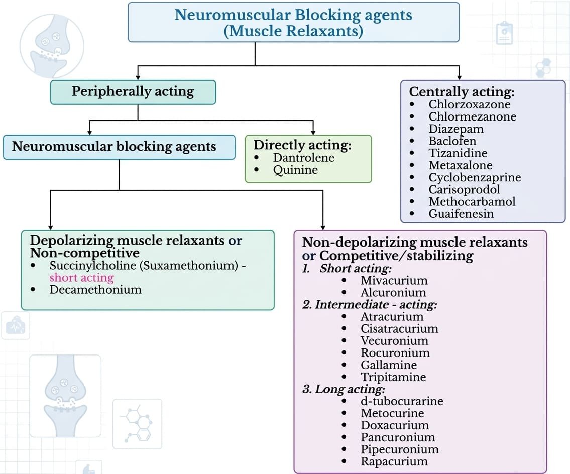
Non-Depolarizing (Competitive) Agents
The botanical prototype is d-tubocurarine. Modern synthetic and semi-synthetic analogs are subclassified by duration of action:
- Short-Acting: Mivacurium (metabolized by plasma cholinesterase; duration ~15–20 minutes).
- Intermediate-Acting: Atracurium, Cisatracurium, Vecuronium, Rocuronium (duration ~30–60 minutes).
- Long-Acting: Pancuronium, Doxacurium, Pipecuronium (duration >60–120 minutes).
Mechanism of Action: Low Dose vs. High Dose
- Low Therapeutic Doses: Act as classical competitive antagonists. They compete directly with ACh for the two α-subunits of the nicotinic NM receptor without stimulating the channel, preventing motor endplate depolarization and subsequent muscle action potentials. This competitive blockade is readily reversed by acetylcholinesterase inhibitors (e.g., neostigmine, edrophonium), which elevate endogenous synaptic ACh levels.
- High Doses: Non-depolarizing molecules enter and physically occlude the central ion channel pore. This non-competitive channel block severely weakens transmission and cannot be reversed by acetylcholinesterase inhibitors.
Pharmacokinetics & Clearance Pathways
- Polar Structure: All NMBDs possess two or more bulky, positively charged quaternary ammonium groups, preventing oral absorption and excluding them from the central nervous system and placental transfer. Must be administered intravenously.
- Elimination Pathways:
- Atracurium & Cisatracurium: Undergo spontaneous non-enzymatic chemical breakdown at physiological pH and temperature (Hofmann elimination) alongside plasma ester hydrolysis. Because clearance is independent of renal or hepatic function, they are the agents of choice in patients with renal failure or hepatic disease. (Atracurium breakdown yields laudanosine, which can trigger seizures at high concentrations; cisatracurium produces significantly less laudanosine).
- Vecuronium & Rocuronium: Undergo biliary and hepatic elimination; duration is prolonged in hepatic failure.
- Pancuronium: Excreted unchanged in urine; duration is significantly prolonged in renal impairment.
Drug Interactions with Non-Depolarizing Blockers
- Cholinesterase Inhibitors: Neostigmine, pyridostigmine, and edrophonium reverse competitive blockade. However, excessive doses can produce a depolarizing block by generating toxic ACh concentrations at the endplate.
- Inhalation Anesthetics: Volatile halogenated hydrocarbons (halothane, isoflurane, sevoflurane) enhance and prolong neuromuscular blockade via central nervous system depression, enhanced local blood flow, and stabilizing actions at the motor endplate.
- Aminoglycoside Antibiotics: Streptomycin, gentamicin, and tobramycin inhibit presynaptic voltage-gated Ca2+ channels, preventing ACh release. They act synergistically with competitive blockers to deepen neuromuscular paralysis.
- Calcium Channel Blockers: Enhance competitive neuromuscular blockade by reducing trans-sarcolemmal calcium influx.
Depolarizing NMBDs (succinylcholine)
The clinical prototype and only available depolarizing neuromuscular blocker is succinylcholine (suxamethonium), a flexible molecule composed of two conjugated acetylcholine molecules.
Biphasic Mechanism of Action
- Phase I Block (Depolarizing Phase): Succinylcholine binds to the nicotinic NM receptor, opening the associated ion channel and depolarizing the motor endplate. Unlike acetylcholine (which is hydrolyzed in microseconds by synaptic acetylcholinesterase), succinylcholine is not metabolized by acetylcholinesterase, persisting in the cleft and producing sustained depolarization. Because voltage-gated sodium channels in the perijunctional sarcolemma require repolarization to reset, they remain trapped in the inactivated state. This manifests as initial, transient muscle fasciculations followed rapidly by complete flaccid paralysis. Phase I blockade is augmented, not reversed, by cholinesterase inhibitors!
- Phase II Block (Desensitization Phase): With continued, prolonged exposure or high cumulative doses, the endplate membrane gradually repolarizes, but the nicotinic receptor becomes desensitized and refractory to acetylcholine. The junction behaves electrophysiologically like a non-depolarizing competitive block, displaying tetanic fade and partial reversibility with acetylcholinesterase inhibitors.
Pharmacokinetics: Ultra-Short Action & Metabolism
- Onset & Duration: Injected intravenously. Produces rapid muscular relaxation within 30 to 60 seconds, with a brief duration of action (5 to 10 minutes) due to rapid redistribution and hydrolysis by circulating plasma cholinesterase (also termed pseudocholinesterase or butyrylcholinesterase) in the liver and blood.
- Clinical Indication: The drug of choice for rapid sequence intubation (RSI) during emergency airway management, full-stomach aspiration risk, and electroconvulsive shock therapy (ECT).
Adverse Effects & Clinical Complications
- 1. Prolonged Apnea: Patients with an inherited deficiency or atypical variant of plasma cholinesterase (quantified by the dibucaine number) cannot hydrolyze succinylcholine, leading to prolonged diaphragmatic paralysis and apnea lasting hours. Requires continued mechanical ventilation.
- 2. Malignant Hyperthermia: When administered alongside volatile halogenated anesthetics (especially halothane), succinylcholine can trigger malignant hyperthermia in patients carrying genetic mutations in the skeletal muscle sarcoplasmic reticulum ryanodine receptor (RyR1) or the dihydropyridine receptor. Uncontrolled calcium efflux triggers muscle rigidity, hypermetabolism, hypercapnia, and dangerous fever. Treatment: Discontinue offending agents, provide active cooling, and administer intravenous dantrolene immediately.
- 3. Hyperkalemia: Sustained endplate depolarization releases intracellular potassium into circulation. In patients with extensive burns, massive crush trauma, spinal cord transection, or denervating neuromuscular disease, upregulation of extrajunctional nicotinic receptors can trigger life-threatening hyperkalemia, cardiac arrhythmias, and cardiac arrest.
- 4. Postoperative Myalgia: Vigorous, unsynchronized fasciculations prior to Phase I paralysis cause postoperative muscle pain, particularly across the neck, back, and abdomen.
- 5. Increased Pressures: Fasciculations cause transient elevations in intraocular pressure (risking globe rupture in open-eye trauma) and intragastric pressure (increasing regurgitation risk).
Centrally & directly acting relaxants (spasmolytics)
In contrast to neuromuscular junction blockers used in anesthesia, spasmolytic muscle relaxants reduce spasticity resulting from upper motor neuron lesions (cerebral palsy, stroke, spinal cord transection, multiple sclerosis) characterized by heightened excitability of α- and γ-motor systems.
1. Baclofen
- Mechanism: An orally active structural analog of γ-aminobutyric acid (GABA) that acts as a selective agonist at GABAB receptors. Activates Gi-coupled pathways in the spinal cord, closing presynaptic Ca2+ channels to inhibit the release of excitatory neurotransmitters (glutamate, aspartate) while increasing postsynaptic K+ conductance, suppressing monosynaptic and polysynaptic stretch reflexes.
- Clinical Uses: Drug of choice for treating spinal cord spasticity and spasticity in multiple sclerosis. Ineffective for supraspinal cortical spasticity. Can be administered intrathecally via an implanted pump for severe, refractory spasticity.
- Pharmacokinetics: Well absorbed orally; lipophilic; penetrates the blood-brain barrier. Approximately 35% is excreted unchanged in urine and feces. Doses are titrated gradually to 100–150 mg/day in four divided doses.
- Adverse Effects: Produces lassitude, nausea, and mental disturbances (confusion, depression). Less sedating than diazepam. Can lower seizure thresholds in patients with epilepsy; abrupt withdrawal can cause seizures and hallucinations.
2. Diazepam (Valium)
- Mechanism: A benzodiazepine that binds as a positive allosteric modulator at GABAA receptors in the brainstem and spinal cord. Increases the frequency of chloride channel opening in the presence of GABA, enhancing presynaptic inhibition of motor tone and suppressing polysynaptic spinal reflexes.
- Clinical Uses: Relieves flexor and extensor spasms, muscle strain, spinal spasticity, and multiple sclerosis.
- Adverse Effects: Significant dose-dependent drowsiness and sedation. Chronic use leads to tolerance, dependence, and potential withdrawal symptoms upon discontinuation.
3. Dantrolene
- Mechanism: A directly acting muscle relaxant that does not work within the central nervous system. Binds to the ryanodine receptor channel (RyR1) on the sarcoplasmic reticulum of skeletal muscle, blocking calcium release into the sarcoplasm and uncoupling electrical excitation from mechanical contraction. Has minimal effect on cardiac or smooth muscle (which express RyR2).
- Clinical Uses: Administered intravenously as the specific, lifesaving antidote for malignant hyperthermia. Administered orally for chronic spasticity due to stroke, spinal cord injury, cerebral palsy, or multiple sclerosis.
- Pharmacokinetics: Incomplete oral absorption; biological half-life ≈ 8.7 hours in adults. Metabolized by hepatic microsomal enzymes and excreted in urine and bile.
- Adverse Effects & Toxicity: Generalized, dose-dependent skeletal muscle weakness is the primary limiting effect. Drowsiness, dizziness, malaise, and diarrhea occur. Most importantly, it causes symptomatic hepatitis in 0.5% and fatal hepatic necrosis in up to 0.2% of patients. Absolutely contraindicated in patients with underlying liver disease or pre-existing respiratory muscle compromise. Regular monitoring of liver function tests (LFTs) is mandatory.
Quick recap
References
The following authoritative medical pharmacology texts and guidelines support this curriculum study note:
- Katzung’s Basic & Clinical Pharmacology: Katzung, B.G. and Vanderah, T.W. (2021) Basic & Clinical Pharmacology. 15th edn. New York: McGraw-Hill Education. [Chapter 8: Cholinoceptor-Blocking Drugs; Chapter 27: Skeletal Muscle Relaxants].
- Goodman & Gilman’s Pharmacological Basis of Therapeutics: Brunton, L.L., Hilal-Dandan, R. and Knollmann, B.C. (2018) Goodman & Gilman’s The Pharmacological Basis of Therapeutics. 13th edn. New York: McGraw-Hill Education. [Chapter 9: Muscarinic Receptor Agonists and Antagonists; Chapter 11: Nicotine and Agents Acting at the Neuromuscular Junction and Autonomic Ganglia].
- Lippincott Illustrated Reviews: Pharmacology: Whalen, K. (2019) Lippincott Illustrated Reviews: Pharmacology. 7th edn. Philadelphia: Wolters Kluwer. [Chapter 5: Cholinergic Antagonists].
- Rang & Dale’s Pharmacology: Ritter, J.M., Flower, R., Henderson, G., Loke, Y.K., MacEwan, D. and Rang, H.P. (2020) Rang & Dale’s Pharmacology. 9th edn. Edinburgh: Elsevier. [Chapter 14: Cholinergic Transmission].
- Uganda Clinical Guidelines: Ministry of Health Uganda (2023) Uganda Clinical Guidelines: National Guidelines for the Management of Common Conditions. Kampala: Ministry of Health. [Section 1.8: Management of Acute Poisoning and Chemical Exposures – Organophosphate Toxicity].