
Pharmacogenetics: genetic variation, drug metabolism & precision therapeutics
Overview & core definitions
Traditional pharmacology has long operated on an empirical, trial-and-error approach. Standard clinical guidelines frequently recommend uniform drug dosages for all adult patients diagnosed with a given illness. However, bedside clinical practice reveals dramatic inter-individual differences: when given the exact same dose of a medication, one patient achieves rapid clinical recovery, another exhibits zero therapeutic benefit, and a third experiences severe or even fatal drug toxicity.
This variability occurs because a patient's pharmacological response is governed by a dynamic interaction between their unique genetic constitution and environmental influences. The field of pharmacogenetics emerged to uncover the biological mechanisms behind these differences.
Pharmacogenetics (PGx)
The study of how individual gene variations influence therapeutic responses, drug disposition, drug safety, tolerability, and clinical efficacy. Historically, pharmacogenetics focused on single-gene (monogenic) variations—such as an inherited defect in a single drug-metabolizing liver enzyme.
Pharmacogenomics
A broader, genome-wide discipline that applies whole-genome technologies, high-throughput sequencing, and bioinformatics to examine the multigenic determinants of drug response. Rather than focusing on a single enzyme, pharmacogenomics evaluates how complex networks of genes, transcription factors, membrane transporters, and biological targets interact across the entire genome to dictate drug outcomes.
In modern clinical practice, these two terms are frequently used interchangeably to describe personalized medicine (or precision medicine): the practice of tailoring medical treatment and drug selection to the individual genetic profile of each patient.
The Paradigm Shift: From Forward to Reverse Genetics
Modern genomics has transformed pharmacological investigation from traditional forward genetics into a systematic reverse genetic framework:
- Classical Forward Genetics (Phenotype-to-Genotype): Clinicians first observed an unusual drug reaction in a patient (such as prolonged muscle paralysis after suxamethonium or acute hemolysis following antimalarial therapy), followed by family pedigrees and biochemical assays to identify the underlying defective gene.
- Modern Reverse Genetics (Genotype-to-Phenotype): High-throughput whole-genome sequencing maps an individual's genetic polymorphisms first. Clinicians and researchers then use these genomic variations to predict how the patient will process, activate, or clear specific pharmacologic agents before the first dose is ever prescribed.
Genomic architecture & terminology
To understand the genetic basis of drug response, we must review the structural architecture of the human genome. The molecular structure of DNA was determined in April 1953 by James Watson and Francis Crick, revealing the double-helical polymer linked by complementary, hydrogen-bonded nucleotide bases.
Fifty years later, in April 2003, the international Human Genome Project completed the sequencing of the entire human euchromatic genome, establishing that human DNA consists of approximately 3 billion nucleotide base pairs containing roughly 20,000 to 25,000 protein-coding genes.
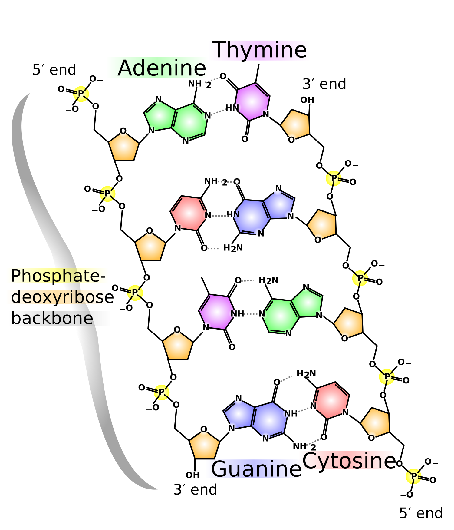
Structural Organization of a Protein-Coding Gene
A gene is the fundamental physical and functional unit of heredity. It carries the molecular blueprint necessary to assemble a specific functional polypeptide chain or structural RNA molecule. A typical eukaryotic gene consists of several distinct structural regions:
- Promoter Region: Located directly upstream (toward the 5′ end) of the structural gene. It contains specialized consensus sequences (such as the TATA box) that serve as recognition and binding sites for RNA polymerase II and essential transcription factors. Polymorphisms in promoter regions can alter transcription factor binding, leading to marked increases or reductions in enzyme expression.
- Exons: Protein-coding nucleotide sequences that are transcribed into pre-mRNA and retained in mature mRNA following RNA splicing. These sequences directly dictate the amino acid order of the resulting protein.
- Introns: Intervening, non-coding nucleotide sequences located between exons. They are transcribed into pre-mRNA but are enzymatically excised during post-transcriptional RNA splicing.
- Flanking Regulatory Regions (5′ and 3′ UTRs): Untranslated regions flanking the coding core that regulate mRNA stability, cytoplasmic export, and translational efficiency.
Essential Genetic Nomenclature in Pharmacogenetics
| Term | Scientific Definition | Practical Clinical Interpretation |
|---|---|---|
| Allele | One of two or more alternative functional forms of a gene located at a specific chromosomal locus. | Because human somatic cells are diploid (containing 22 pairs of autosomes and 1 pair of sex chromosomes), every individual carries two alleles for each autosomal gene: one inherited maternally and one paternally. |
| Star (*) Allele Notation | The standardized naming system used to designate specific gene variations and polymorphisms. | The normal, fully functional reference form of an enzyme is designated as *1 (the "wild-type" allele, e.g., CYP2D6*1). Variant alleles with altered or abolished function are assigned sequential numbers (e.g., CYP2D6*2, *3, *4, *5). |
| Genotype | The precise combination of alleles present at a specific genetic locus in an individual's DNA. | Reflects the underlying genetic code (e.g., a patient carrying one wild-type allele and one non-functional allele has the genotype CYP2C9*1/*3). |
| Phenotype | The observable physiological, biochemical, or clinical trait expressed by an individual. | Reflects real-world drug handling—such as the rate of drug clearance, measured drug serum levels, or clinical responsiveness (e.g., poor metabolizer vs. ultrarapid metabolizer). |
| Diplotype | The specific pair of alleles present on both homologous chromosomes for a single gene. | Used in clinical lab reports to convey a patient's two-allele status (e.g., CYP2D6*3/*5 indicates the patient carries the non-functional *3 allele on one chromosome and a total gene deletion *5 on the other). |
| Haplotype | A specific linear cluster of linked polymorphic alleles located closely together on the same physical chromosome that tend to be inherited together as a unit. | Important for understanding multi-variant genes where multiple mutations occur within the same gene locus (e.g., linked variants across ABCB1). |
| Mitochondrial & Y-DNA | mtDNA is circular DNA located inside mitochondria; Y-DNA is the non-recombining portion of the Y chromosome. | mtDNA is inherited exclusively through maternal lineage (egg cytoplasm); Y-DNA is passed strictly through paternal lineage from father to son. |
| GWAS | Genome-Wide Association Study: a population-scale genomic survey comparing hundreds of thousands of genetic markers between groups. | Identifies statistical links between specific allelic variants and clinical phenotypes (such as susceptibility to severe drug-induced liver injury or Stevens-Johnson syndrome). |
Types of genetic variants
Any two unrelated individuals differ from one another by approximately 1 nucleotide base pair every 300 to 1,000 base pairs across the genome. In total, an estimated 3.2 million single nucleotide variations exist across the human population. Understanding how these variations are categorized is essential for interpreting pharmacogenetic profiles.
Polymorphism vs. Mutation: The 1% Frequency Threshold
The distinction between a genetic polymorphism and a mutation is based on population genetics and allele frequency:
- Polymorphism: A stable DNA sequence variation that occurs at an allele frequency of ≥ 1% in a general population. Because they are common, polymorphisms account for the widespread variability in drug handling seen across diverse human populations.
- Mutation: A rare sequence variation that occurs at an allele frequency of < 1% in the population. Mutations are typically associated with monogenic inherited diseases (such as cystic fibrosis, sickle cell anemia, or Crigler-Najjar syndrome), where a single defective allele causes clear biological pathology.
Single Nucleotide Polymorphisms (SNPs)
A single base-pair substitution where one nucleotide is swapped for another (e.g., cytosine replaced by thymine). SNPs are the most common form of genetic variation in humans.
- Synonymous (Silent) SNPs: The nucleotide substitution alters a codon, but due to the redundancy of the genetic code, it still codes for the same amino acid. The primary polypeptide structure remains unchanged. These variants are found at higher frequencies across the genome because they are subject to less selective pressure.
- Nonsynonymous (Missense) SNPs: The nucleotide substitution changes the codon so that it codes for a different amino acid. This alterative residue can disrupt tertiary folding, substrate binding, or catalytic activity, leading to altered enzyme function.
- Nonsense SNPs: The substitution creates a premature stop codon (UAA, UAG, or UGA), resulting in truncated, non-functional protein fragments that are rapidly degraded.
Insertions & Deletions (Indels)
The insertion or deletion of one or more nucleotide bases within a gene sequence.
- Frameshift Indels: Occur when the number of inserted or deleted bases is not a multiple of three. This shifts the downstream reading frame, scrambling all subsequent amino acids and typically resulting in a non-functional protein.
- Promoter/Non-coding Indels: Indels within non-coding regulatory sequences can alter transcription rates. For example, the UGT1A1*28 allele involves a dinucleotide repeat insertion in the promoter that decreases enzyme expression.
Copy Number Variations (CNVs)
Structural genomic rearrangements where large sections of DNA—including entire, intact genes—are duplicated, multiplied, or completely deleted on a chromosome.
- Designated in allele notation as *1xN or *2xN, where N represents the total number of gene copies present on that chromosome.
- Gene Deletion (*5): Complete absence of the functional gene sequence, resulting in total loss of enzyme production.
- Gene Duplication (*1xN): Presence of multiple functional gene copies (up to 13 copies in some individuals), causing massive enzyme overproduction and creating an ultrarapid metabolizer phenotype.
Causative vs. Linked Polymorphisms
Not every identified polymorphism directly impacts drug action:
- Causative (Functional) Polymorphism: The specific genetic alteration that directly produces the altered phenotype. For example, a missense SNP in an evolutionarily conserved catalytic pocket that reduces substrate metabolism.
- Linked (Marker) Polymorphism: A neutral polymorphism that does not alter protein function itself, but resides physically close to a causative mutation on the same chromosome. Because it is co-inherited with the causative variant through linkage disequilibrium, it can serve as a diagnostic marker in genetic testing.
Determinants of drug response
A patient's pharmacological response is governed by three primary physiological factors:
- Presence of Functional Drug Receptors: The presence and structural integrity of target proteins that bind the drug to trigger a therapeutic response.
- Physiological Traits & Host Factors: Baseline organ function, age, renal clearance, hepatic perfusion, and physiological sensitivity.
- Pharmacokinetic Processing (ADME): The body's biological handling of the drug, encompassing Absorption, Distribution, Metabolism, and Excretion.

Receptor Presence & Drug Recognition
If a patient's cells lack the specific receptor targeted by a drug, that drug cannot produce a pharmacological effect. Two clinical examples illustrate this principle:
HER2 Receptors in Breast Oncology
Human Epidermal Growth Factor Receptor 2 (HER2, encoded by ERBB2) is a transmembrane tyrosine kinase receptor. In approximately 20% to 25% of breast cancers, the HER2 gene is amplified, leading to receptor overexpression on the malignant cell surface.
- HER2-Positive (HER2+) Tumors: Express abundant HER2 receptors. Targeted monoclonal antibodies (such as trastuzumab) bind these receptors, blocking oncogenic downstream signaling and arresting tumor growth.
- HER2-Negative (HER2-) Tumors: Lack this receptor. In these patients, trastuzumab has no target, provides zero clinical benefit, and exposes the patient to potential cardiotoxicity without therapeutic upside.
PTC Bitter Taste Receptor Phenotypes
The ability to taste the synthetic chemical phenylthiocarbamide (PTC) is controlled by the TAS2R38 gene, which encodes a G-protein-coupled bitter taste receptor on the tongue. This trait serves as an educational model for inherited receptor responsiveness:
- Homozygous Dominant (TT) — Supertasters: Express high-affinity receptors and possess high taste bud density; perceive PTC as intensely bitter.
- Heterozygous (Tt) — Tasters: Express moderate-affinity receptors; perceive PTC as moderately bitter.
- Homozygous Recessive (tt) — Non-tasters: Carry non-functional receptor variants; perceive PTC as completely tasteless.
Gene-by-Environment Interactions
While an individual's genetic code provides the baseline potential for enzyme activity, environmental exposures can alter that baseline. For instance, concurrent administration of phenobarbital or rifampicin strongly induces hepatic cytochrome P450 enzymes via nuclear receptors (such as PXR and CAR). This environmental induction can overwhelm baseline genetic differences, dramatically accelerating drug metabolism regardless of a patient's underlying genotype.
Clinical case: nortriptyline metabolism
The practical value of pharmacogenetics is well illustrated by examining how different patients process the tricyclic antidepressant nortriptyline. Consider three adult female patients who share the same age, body weight, height, and ancestral background, all diagnosed with major depressive disorder and prescribed an identical standard dose of 100 mg daily:
Patient A: The Poor Metabolizer (PM)
Clinical Presentation: Suffers severe adverse drug reactions: dry mouth, blurred vision, urinary retention, cardiac arrhythmias, and excessive sedation.
Underlying Pharmacokinetics: Carries two non-functional alleles for the hepatic enzyme CYP2D6 (*4/*4). The liver cannot metabolize the drug via hydroxylation. Nortriptyline accumulates to toxic plasma levels (95 mg active drug circulating).
Clinical Action: Substantially reduce the dose (e.g., to 20% of standard), or select an alternative antidepressant not dependent on CYP2D6.
Patient B: The Ultrarapid Metabolizer (UM)
Clinical Presentation: Experiences zero symptom improvement; depression remains untreated despite confirmed adherence to therapy.
Underlying Pharmacokinetics: Carries duplicated functional CYP2D6 gene copies (*1xN). The liver metabolizes the drug at an accelerated rate, clearing it before it can reach steady-state therapeutic levels (only 5 mg active drug circulating).
Clinical Action: Increase the daily dose under therapeutic drug monitoring, or switch to an agent that bypasses the CYP2D6 pathway.
Patient C: The Normal / Extensive Metabolizer (NM/EM)
Clinical Presentation: Achieves clinical remission of depressive symptoms with minimal, tolerable side effects.
Underlying Pharmacokinetics: Carries two wild-type alleles (*1/*1). Hepatic enzyme capacity matches standard dosing assumptions, maintaining balanced steady-state serum levels (50 mg active drug circulating).
Clinical Action: Maintain current standard dosing regimen.
The cytochrome P450 superfamily
The Cytochrome P450 (CYP) superfamily consists of heme-containing monooxygenase enzymes anchored primarily to the smooth endoplasmic reticulum in hepatocytes and enterocytes. They are responsible for Phase I functionalization reactions (introducing or exposing polar functional groups via oxidation, reduction, or hydrolysis).
Together, CYP enzymes are responsible for the metabolic clearance of roughly 75% to 80% of all clinically prescribed medications. Among the dozens of human CYP isoforms, four families demonstrate prominent, clinically significant genetic polymorphisms:
| Isoenzyme | Fraction of All Drugs Metabolized | Common Defective Alleles | Primary Clinical Drug Substrates |
|---|---|---|---|
| CYP2D6 | ~20% to 25% | *3, *4, *5 (null activity); *10, *17 (reduced); *1xN, *2xN (ultrarapid) | Codeine, tramadol, tamoxifen, beta-blockers (metoprolol), tricyclic antidepressants, SSRIs, antiarrhythmics. |
| CYP2C9 | ~10% to 15% | *2, *3, *5 (substantially reduced catalytic function) | S-warfarin, phenytoin, antidiabetic sulfonylureas (glimepiride, glipizide), NSAIDs (celecoxib, ibuprofen). |
| CYP2C19 | ~10% to 15% | *2, *3 (null activity); *17 (increased transcription, ultrarapid) | Clopidogrel (prodrug activation), proton pump inhibitors (omeprazole), voriconazole, diazepam. |
| CYP3A4 / CYP3A5 | ~40% to 50% | CYP3A5*3, *6, *7 (splice defects; non-expressors) | Tacrolimus, cyclosporine, statins (atorvastatin), dihydropyridine calcium channel blockers, protease inhibitors. |
CYP2D6 genotypes & phenotypes
The CYP2D6 gene locus on chromosome 22q13 is the most extensively characterized polymorphic drug-metabolizing gene in medicine. More than 100 allelic variants have been identified, spanning the entire functional range from complete gene absence to multi-copy duplication.
The Four Clinical Metabolizer Phenotypes
Based on their combined diplotype, patients fall into one of four distinct functional categories:
- Poor Metabolizers (PM): Individuals who carry two non-functional (null) alleles (e.g., *4/*4, *4/*5, *3/*4). They produce no active CYP2D6 enzyme. They cannot clear active drug substrates normally, leading to elevated plasma levels, and cannot convert CYP2D6-dependent prodrugs into their active forms.
- Intermediate Metabolizers (IM): Individuals who are heterozygous for one functional and one null allele (e.g., *1/*4), or who carry two reduced-function alleles (e.g., *10/*17). They possess decreased metabolic capacity compared to the general population.
- Extensive / Normal Metabolizers (NM/EM): Individuals who carry two fully functional wild-type alleles (e.g., *1/*1, *1/*2). They represent the majority of the population and metabolize drugs at standard expected rates.
- Ultrarapid Metabolizers (UM): Individuals who carry duplicated or multiduplicated functional gene copies on one or both chromosomes (designated *1xN or *2xN, where N ≥ 2). They exhibit markedly elevated metabolic capacity, clearing substrates unusually fast and converting prodrugs into active metabolites with unexpected speed.
Key clinically actionable polymorphisms
The following table synthesizes the most important, clinically actionable pharmacogenetic variants across Phase I enzymes, Phase II enzymes, drug transporters, and pharmacological receptors:
| Functional Category | Gene / Target | Major Alleles or Variants | Clinical Consequences & Actionable Drugs |
|---|---|---|---|
| Phase I Oxidation | CYP2C9 | *2, *3 (decreased activity) | Impaired clearance of narrow-therapeutic-index drugs: S-Warfarin (requires lower maintenance doses to avoid life-threatening bleeding), Phenytoin (risk of ataxia, nystagmus, neurotoxicity), and sulfonylureas (risk of severe hypoglycemia). |
| Phase I Oxidation | CYP2C19 | *2, *3 (loss of function); *17 (gain of function) | Clopidogrel: Poor metabolizers (*2/*2) cannot convert clopidogrel into its active antiplatelet thiol metabolite, leading to stent thrombosis and recurrent myocardial infarction. Guidelines recommend alternative antiplatelets (prasugrel or ticagrelor). |
| Phase I Oxidation | CYP2D6 | *1xN, *2xN (gain of function); *3, *4, *5 (loss of function) | Codeine / Tramadol: Life-threatening respiratory depression in ultrarapid metabolizers due to rapid morphine conversion; zero analgesia in poor metabolizers. |
| Phase I Oxidation | DPYD (Dihydropyrimidine dehydrogenase) | DPYD*2A, *13, rs67376798 (severely reduced or null) | DPD catabolizes >80% of fluoropyrimidine chemotherapies (5-Fluorouracil / 5-FU, Capecitabine). Deficient patients develop life-threatening pancytopenia, severe mucositis, and sepsis from standard chemotherapy doses. |
| Phase II Conjugation | UGT1A1 (UDP-glucuronosyltransferase) | UGT1A1*28 (promoter TA repeat expansion: [TA]7 instead of [TA]6) | Reduced glucuronidation of SN-38 (active metabolite of irinotecan), leading to severe dose-limiting neutropenia and life-threatening diarrhea. Also responsible for benign unconjugated hyperbilirubinemia in Gilbert syndrome. |
| Phase II Conjugation | TPMT (Thiopurine methyltransferase) | *2, *3A, *3C (markedly reduced or absent) | TPMT metabolizes cytotoxic thiopurines (Azathioprine, 6-Mercaptopurine, 6-Thioguanine). In homozygous deficient individuals (1 in 300), thiopurine drugs are shunted into toxic thioguanine nucleotides (TGNs), causing fatal bone marrow suppression unless the dose is reduced by 90%. |
| RBC Protection Enzyme | G6PD (Glucose-6-phosphate dehydrogenase) | G6PD A- (African variant), G6PD Mediterranean, Canton | Decreased NADPH production leaves erythrocytes vulnerable to oxidative stress. Exposure to oxidative drugs (Primaquine, Dapsone, Rasburicase, Nitrofurantoin) causes acute hemolytic anemia. Confers evolutionary resistance to severe falciparum malaria. |
| Drug Transporter | SLCO1B1 (OATP1B1) | rs4149056 (521T>C; decreased hepatic uptake) | Reduces hepatic uptake of statins (Simvastatin), causing elevated statin concentrations in systemic circulation and muscle tissue, markedly increasing the risk of statin-induced myopathy and rhabdomyolysis. |
| Pharmacological Receptor | ADRB1 (β1-adrenoceptor) | ADRB1 Arg389Gly | Patients homozygous for Arg389 demonstrate significantly greater reductions in blood pressure and heart rate in response to Metoprolol compared to Gly389 carriers. |
| One-Carbon Metabolism | MTHFR (Methylenetetrahydrofolate reductase) | C677T, A1298C | Reduces 5,10-methylenetetrahydrofolate to 5-methyltetrahydrofolate. Influences cellular folate pools, homocysteine-methionine cycling, pyrimidine/purine synthesis, and clinical sensitivity to Methotrexate toxicity. |
Drug discovery & personalized medicine
Pharmacogenomics is reshaping clinical trial design and drug development. Historically, pharmaceutical candidates with narrow therapeutic windows were frequently abandoned during late-stage clinical trials due to unpredictable toxicity or variable efficacy across unselected patient cohorts.
Genotype-Stratified Clinical Trials
Modern clinical development incorporates genetic screening into trial design:
- Targeted Patient Selection: Clinical trials enroll patients whose genetic profiles indicate a high probability of response, maximizing observed efficacy.
- Exclusion of At-Risk Subsets: Patients with known risk genotypes (such as poor metabolizers of an active compound with narrow safety margins) are excluded, minimizing preventable drug toxicities.
- Genotype-Guided Dosing Regimens: Developing companion diagnostic assays alongside new medications allows manufacturers to establish tailored dosing guidelines before regulatory approval.
Somatic vs. Germline Variations in Cancer Therapeutics
Oncological pharmacogenomics differs fundamentally from other therapeutic areas because treatment selection must account for two distinct genomes:
Germline Genome (Host Genetics)
The patient's inherited DNA, present in every non-cancerous cell. It determines systemic drug clearance, hepatic metabolism, and vulnerability to adverse drug reactions (e.g., a patient's DPYD status before receiving fluorouracil, or UGT1A1 status before irinotecan).
Somatic Genome (Tumor Genetics)
Acquired mutations found exclusively within the malignant tumor cells. These mutations drive tumor proliferation and determine whether targeted antineoplastic therapies will work.
Current Barriers to Widespread Clinical Implementation
Despite compelling research evidence, pharmacogenetic testing has not yet become universal routine practice. Several practical hurdles remain:
- Clinical Complexity: In real-world medicine, patients frequently take multiple medications concurrently for comorbid conditions. Predicting the combined impact of multiple genetic polymorphisms alongside drug-drug interactions creates substantial clinical complexity.
- Evidence Requirements: Professional guidelines require rigorous evidence before endorsing mandatory testing: analytical validity (test accuracy), clinical validity (consistency in predicting response), and clinical utility (proof that testing improves patient outcomes or reduces costs).
- Infrastructure Limitations: Many clinical settings, particularly in resource-constrained environments, lack the rapid sequencing platforms, laboratory turnaround times, and electronic clinical decision support systems needed to deliver genomic guidance at the point of care.
Clinical review cases & explanations
The following case scenarios examine the core clinical concepts covered in this note: