The Structure of a Joint
A Comprehensive Guide to Synovial Joint Anatomy for medical students.
2.1 General Structure of a Synovial Joint (Most Common Type)
Synovial joints are the most complex and clinically significant type of joint in the human body. They are characterized by the presence of a joint cavity filled with synovial fluid, which allows for smooth, friction-free movement between articulating bones. Functionally, all synovial joints are classified as diarthroses (freely movable joints). They are the body's main functional joints and are essential for locomotion and manipulation.
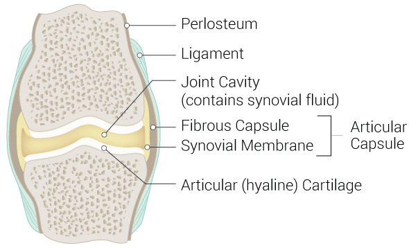
A. Articular (Hyaline) Cartilage
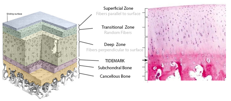
Articular cartilage is a specialized connective tissue that covers the articulating surfaces of bones within synovial joints. It is composed primarily of type II collagen and proteoglycans (mainly aggrecan), which give it unique mechanical properties. The cartilage is organized into four distinct histological zones:
| Zone | Description | Function |
|---|---|---|
| Superficial (Tangential) | Thin collagen fibers parallel to surface; flattened chondrocytes. | Resists shear forces; provides smooth gliding surface. |
| Transitional (Intermediate) | Random fiber orientation; rounded chondrocytes. | Transition between shear and compressive resistance. |
| Deep (Radial) | Collagen fibers perpendicular to surface; chondrocytes in columns. | Resists compressive forces; anchors to subchondral bone. |
| Calcified | Mineralized cartilage at the tidemark. | Anchors cartilage to subchondral bone. |
Key Properties: Articular cartilage is avascular (no blood vessels) and aneural (no nerves). It receives nutrition entirely through diffusion from synovial fluid. This avascular nature is a critical clinical consideration—without a direct blood supply, cartilage has a very limited capacity for self-repair.
Poor Healing Capacity
Because articular cartilage lacks blood supply, it has poor healing capacity. Cartilage defects—whether from trauma, degeneration, or osteochondritis dissecans—often require surgical intervention. Treatment options include microfracture (to stimulate fibrocartilage formation), autologous chondrocyte implantation (ACI), and osteochondral autograft transfer (OATS).
B. Joint Capsule
The joint capsule (articular capsule) is a fibrous connective tissue structure that surrounds the joint and is continuous with the periosteum of the articulating bones. It consists of two distinct layers:
- Outer Fibrous Layer: Composed of dense irregular connective tissue (white fibrous tissue). This layer provides mechanical strength and stability. It may be reinforced by localized thickenings called intrinsic ligaments (e.g., glenohumeral ligaments) or by extrinsic ligaments located outside the capsule.
- Inner Synovial Membrane (Synovium): A highly vascularized and innervated layer of serous connective tissue. It secretes synovial fluid and mediates nutrient exchange. The synovium consists of a cellular intima (lining layer) and a subintima (supportive layer containing vessels and nerves). The membrane contains two cell types: Type A synoviocytes (macrophagic; remove debris) and Type B synoviocytes (fibroblastic; manufacture hyaluronan and lubricin).
Synovitis
Synovitis—inflammation of the synovial membrane—is a hallmark of many joint diseases, including Rheumatoid Arthritis. It causes joint swelling, warmth, and pain. Persistent synovitis can lead to cartilage and bone destruction through the release of inflammatory cytokines and proteolytic enzymes.
C. Joint Cavity
The joint cavity is a potential space between articulating bones, normally containing <3 mL of viscous, clear synovial fluid. It is enclosed by the joint capsule and lined by the synovial membrane. The cavity is maintained at negative pressure relative to atmospheric pressure, which helps stabilize the joint and resist dislocation.
Arthrocentesis
Joint aspiration (arthrocentesis) is a critical diagnostic and therapeutic procedure. Analysis of synovial fluid can diagnose:
— Septic Arthritis: Elevated WBC, positive Gram stain.
— Gout: Negatively birefringent monosodium urate crystals.
— Pseudogout: Positively birefringent calcium pyrophosphate crystals.
— Hemarthrosis: Bloody fluid, often due to trauma or bleeding disorders.
D. Synovial Fluid
A viscous, straw-colored fluid with a composition similar to blood plasma but with high-molecular-weight components.
| Component | Function |
|---|---|
| Hyaluronic acid | Provides viscosity and lubrication; secreted by Type B synoviocytes. |
| Lubricin (PRG4) | Boundary lubricant; reduces friction between cartilage surfaces. |
| Phospholipids | Surface-active lubrication. |
| Albumin & Globulins | Nutrient transport and immunological functions. |
Primary Functions: (1) Lubrication; (2) Nutrient delivery to avascular cartilage; (3) Shock absorption; (4) Waste removal of metabolic byproducts.
Normal synovial fluid is clear and contains <200 WBCs/µL. Inflammatory conditions show counts >2,000/µL, while septic arthritis typically shows >50,000–100,000 WBCs/µL with neutrophil predominance.
E. Accessory Structures
| Structure | Description | Example |
|---|---|---|
| Ligaments | Dense regular CT connecting bone to bone; resists abnormal movement. | ACL, PCL in knee; Collateral ligaments. |
| Tendons | Connect muscle to bone; provide dynamic stability. | Rotator cuff tendons; Quadriceps tendon. |
| Bursae | Synovial fluid-filled sacs reducing friction between moving structures. | Subacromial bursa; Prepatellar bursa. |
| Menisci / Articular Discs | Fibrocartilage structures improving congruence and shock absorption. | Medial/Lateral meniscus; TMJ disc. |
| Fat Pads | Adipose tissue filling spaces and providing cushioning. | Hoffa's fat pad (infrapatellar). |
| Labra | Fibrocartilaginous rings deepening sockets. | Glenoid labrum; Acetabular labrum. |
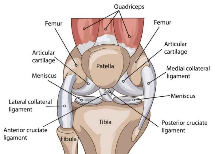
The knee contains approximately 12 bursae, some of which communicate with the joint cavity (e.g., suprapatellar bursa). The infrapatellar fat pad (Hoffa's fat pad) can become impinged, causing severe anterior knee pain.
2.2 Blood Supply
Blood supply is derived from articular arteries that arise from vessels surrounding the joint. These form a periarticular anastomosis—a network of communicating vessels that ensures continuous blood flow regardless of joint position. These are located primarily within the joint capsule and synovial membrane.
- Articular veins: Accompany the arteries and are found in the synovial membrane to facilitate drainage.
- Subchondral bone: Unlike cartilage, the bone beneath does have a blood supply; damage to it (via microfracture) can stimulate repair.
Intra-articular fractures carry a high risk of Avascular Necrosis (AVN) because they may disrupt the delicate blood supply to the subchondral bone. This is critical in fractures of the femoral neck, scaphoid, and talus. Furthermore, the rich vascularity of the synovium makes joints susceptible to hematogenous spread of infection (bacteremia seeding the joint).
2.3 Nerve Supply
Hilton's Law
Named after John Hilton, this law states: "The nerves supplying a joint also supply the muscles moving the joint and the skin covering their distal attachments." This explains why joint pain is often referred to specific dermatomes and why muscles around an injured joint may enter protective spasm.
Key Innervation Facts:
- The capsule, ligaments, and synovium are richly innervated with nociceptive (pain) and proprioceptive (position) fibers.
- Articular cartilage has NO nerve supply—pain in joint disease arises from the capsule, synovium, bone, or periosteum.
| Joint | Nerve Supply |
|---|---|
| Shoulder | Axillary nerve, suprascapular nerve. |
| Hip | Femoral, obturator, sciatic (tibial division) nerves. |
| Knee | Femoral, obturator, sciatic (tibial & common fibular) nerves. |
2.4 Classification of Synovial Joints
| Type | Movement | Description | Examples |
|---|---|---|---|
| Hinge | Uniaxial | Flexion & extension only; convex fits into concave. | Elbow, knee, ankle, IP joints. |
| Ball-and-Socket | Multiaxial | Greatest range of motion; spherical head in cup. | Shoulder, hip. |
| Condyloid | Biaxial | Flexion, extension, abduction, adduction. | MCP joints (knuckles), radiocarpal. |
| Saddle | Biaxial | Modified ellipsoid; each surface concave & convex. | Thumb CMC, sternoclavicular. |
| Pivot | Uniaxial | Rotation around a central axis. | Atlantoaxial, proximal radioulnar. |
| Plane (Gliding) | Non-axial | Sliding/gliding movements. | Intercarpal, intertarsal. |
2.5 Clinical Conditions Affecting Joints
Osteoarthritis (OA)
The most common form of arthritis. results from progressive wear of articular cartilage. Features include subchondral bone exposure, osteophyte (bone spur) formation, and joint space narrowing. It predominantly affects weight-bearing joints (hips, knees).
Septic Arthritis
Bacterial infection of the cavity, most commonly Staphylococcus aureus. The highly vascular synovium allows rapid hematogenous spread. It is a medical emergency presenting with monoarthritis, fever, and WBC > 50,000/µL.
Gout and Pseudogout
- Gout: Deposition of monosodium urate crystals due to hyperuricemia. Negatively birefringent needle-shaped crystals. Classically affects the 1st MTP joint (podagra).
- Pseudogout (CPPD): Calcium pyrophosphate dihydrate crystal deposition. Positively birefringent rhomboid crystals. Common in the knee and wrist.
Meniscal and Ligamentous Injuries
- Medial Meniscus: Less mobile and more commonly torn due to its attachment to the MCL. Clinical signs: locking, catching, and positive McMurray's test.
- ACL: Most commonly injured knee ligament via non-contact pivoting. Usually requires surgical reconstruction in active individuals.
- MCL: Has a better blood supply than the ACL and often heals with conservative management.
