Common Disorders of the Urinary System
Exhaustive medical notes covering Urinary Tract Obstruction, Renal Failure (AKI and CKD), Renal Stones (Nephrolithiasis), and Urinary Tract Infections (UTIs).
1. OVERVIEW
The urinary system comprises the kidneys, ureters, bladder, and urethra. Its primary functions include the excretion of metabolic waste, maintenance of fluid and electrolyte balance, acid-base homeostasis, and blood pressure regulation. Disorders of this system range from obstructive uropathy and renal failure to nephrolithiasis and infectious processes.
2. URINARY TRACT OBSTRUCTION
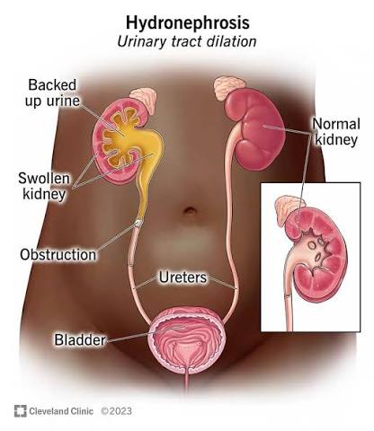
Urinary tract obstruction (obstructive uropathy) refers to any condition that impedes the flow of urine from the kidneys to the urethral meatus. If left untreated, it leads to hydronephrosis, permanent renal parenchymal damage, and ultimately renal failure.
2.1 Etiology and Classification
Upper Tract Obstruction:
- Intrinsic causes: Ureteral stones (most common), blood clots, sloughed papillae (papillary necrosis), fungal balls, ureteral strictures, ureteroceles, and congenital anomalies (e.g., posterior urethral valves in males).
- Extrinsic causes: Retroperitoneal fibrosis, abdominal/pelvic tumors (cervical, colorectal, prostate), endometriosis, aortic aneurysm, and gravid uterus (physiological hydronephrosis of pregnancy).
Lower Tract Obstruction:
- Bladder outlet obstruction: Benign prostatic hyperplasia (BPH)—most common in men >50 years, prostate cancer, bladder neck contracture, and urethral strictures.
- Urethral obstruction: Urethral strictures (post-infectious, post-traumatic, iatrogenic), phimosis, paraphimosis, and posterior urethral valves.
- Neurogenic bladder: Spinal cord injury, multiple sclerosis, diabetes mellitus (autonomic neuropathy), Parkinson disease, and cauda equina syndrome.
2.2 Pathophysiology
- Acute obstruction: Sudden blockage causes a rapid increase in intraluminal pressure proximal to the obstruction. The renal pelvis and calyces dilate (hydronephrosis), and the glomerular filtration rate (GFR) declines.
- Chronic obstruction: Prolonged blockage leads to progressive tubular atrophy, interstitial fibrosis, and glomerulosclerosis. The kidney becomes thin-walled and non-functional. Bilateral chronic obstruction results in chronic kidney disease (CKD) or end-stage renal disease (ESRD).
- Post-obstructive diuresis: After relief of bilateral chronic obstruction, massive diuresis occurs due to accumulated urea (osmotic diuresis) and impaired concentrating ability. This can cause severe volume depletion and electrolyte disturbances.
2.3 Clinical Features
- Acute: Severe colicky flank pain (renal colic) radiating to the groin, nausea, vomiting, hematuria (microscopic or gross), and urinary urgency.
- Chronic: Often insidious and asymptomatic until advanced. May present with dull flank pain, nocturia, and polyuria.
- Physical Exam: Palpable enlarged kidney (hydronephrosis), distended bladder (acute retention), and enlarged prostate on DRE (BPH).
2.4 Investigations & Management
Investigations: Urinalysis (hematuria/pyuria), Serum creatinine/BUN (elevated in bilateral), Ultrasonography (first-line imaging), and CT urography (non-contrast CT is the gold standard for stones).
Immediate Relief: Analgesia (NSAIDs are first-line), medical expulsive therapy (tamsulosin 0.4 mg daily), or Nephrostomy (percutaneous tube placement) for infected or obstructed systems.
Pyonephrosis
The combination of fever, flank pain, and an obstructing stone constitutes a urological emergency. Emergency decompression via nephrostomy or ureteral stenting is life-saving and must precede definitive stone management.
3. RENAL FAILURE
Renal failure is the inability of the kidneys to adequately filter waste products from the blood. It is classified as Acute Kidney Injury (AKI) or Chronic Kidney Disease (CKD) based on duration and reversibility.
3.1 Acute Kidney Injury (AKI)
AKI is a sudden decline in renal function (hours to days) characterized by an increase in serum creatinine and/or reduction in urine output.
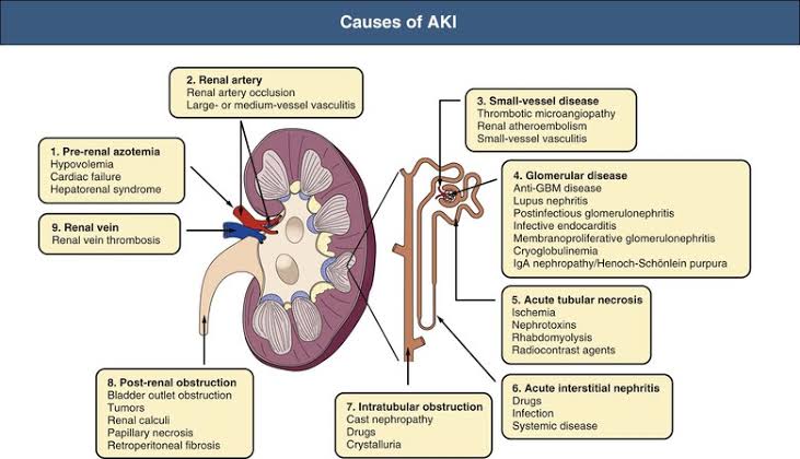
Etiology (RIFLE/AKIN Classification by Site):
- Pre-renal (55-60%): Reduced renal perfusion. Causes: hypovolemia (hemorrhage, burns, dehydration), heart failure, sepsis, and use of NSAIDs or ACE inhibitors.
- Intrinsic renal (35-40%): Direct damage to parenchyma. Most common is Acute Tubular Necrosis (ATN) due to ischemia or nephrotoxins (aminoglycosides, contrast agents). Others include acute interstitial nephritis and rhabdomyolysis.
- Post-renal (5-10%): Obstruction of urine outflow. Causes: bilateral ureteral obstruction, BPH, or bladder outlet obstruction.
Management of AKI:
- Volume resuscitation for pre-renal causes.
- Hyperkalemia management: Calcium gluconate (cardiac stabilization), insulin with dextrose, and sodium bicarbonate.
- Renal replacement therapy (Dialysis): Indicated for refractory hyperkalemia, severe metabolic acidosis, or fluid overload with pulmonary edema.
3.2 Chronic Kidney Disease (CKD)
CKD is defined as abnormalities of kidney structure or function present for >3 months. It is primarily caused by Diabetes Mellitus and Hypertension.
The KDIGO CKD staging combines GFR categories (G1-G5) with albuminuria categories (A1-A3). A patient with G3bA3 (eGFR 30-44 with ACR >300 mg/g) has a much higher risk of cardiovascular events and progression to ESRD than G3bA1.
4. RENAL STONES (NEPHROLITHIASIS)
Nephrolithiasis affects 10-15% of the population. Stones form from the supersaturation of urine coupled with a deficiency of stone inhibitors.
4.1 Types of Renal Stones
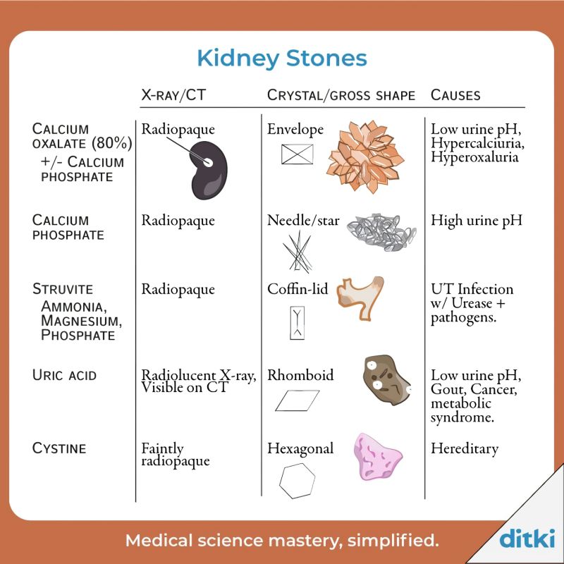
| Stone Type | Prevalence | X-Ray/CT Appearance | Crystal Shape |
|---|---|---|---|
| Calcium Oxalate | 70-80% | Radiopaque | Envelope |
| Calcium Phosphate | 10-15% | Radiopaque | Needle/Star |
| Struvite (Infection) | 5-10% | Radiopaque | Coffin-lid |
| Uric Acid | 5-10% | Radiolucent (visible on CT) | Rhomboid |
| Cystine | 1-2% | Faintly radiopaque | Hexagonal |
4.3 Clinical Features & Management
Renal Colic: Sudden, severe, colicky flank pain radiating to the groin. Associated with nausea, vomiting, and diaphoresis.
Surgical Management:
— ESWL: Shock waves for stones <2 cm.
— Ureteroscopy (URS): Laser fragmentation for ureteral stones.
— PCNL: Minimally invasive surgery for large (>2 cm) or complex staghorn calculi.
5. URINARY TRACT INFECTIONS (UTIs)
Defined as the presence of microorganisms in the urinary tract. UTIs are significantly more common in women (8:1 ratio) due to a shorter urethra.
5.1 Classification
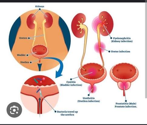
- Cystitis (Lower UTI): Infection of the bladder. Symptoms: Dysuria, frequency, urgency, and suprapubic pain.
- Pyelonephritis (Upper UTI): Infection of the renal pelvis and parenchyma. Symptoms: Fever (>38°C), chills, and costovertebral angle tenderness.
- Complicated UTI: Occurs in the presence of functional or structural abnormalities (obstruction, catheterization, pregnancy, immunosuppression).
5.2 Etiology & Pathophysiology
Causative Agents: Escherichia coli (80-90%), Staphylococcus saprophyticus (5-15%), and Klebsiella pneumoniae.
Pathophysiology: The vast majority are ascending infections caused by enteric flora migrating from the fecal reservoir into the urethra and bladder.
5.6 Management (Non-Pregnant Women)
- Uncomplicated Cystitis: Nitrofurantoin 100 mg twice daily for 5 days OR TMP-SMX 160/800 mg twice daily for 3 days.
- Uncomplicated Pyelonephritis: Oral fluoroquinolone (Ciprofloxacin 500 mg twice daily) for 7 days.
Asymptomatic bacteriuria in pregnancy must always be treated as it carries a high risk of progression to pyelonephritis and preterm labor. Fluoroquinolones and tetracyclines are contraindicated.
6. MASTER SUMMARY TABLE
| Condition | Key Etiology | Clinical Features | Key Investigation |
|---|---|---|---|
| Obstruction | Stones, BPH, tumors | Colicky flank pain, hydronephrosis | Ultrasound, CT KUB |
| AKI | Hypovolemia (Pre), ATN (Intrinsic) | Oliguria, uremic symptoms | Creatinine, FENa, Urinalysis |
| Renal Stones | Hypercalciuria, low urine volume | Flank-to-groin pain, hematuria | Non-contrast CT KUB |
| UTI | E. coli (ascending) | Dysuria, frequency, CVA tenderness | Dipstick (Nitrite+), Culture |
