Muscles of the Lower Limb
The powerful muscles of the lower limb are designed for stability, locomotion, and maintaining an upright posture. We will cover them regionally, starting with the hip and gluteal region.
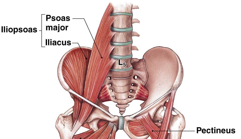
Hip Muscles: The Iliopsoas Group
The Iliopsoas is the strongest hip flexor in the body. It's a composite muscle formed by the Psoas Major and Iliacus, which merge to insert on the lesser trochanter of the femur.
- Psoas Major: Originates from the lumbar vertebrae.
- Iliacus: Originates from the iliac fossa.
- Main Actions: As the main flexor of the hip, it is essential for walking, running, and lifting the leg.
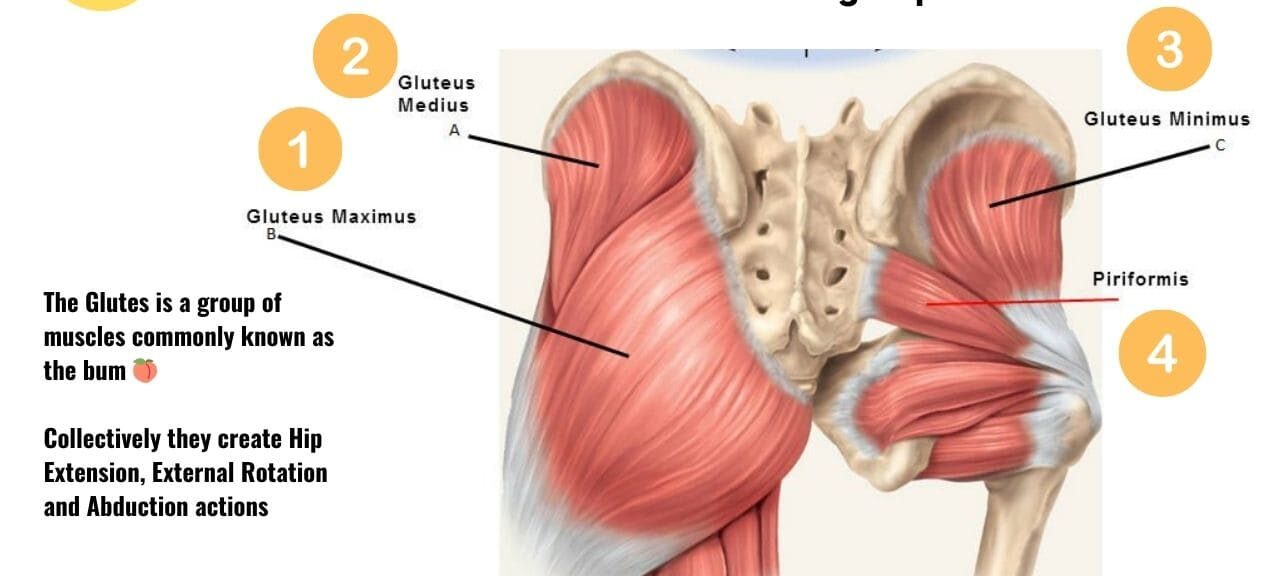
1. Muscles of the Gluteal Region (Buttocks)
These muscles are essential for hip movement, stability, and posture, divided into superficial and deep layers.
Superficial Gluteal Muscles
Gluteus Maximus
The largest and most superficial gluteal muscle. It is the main extensor of the thigh (crucial for climbing stairs or standing up) and a lateral rotator.
Gluteus Medius
Lies deep to Gluteus Maximus. It is the main abductor and a medial rotator of the thigh. It is crucial for stabilizing the pelvis during walking to prevent the hip from dropping on the unsupported side (Trendelenburg sign).
Gluteus Minimus
The smallest and deepest gluteal muscle. It works with the Gluteus Medius to abduct and medially rotate the thigh and stabilize the pelvis.
Tensor Fasciae Latae (TFL)
A small anterolateral muscle that flexes, abducts, and medially rotates the thigh. It tenses the iliotibial (IT) tract, which helps to stabilize the knee in extension.
Deep Gluteal Muscles (Short External Rotators)
This group of six smaller muscles lies deep to the gluteus maximus. They collectively function as powerful lateral rotators of the thigh and help stabilize the head of the femur in the acetabulum.
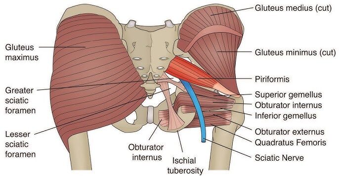
Piriformis
- Origin: Anterior surface of sacrum.
- Insertion: Superior border of greater trochanter.
- Innervation: Nerve to Piriformis (S1, S2).
- Actions: Laterally rotates, abducts (when hip is flexed), and extends the thigh.
Superior Gemellus
- Origin: Ischial spine.
- Insertion: Medial surface of greater trochanter (with Obturator Internus tendon).
- Innervation: Nerve to Obturator Internus (L5, S1).
- Actions: Laterally rotates and abducts the thigh.
Obturator Internus
- Origin: Pelvic surface of obturator membrane.
- Insertion: Medial surface of greater trochanter.
- Innervation: Nerve to Obturator Internus (L5, S1).
- Actions: Laterally rotates and abducts the thigh.
Inferior Gemellus
- Origin: Ischial tuberosity.
- Insertion: Medial surface of greater trochanter (with Obturator Internus tendon).
- Innervation: Nerve to Quadratus Femoris (L4, L5, S1).
- Actions: Laterally rotates and abducts the thigh.
Obturator Externus
- Origin: External surface of obturator membrane.
- Insertion: Trochanteric fossa of femur.
- Innervation: Obturator Nerve (L3, L4).
- Actions: Laterally rotates and adducts the thigh.
Quadratus Femoris
- Origin: Lateral border of ischial tuberosity.
- Insertion: Quadrate tubercle on intertrochanteric crest.
- Innervation: Nerve to Quadratus Femoris (L4, L5, S1).
- Actions: A powerful lateral rotator and adductor of the thigh.
Summary Table of Gluteal Muscles
| Muscle | Origin | Insertion | Innervation | Main Actions |
|---|---|---|---|---|
| Gluteus Maximus | Ilium, sacrum, coccyx | IT tract, gluteal tuberosity | Inferior Gluteal N. | Extends & laterally rotates thigh |
| Gluteus Medius | External surface of ilium | Greater trochanter | Superior Gluteal N. | Abducts & medially rotates thigh; stabilizes pelvis |
| Gluteus Minimus | External surface of ilium | Greater trochanter | Superior Gluteal N. | Abducts & medially rotates thigh; stabilizes pelvis |
| Tensor Fasciae Latae | ASIS, iliac crest | IT tract | Superior Gluteal N. | Flexes, abducts, medially rotates thigh |
| Piriformis | Anterior sacrum | Greater trochanter | N. to Piriformis | Laterally rotates & abducts thigh |
| Obturator Internus | Obturator membrane | Greater trochanter | N. to Obturator Internus | Laterally rotates & abducts thigh |
| Gemelli (Sup & Inf) | Ischial spine/tuberosity | Greater trochanter | Varies | Laterally rotate & abduct thigh |
| Quadratus Femoris | Ischial tuberosity | Intertrochanteric crest | N. to Quadratus Femoris | Powerful lateral rotator of thigh |
2. Muscles of the Thigh
The powerful muscles of the thigh are divided into three compartments: anterior (extensors), medial (adductors), and posterior (flexors/hamstrings).
Anterior Compartment of the Thigh (Extensors)
- Innervation: Femoral Nerve (L2, L3, L4)
- Main Actions: Primarily extension of the knee; some flexion of the hip.
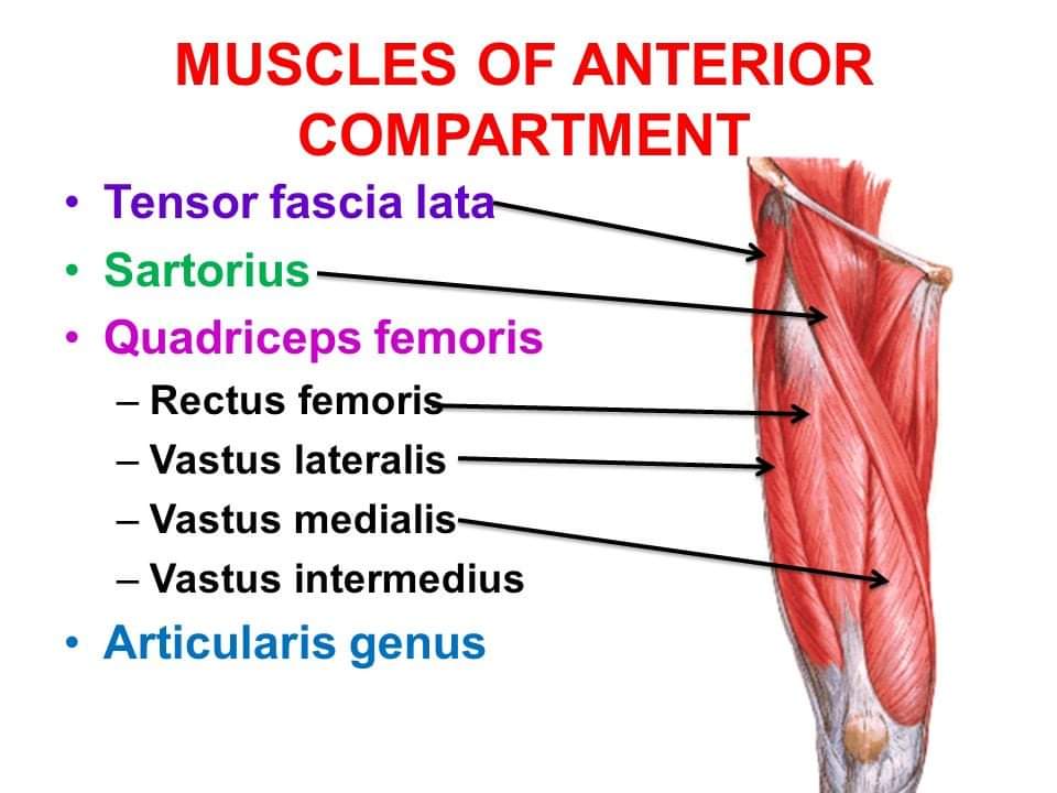
Quadriceps Femoris
A group of four muscles (Rectus Femoris, Vastus Lateralis, Vastus Medialis, Vastus Intermedius) that converge on the patellar tendon. It is the powerful extensor of the knee. The Rectus Femoris is unique as it also flexes the hip.
Sartorius
The longest muscle in the body. It flexes, abducts, and laterally rotates the thigh, and also flexes the knee (the "tailor's muscle" for crossing legs).
Medial Compartment of the Thigh (Adductors)
- Innervation: Mostly Obturator Nerve (L2, L3, L4).
- Main Actions: Primarily adduction of the thigh.
This group includes the Pectineus, Adductor Longus, Adductor Brevis, the powerful Adductor Magnus (which has both an adductor and a hamstring part), and the long, strap-like Gracilis.
Posterior Compartment of the Thigh (Hamstrings)
- Innervation: Sciatic Nerve (Tibial portion), except for the short head of Biceps Femoris.
- Main Actions: Primarily flexion of the knee and extension of the hip.
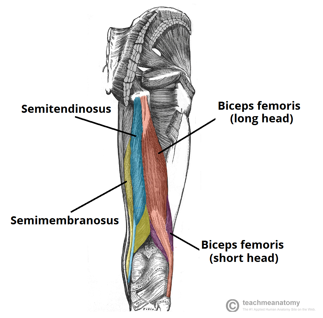
Biceps Femoris
The lateral hamstring muscle, with a long and a short head. It flexes the knee and laterally rotates the leg.
Semitendinosus
A medial hamstring muscle. It flexes the knee and medially rotates the leg.
Semimembranosus
A medial hamstring muscle, deep to the Semitendinosus. It flexes the knee and medially rotates the leg.
Summary Table of Thigh Muscles
| Muscle | Origin | Insertion | Innervation | Main Actions |
|---|---|---|---|---|
| ANTERIOR COMPARTMENT | ||||
| Rectus Femoris | AIIS | Patella & Tibial Tuberosity | Femoral N. | Extends knee, flexes hip |
| Vastus Lateralis | Greater trochanter, linea aspera | Extends knee | ||
| Vastus Medialis | Intertrochanteric line, linea aspera | Extends knee | ||
| Vastus Intermedius | Femoral shaft | Extends knee | ||
| Sartorius | ASIS | Medial tibia (Pes Anserinus) | Femoral N. | Flexes, abducts, lat. rotates thigh; flexes knee |
| MEDIAL COMPARTMENT | ||||
| Adductor Longus/Brevis/Magnus | Pubis, Ischial ramus | Femur (linea aspera) | Obturator N. (Magnus also Sciatic N.) | Adduct thigh; Magnus also extends thigh |
| Gracilis | Pubic symphysis | Medial tibia (Pes Anserinus) | Obturator N. | Adducts thigh, flexes knee |
| POSTERIOR COMPARTMENT (HAMSTRINGS) | ||||
| Biceps Femoris | Long: Ischial tuberosity; Short: Linea aspera | Head of fibula | Sciatic N. (Tibial & Common Fibular) | Flexes knee, extends hip, lat. rotates leg |
| Semitendinosus | Ischial tuberosity | Medial tibia (Pes Anserinus) | Sciatic N. (Tibial) | Flexes knee, extends hip, med. rotates leg |
| Semimembranosus | Ischial tuberosity | Medial condyle of tibia | Sciatic N. (Tibial) | Flexes knee, extends hip, med. rotates leg |
3. Muscles of the Leg
The muscles of the leg are divided into four compartments by interosseous membrane and fascial septa: anterior, lateral, posterior superficial, and posterior deep.
Anterior Compartment of the Leg
- Innervation: Deep Fibular (Peroneal) Nerve (L4, L5, S1)
- Main Actions: Primarily dorsiflexion of the ankle and extension of the toes.

This compartment includes the Tibialis Anterior (main dorsiflexor and invertor), Extensor Digitorum Longus (extends lateral four toes), Extensor Hallucis Longus (extends great toe), and Fibularis Tertius (dorsiflexes and everts).
Lateral Compartment of the Leg
- Innervation: Superficial Fibular (Peroneal) Nerve (L5, S1, S2)
- Main Actions: Primarily eversion of the foot; some plantarflexion.
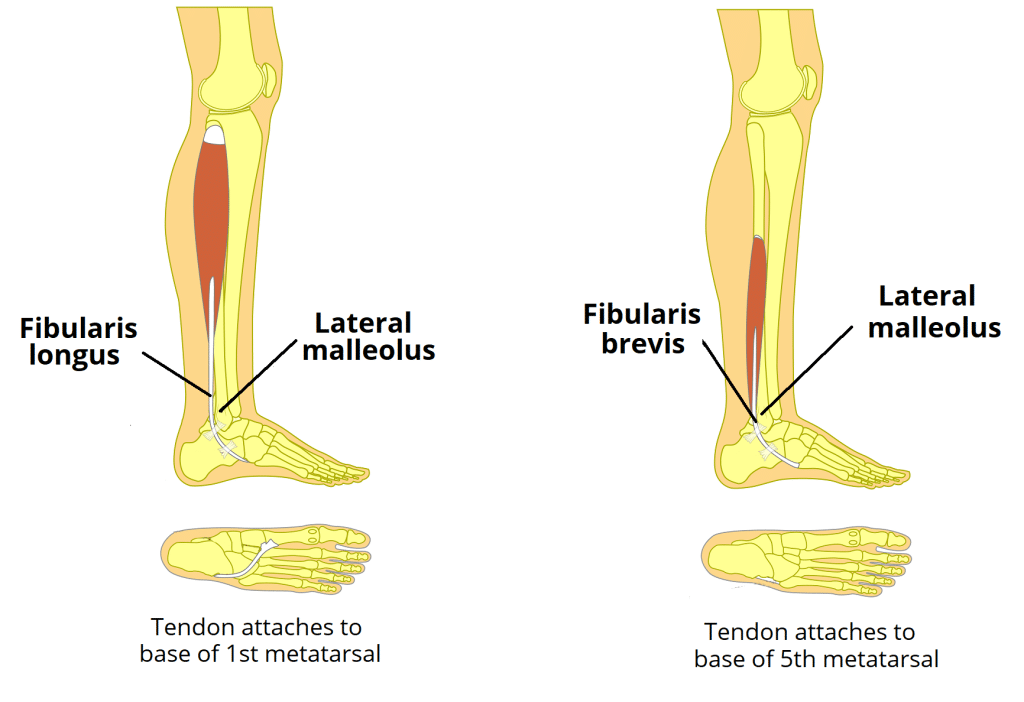
This compartment contains two muscles: Fibularis (Peroneus) Longus and Fibularis (Peroneus) Brevis. Together, they are the main everters of the foot.
Posterior Compartment of the Leg
- Innervation: Tibial Nerve (L4-S2)
- Main Actions: Primarily plantarflexion of the ankle, inversion of the foot, and flexion of the toes.
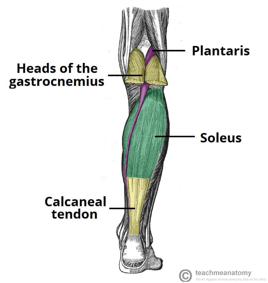
Superficial Group
This group forms the bulk of the calf and inserts via the calcaneal (Achilles) tendon. It includes the two-headed Gastrocnemius, the powerful underlying Soleus, and the small Plantaris. Together, Gastrocnemius and Soleus are known as the Triceps Surae and are powerful plantarflexors.
Deep Group
These muscles lie deep to the superficial group. They include the Popliteus (unlocks the knee), Flexor Digitorum Longus (flexes lateral four toes), Flexor Hallucis Longus (flexes the great toe), and the Tibialis Posterior (the main invertor of the foot).

Summary Table of Leg Muscles
| Muscle | Origin | Insertion | Innervation | Main Actions |
|---|---|---|---|---|
| ANTERIOR COMPARTMENT | ||||
| Tibialis Anterior | Lateral tibia | Medial cuneiform, 1st metatarsal | Deep Fibular N. | Main dorsiflexor; inverts foot |
| Extensor Digitorum Longus | Tibia, fibula | Distal phalanges of digits 2-5 | Deep Fibular N. | Extends lateral four toes |
| Extensor Hallucis Longus | Fibula | Distal phalanx of great toe | Deep Fibular N. | Extends great toe |
| LATERAL COMPARTMENT | ||||
| Fibularis (Peroneus) Longus | Head of fibula | 1st metatarsal, medial cuneiform | Superficial Fibular N. | Everts foot; plantarflexes ankle |
| Fibularis (Peroneus) Brevis | Lateral fibula | Base of 5th metatarsal | Superficial Fibular N. | Everts foot; plantarflexes ankle |
| POSTERIOR COMPARTMENT (SUPERFICIAL) | ||||
| Gastrocnemius | Femoral condyles | Calcaneus via Achilles tendon | Tibial N. | Plantarflexes ankle, flexes knee |
| Soleus | Tibia, fibula | Powerful plantarflexor | ||
| POSTERIOR COMPARTMENT (DEEP) | ||||
| Tibialis Posterior | Tibia, fibula | Navicular, cuneiforms, etc. | Tibial N. | Main invertor of foot |
| Flexor Digitorum Longus | Posterior tibia | Distal phalanges of digits 2-5 | Flexes lateral four toes | |
| Flexor Hallucis Longus | Posterior fibula | Distal phalanx of great toe | Flexes great toe | |
4. Muscles of the Foot
The intrinsic muscles of the foot are divided into dorsal (top) and plantar (sole) groups, responsible for fine motor control, supporting the arches, and assisting in locomotion.
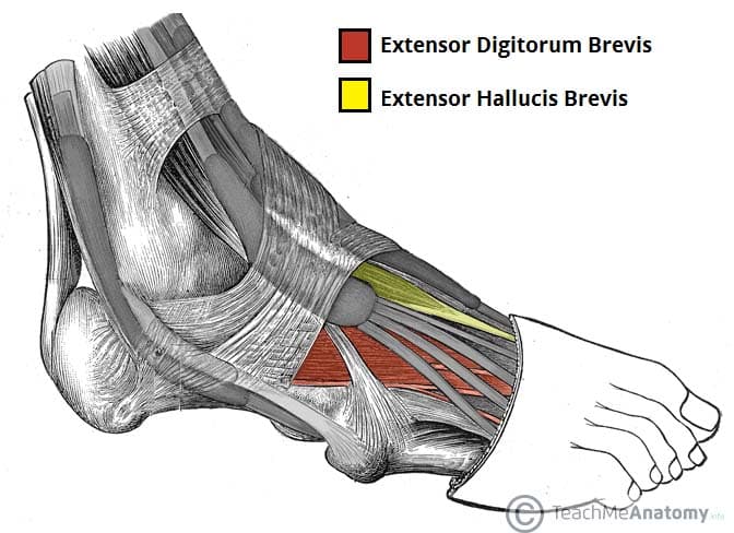
Dorsal Muscles of the Foot
Extensor Digitorum Brevis
Originates from the calcaneus and helps extend toes 2-4.
Extensor Hallucis Brevis
Originates from the calcaneus and helps extend the great toe.
Plantar Muscles of the Foot
These muscles are organized into four layers from superficial to deep. They are primarily innervated by the Medial and Lateral Plantar Nerves (branches of the Tibial Nerve) and are crucial for supporting the arches and controlling fine movements of the toes.
Layer 1 (Superficial)

Abductor Hallucis
Abducts and flexes the great toe. (Innervated by Medial Plantar N.)
Flexor Digitorum Brevis
Flexes the lateral four toes at the PIP joints. (Innervated by Medial Plantar N.)
Abductor Digiti Minimi
Abducts and flexes the little toe. (Innervated by Lateral Plantar N.)
Layer 2

Quadratus Plantae
Assists the Flexor Digitorum Longus (FDL) tendon in flexing the toes by straightening its line of pull. (Innervated by Lateral Plantar N.)
Lumbricals (4)
Flex the MTP joints and extend the IP joints of the lateral four toes. (Innervated by both Medial and Lateral Plantar Nerves).
Layer 3
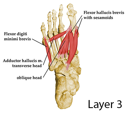
Flexor Hallucis Brevis
Flexes the great toe at the MTP joint. (Innervated by Medial Plantar N.)
Adductor Hallucis
Has two heads (oblique and transverse); adducts the great toe. (Innervated by Lateral Plantar N.)
Flexor Digiti Minimi Brevis
Flexes the little toe. (Innervated by Lateral Plantar N.)
Layer 4 (Deepest)

Plantar Interossei (3)
Adduct toes 3-5 (PAD - Plantar Adduct). (Innervated by Lateral Plantar N.)
Dorsal Interossei (4)
Abduct toes 2-4 (DAB - Dorsal Abduct). (Innervated by Lateral Plantar N.)
Summary Table of Foot Muscles
| Layer/Group | Muscle | Origin | Insertion | Innervation | Main Actions |
|---|---|---|---|---|---|
| DORSAL MUSCLES | |||||
| Dorsal | Extensor Digitorum Brevis | Calcaneus | Extensor expansions 2-4 | Deep Fibular N. | Extends toes 2-4 |
| Extensor Hallucis Brevis | Calcaneus | Prox. phalanx of great toe | Extends great toe | ||
| PLANTAR MUSCLES | |||||
| Layer 1 | Abductor Hallucis | Calcaneus | Prox. phalanx of great toe | Medial Plantar N. | Abducts & flexes great toe |
| Flexor Digitorum Brevis | Calcaneus | Middle phalanges 2-5 | Medial Plantar N. | Flexes toes 2-5 (PIP) | |
| Abductor Digiti Minimi | Calcaneus | Prox. phalanx of 5th toe | Lateral Plantar N. | Abducts & flexes 5th toe | |
| Layer 2 | Quadratus Plantae | Calcaneus | Tendon of FDL | Lateral Plantar N. | Assists FDL in flexing |
| Lumbricals (4) | Tendons of FDL | Extensor expansions 2-5 | Medial & Lateral Plantar N. | Flex MTPs, Extend IPs | |
| Layer 3 | Flexor Hallucis Brevis | Cuboid, cuneiforms | Prox. phalanx of great toe | Medial Plantar N. | Flexes great toe |
| Adductor Hallucis | Metatarsals 2-4 | Prox. phalanx of great toe | Lateral Plantar N. | Adducts great toe | |
| Flexor Digiti Minimi Brevis | Base of 5th metatarsal | Prox. phalanx of 5th toe | Lateral Plantar N. | Flexes little toe | |
| Layer 4 | Plantar Interossei (3) | Metatarsals 3-5 | Prox. phalanges 3-5 | Lateral Plantar N. | Adduct toes (PAD) |
| Dorsal Interossei (4) | Adjacent metatarsals | Prox. phalanges 2-4 | Lateral Plantar N. | Abduct toes (DAB) | |