I. Introduction to Body Cavities
Body cavities are enclosed, fluid-filled spaces within the human body that contain and protect internal organs. They are crucial for:
- Protection: Cushioning delicate organs from shocks and impacts.
- Support: Providing a stable environment for organs.
- Permitting organ movement: Allowing organs to change size and shape (e.g., heart beating, lungs expanding, stomach distending) without friction or damage to surrounding tissues.
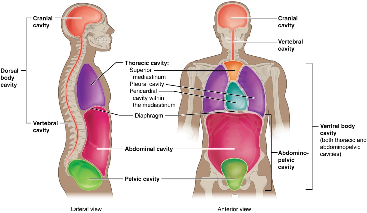
The human body possesses two main sets of internal cavities: the Dorsal Body Cavity and the Ventral Body Cavity. These cavities are formed during embryonic development and house organs of the nervous, circulatory, respiratory, digestive, urinary, and reproductive systems.
II. The Dorsal Body Cavity
The dorsal body cavity is located posteriorly and protects the fragile organs of the central nervous system. It has two continuous subdivisions:
A. Cranial Cavity
- Definition: The space enclosed by the cranium (skull).
- Boundaries:
- Superior, Lateral, Posterior: Formed by the cranial bones (frontal, parietal, temporal, occipital, sphenoid, ethmoid).
- Inferior: Formed by the floor of the cranium, which contains the foramen magnum (a large opening through which the brainstem connects to the spinal cord).
- Contents:
- Brain: The primary organ of the central nervous system, responsible for thought, sensation, and coordination.
- Meninges: Three protective membranes (dura mater, arachnoid mater, pia mater) that surround the brain and spinal cord.
- Cerebrospinal Fluid (CSF): A clear fluid that circulates within the meninges and ventricles of the brain, providing cushioning and nutrient transport.
- Blood Vessels: Arteries, veins, and venous sinuses that supply and drain blood from the brain.
- Cranial Nerves: Twelve pairs of nerves that emerge directly from the brain.
B. Vertebral (Spinal) Cavity
- Definition: The space formed by the vertebral column, extending from the foramen magnum to the sacrum.
- Boundaries:
- Anterior, Lateral, Posterior: Formed by the vertebral arches of the individual vertebrae, which collectively create the vertebral canal.
- Superior: Continuous with the cranial cavity at the foramen magnum.
- Inferior: Ends at the sacrum.
- Contents:
- Spinal Cord: A long, delicate structure that extends from the brainstem, transmitting nerve signals throughout the body.
- Meninges: (Dura mater, arachnoid mater, pia mater) that continue from the brain, enclosing the spinal cord.
- Cerebrospinal Fluid (CSF): Circulates within the subarachnoid space around the spinal cord.
- Spinal Nerves: Nerves that branch off the spinal cord at each vertebral level.
- Blood Vessels: Supplying and draining the spinal cord.
III. The Ventral Body Cavity
The ventral body cavity is much larger than the dorsal cavity and is located anteriorly. It houses a wide range of visceral organs (organs of the digestive, urinary, respiratory, and reproductive systems) and is subdivided by the diaphragm into two main parts: the Thoracic Cavity (superior) and the Abdominopelvic Cavity (inferior).
A. Thoracic Cavity:
- Definition: The superior subdivision of the ventral body cavity, enclosed by the rib cage.
- Boundaries:
- Superior: Thoracic inlet (formed by the first thoracic vertebra, first pair of ribs, and manubrium of the sternum).
- Inferior: Diaphragm (a large, dome-shaped muscle that separates the thoracic and abdominopelvic cavities).
- Anterior: Sternum and costal cartilages.
- Posterior: Thoracic vertebrae.
- Lateral: Ribs and intercostal muscles.
- Subdivisions within the Thoracic Cavity:
- Pleural Cavities (x2):
- Definition: Two lateral compartments, each surrounding a lung. These are potential spaces between the parietal and visceral pleura.
- Contents: Lungs.
- Mediastinum:
- Definition: The central compartment of the thoracic cavity, located between the two pleural cavities. It extends from the sternum anteriorly to the vertebral column posteriorly, and from the thoracic inlet superiorly to the diaphragm inferiorly.
- Contents:
- Heart: Enclosed within the pericardial cavity.
- Great Vessels: Aorta, pulmonary trunk, superior and inferior vena cava.
- Trachea: Windpipe.
- Esophagus: Food pipe.
- Thymus Gland: Located anteriorly in the superior mediastinum (larger in children, atrophies in adults).
- Lymph Nodes, Nerves: (e.g., vagus, phrenic), Major Bronchi.
- Pleural Cavities (x2):
B. Abdominopelvic Cavity:
- Definition: The inferior subdivision of the ventral body cavity, located inferior to the diaphragm. It is generally described as having two indistinct parts: the abdominal cavity and the pelvic cavity, as there is no physical barrier separating them.
- Boundaries:
- Superior: Diaphragm.
- Inferior: Pelvic floor (pelvic diaphragm), formed by muscles and fascia.
- Anterior/Lateral: Abdominal wall muscles.
- Posterior: Lumbar vertebrae and associated muscles.
- Subdivisions within the Abdominopelvic Cavity:
- Abdominal Cavity:
- Definition: The superior and larger portion of the abdominopelvic cavity.
- Contents:
- Digestive Organs: Stomach, small intestine, most of the large intestine, liver, gallbladder, pancreas, spleen.
- Kidneys and Adrenal Glands: Located retroperitoneally (behind the peritoneum).
- Portions of Ureters.
- Many major blood vessels: (e.g., abdominal aorta, inferior vena cava).
- Pelvic Cavity:
- Definition: The inferior and smaller portion of the abdominopelvic cavity, located within the bony pelvis.
- Boundaries: Formed by the bony pelvis (ilium, ischium, pubis, sacrum, coccyx) and the muscles of the pelvic floor.
- Contents:
- Urinary Bladder.
- Sigmoid Colon and Rectum: (terminal part of the large intestine).
- Reproductive Organs:
- Females: Uterus, ovaries, fallopian tubes, vagina.
- Males: Prostate gland, seminal vesicles.
- Abdominal Cavity:
Summary Table of Major Body Cavities:
| Cavity Name | Subdivisions | Major Boundaries | Key Contents |
|---|---|---|---|
| Dorsal Body Cavity | Cranial Cavity | Cranium | Brain, Meninges, CSF |
| Vertebral Cavity | Vertebral Column | Spinal Cord, Meninges, CSF | |
| Ventral Body Cavity | Thoracic Cavity | Rib Cage, Sternum, Thoracic Vertebrae, Diaphragm (inferior) | Lungs (in pleural cavities), Heart (in pericardial cavity), Trachea, Esophagus, Thymus |
| Pleural Cavities (x2) | Within Thoracic Cavity, surrounding lungs | Lungs | |
| Mediastinum | Central compartment of Thoracic Cavity | Heart, Great Vessels, Trachea, Esophagus, Thymus | |
| Abdominopelvic Cavity | (Full Cavity) | Diaphragm (superior), Pelvic Floor (inferior), Abdominal Muscles, Lumbar Vertebrae | Digestive Organs (stomach, intestines, liver, etc.), Kidneys, Bladder, Reproductive Organs |
| Abdominal Cavity | Superior portion of Abdominopelvic Cavity | Stomach, Small/Large Intestines, Liver, Spleen, Pancreas, Kidneys | |
| Pelvic Cavity | Inferior portion of Abdominopelvic Cavity, within bony pelvis | Bladder, Rectum, Reproductive Organs |
IV. Protective Functions of Body Cavities
Body cavities provide much more than just space for organs; they are integral to their protection and optimal function.
A. Mechanical Protection:
- Cushioning: The fluid within cavities (like CSF in the dorsal cavity, or serous fluid in the ventral cavity) and the surrounding structures (bone, muscle) help absorb shock and impact, protecting delicate organs from external trauma.
- Containment: The rigid bony structures surrounding the dorsal cavity (cranium, vertebral column) and parts of the ventral cavity (rib cage, bony pelvis) offer robust protection against physical injury.
- Isolation: Cavities isolate organs from external forces and, to some extent, from infections originating in other body regions.
B. Facilitating Organ Movement and Reducing Friction:
This is where serous membranes play a critical role, primarily in the ventral body cavity.
1. Serous Membranes (Serosa):
- Definition: Thin, double-layered membranes that line the walls of the ventral body cavity and cover the surfaces of the organs within it. They are composed of a layer of simple squamous epithelium (mesothelium) overlying a thin layer of areolar connective tissue.
- Structure: Each serous membrane consists of two layers:
- Parietal Layer: Lines the walls of the body cavity (e.g., parietal pleura lines the thoracic wall).
- Visceral Layer: Covers the external surface of the organs within the cavity (e.g., visceral pleura covers the surface of the lungs).
- Serous Cavity: The potential space between the parietal and visceral layers. This space is not empty but contains a small amount of serous fluid.
- Serous Fluid: A thin, watery lubricating fluid secreted by both layers of the membrane.
- Function: Reduces friction between the moving visceral organs and the body wall. This allows organs like the heart, lungs, and intestines to expand, contract, and slide past one another with minimal wear and tear.
2. Examples of Serous Membranes:
Pleura:
- Location: Thoracic cavity, associated with the lungs.
- Parietal Pleura: Lines the chest wall and superior surface of the diaphragm.
- Visceral Pleura: Covers the surface of the lungs.
- Pleural Cavity: Contains pleural fluid, reducing friction during breathing.
Pericardium:
- Location: Thoracic cavity, associated with the heart (within the mediastinum).
- Parietal Pericardium: Forms the outer layer of the pericardial sac.
- Visceral Pericardium (Epicardium): Covers the surface of the heart.
- Pericardial Cavity: Contains pericardial fluid, reducing friction during heartbeats.
Peritoneum:
- Location: Abdominopelvic cavity, associated with abdominal organs.
- Parietal Peritoneum: Lines the walls of the abdominal and pelvic cavities.
- Visceral Peritoneum: Covers the surface of most abdominal organs.
- Peritoneal Cavity: Contains peritoneal fluid, allowing digestive organs to slide against each other.
- Mesenteries: Folds of peritoneum that connect organs to the posterior abdominal wall, providing routes for blood vessels, nerves, and lymphatic vessels, and holding organs in place.
V. Clinical Relevance of Body Cavities
Understanding body cavities is fundamental for diagnosing and treating a wide range of medical conditions.
A. Fluid Accumulation (Effusions):
Pathology: An abnormal increase in serous fluid within a body cavity. This can impair organ function.
- Pleural Effusion: Excess fluid in the pleural cavity (e.g., due to heart failure, pneumonia, cancer). Can compress the lungs, making breathing difficult.
- Pericardial Effusion: Excess fluid in the pericardial cavity (e.g., due to inflammation, trauma). Can compress the heart, leading to cardiac tamponade (a life-threatening condition).
- Ascites: Excess fluid in the peritoneal cavity (e.g., due to liver cirrhosis, cancer, heart failure). Can cause abdominal distension and discomfort.
Procedures:
Thoracentesis: A procedure to remove pleural fluid using a needle.Pericardiocentesis: A procedure to remove pericardial fluid.Paracentesis: A procedure to remove peritoneal fluid (ascites).
B. Organ Displacement and Herniation:
Pathology: Organs can move from their normal position into another cavity or through a weakened area in the body wall.
- Hiatal Hernia: Part of the stomach pushes upward through the diaphragm into the thoracic cavity.
- Inguinal Hernia: A portion of the intestine protrudes through a weak spot in the abdominal wall, often into the inguinal canal.
- Diaphragmatic Hernia: Abdominal organs herniate into the thoracic cavity through a defect in the diaphragm (can be congenital or acquired).
C. Infections and Inflammation:
Pathology: Infection or inflammation of the serous membranes.
- Pleurisy (Pleuritis): Inflammation of the pleura, causing sharp chest pain during breathing.
- Pericarditis: Inflammation of the pericardium, causing chest pain.
- Peritonitis: Inflammation of the peritoneum, usually due to bacterial infection (e.g., ruptured appendix, bowel perforation). This is a serious condition.
D. Surgical Approaches:
- Surgeons must have a detailed understanding of cavity anatomy to plan safe and effective surgical approaches, minimize damage to surrounding structures, and prevent complications.
- Laparotomy: Surgical incision into the abdominal cavity.
- Thoracotomy: Surgical incision into the thoracic cavity.
- Craniotomy: Surgical incision into the cranium to access the brain.
E. Imaging and Diagnostics:
- X-rays, CT scans, MRI, Ultrasound: Imaging techniques rely on the distinct characteristics and relationships of organs within cavities to visualize pathologies. For example, fluid appears differently than solid tissue on scans.
VI. Utilizing Appropriate Anatomical Terminology
Accurate and consistent use of anatomical terminology is essential for clear communication in healthcare.
A. Key Terms and Their Usage:
- Always specify the cavity and subdivision when describing organ location (e.g., "The heart is located in the pericardial cavity, within the mediastinum of the thoracic cavity").
- Distinguish between parietal (lining the wall) and visceral (covering the organ) layers of serous membranes.
- Use directional terms precisely (e.g., "The liver is superior to the stomach in the abdominal cavity," "The spinal cord is inferior to the brain within the dorsal cavity").
- Be aware of terms like retroperitoneal (e.g., kidneys, pancreas, parts of duodenum, aorta, IVC) for organs located behind the peritoneum.
B. Practice and Application:
- Clinical Case Discussions: Describe organ pathologies and surgical interventions using proper cavity terminology.
- Patient Handoffs: Clearly communicate the location of findings or concerns related to body cavities.
- Documentation: Ensure all clinical notes and reports accurately reflect anatomical positions and relationships.
Source: https://doctorsrevisionuganda.com | Whatsapp: 0726113908
Anatomy: Body Cavities
Test your knowledge with these 20 questions.
Body Cavities Quiz
Question 1/20
Quiz Complete!
Here are your results, .
Your Score
18/20
90%