Nervous Tissue
Nervous tissue is the master controller and communication system of the body. It forms the brain, spinal cord, and peripheral nerves, and its primary function is to regulate and integrate all body functions by rapidly transmitting electrical signals.
The Two Main Cell Types
The nervous system is comprised of two principal types of cells that work in concert.
Neurons (Nerve Cells)
These are the primary functional cells that are specialized to transmit electrical signals (nerve impulses). They send and receive messages using chemical signals called neurotransmitters across junctions known as synapses.
Neuroglia (Glial Cells)
These are the non-excitable, supporting cells of the nervous system. They provide physical and metabolic support, insulation (myelin), and immune defense for the neurons. Examples include Astrocytes, Oligodendrocytes, and Schwann Cells.
General Characteristics
- Primary Function: To receive stimuli, transmit electrical impulses, and process information to control the body's responses.
- Location: Makes up the Central Nervous System (CNS)—the brain and spinal cord—and the Peripheral Nervous System (PNS)—the peripheral nerves.
Key Properties of Neurons
Excitability
The ability to respond to a stimulus by generating an electrical change across its membrane (membrane potential).
Conductivity
The ability to propagate these electrical signals (nerve impulses or action potentials) rapidly along the cell membrane.
The Neuron (Nerve Cell): The Signaling Unit
Neurons are the excitable cells responsible for transmitting electrical signals. They are typically long-lived, amitotic (do not divide in their mature form), and have a very high metabolic rate, requiring a continuous supply of oxygen and glucose to function.
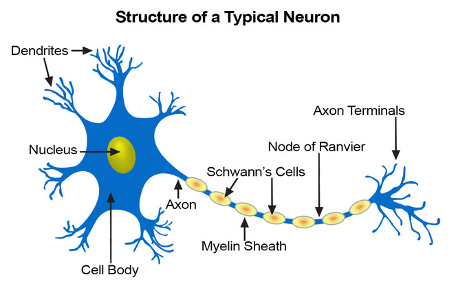
1. Structural Components of a Typical Neuron
Cell Body (Soma)
The neuron's main nutritional and metabolic center. It contains the nucleus, most organelles, and prominent Nissl bodies (rough ER), reflecting its high rate of protein synthesis.
Dendrites
Numerous, short, highly branched processes that act as the main receptive regions. They receive incoming signals from other neurons and convey them towards the cell body.
Axon
A single, long process that acts as the conducting region, generating and transmitting nerve impulses (action potentials) away from the cell body. It terminates in branches called axon terminals.
The Myelin Sheath
Many axons are covered by a fatty, insulating layer called the myelin sheath, which is formed by glial cells (Schwann cells in the PNS and oligodendrocytes in the CNS). This sheath dramatically speeds up nerve impulse transmission. The gaps between the myelin segments are called Nodes of Ranvier, where the action potential "jumps" from node to node (saltatory conduction).
2. Functional Classification of Neurons
Sensory (Afferent)
Transmit impulses from sensory receptors towards the CNS.
Motor (Efferent)
Transmit impulses from the CNS to effector organs (muscles/glands).
Interneurons
Lie between sensory and motor neurons within the CNS to integrate information. Most neurons are interneurons.
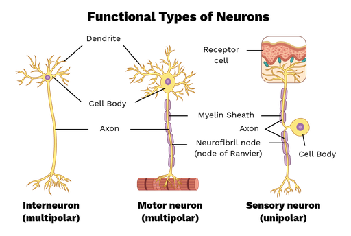
3. Structural Classification of Neurons
Multipolar
Three or more processes (one axon, many dendrites). The most common type in the CNS.
Bipolar
Two processes (one axon, one dendrite). Rare; found in special sense organs like the retina.
Unipolar
A single, short process that divides T-like. Found in most sensory neurons in the PNS.
Neuroglia (Glial Cells): The Supporting Cast
Neuroglia are non-excitable cells that surround, support, insulate, and protect neurons. They are far more numerous than neurons and can divide throughout life. There are six types of neuroglia: four in the CNS and two in the PNS.
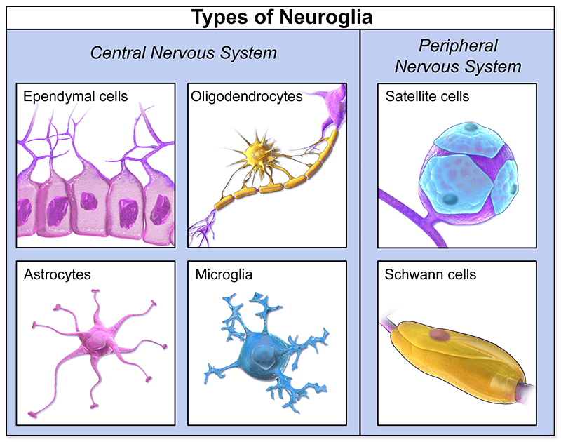
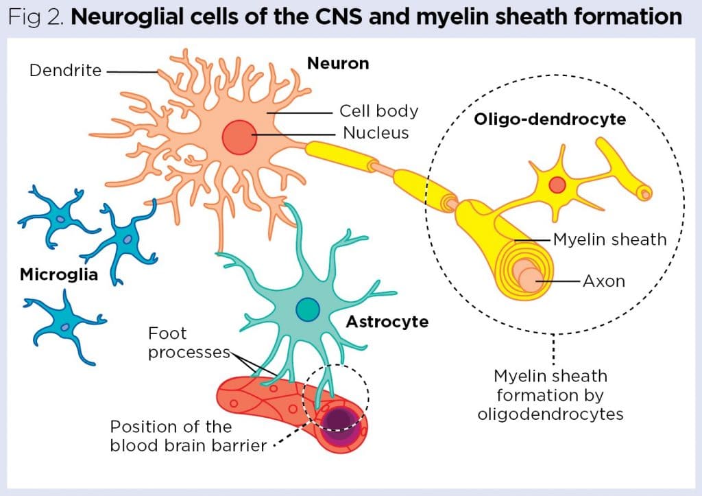
A. Neuroglia of the Central Nervous System (CNS)
Astrocytes (Star Cells)
Most abundant and versatile. Anchor neurons to blood vessels (form the blood-brain barrier) and regulate the chemical environment.
Microglial Cells
The resident macrophages of the CNS. They monitor neuron health and phagocytize microorganisms and debris.
Ependymal Cells
Line the central cavities of the brain and spinal cord. Their cilia help circulate cerebrospinal fluid (CSF).
Oligodendrocytes
Form the myelin sheaths around axons in the CNS. One oligodendrocyte can myelinate several axons.
B. Neuroglia of the Peripheral Nervous System (PNS)
Satellite Cells
Surround neuron cell bodies in PNS ganglia, providing support and regulating the chemical environment.
Schwann Cells
Form the myelin sheaths around thicker axons in the PNS. One Schwann cell myelinates one segment of one axon. Crucial for regeneration.
Nerve Impulse (Action Potential) Generation and Transmission
The ability of neurons to communicate relies on their ability to generate and transmit electrical signals, a process that involves several key stages.
1. Resting Membrane Potential
A neuron at rest has a voltage difference across its membrane of about -70mV. This is maintained by the sodium-potassium pump and ion leak channels.
2. Graded Potentials
Short-lived, localized changes in membrane potential. If a graded potential is strong enough to reach the threshold potential (~ -55mV) at the axon hillock, it triggers an action potential.
3. Action Potential (Nerve Impulse)
A brief, rapid, all-or-none electrical impulse that travels down the axon. It involves a depolarization phase (Na⁺ rushes in) followed by a repolarization phase (K⁺ rushes out).
4. Synapses
The junction where information is transferred. An arriving action potential causes the release of chemical messengers called neurotransmitters across a tiny gap (the synaptic cleft), which then bind to the next cell.