Common Disorders of the Skin
A comprehensive anatomical and clinical study of major integumentary pathologies including Albinism, Thermal Injuries (Burns), and Neoplastic transformations (Skin Tumors).
1. OVERVIEW
The skin is susceptible to a wide range of disorders arising from genetic defects, environmental insults, infectious agents, and neoplastic transformation. This section covers three major categories of skin disorders relevant to clinical practice: albinism (genetic pigmentary disorder), burns (thermal injury), and skin tumors (benign and malignant neoplasms).
2. ALBINISM
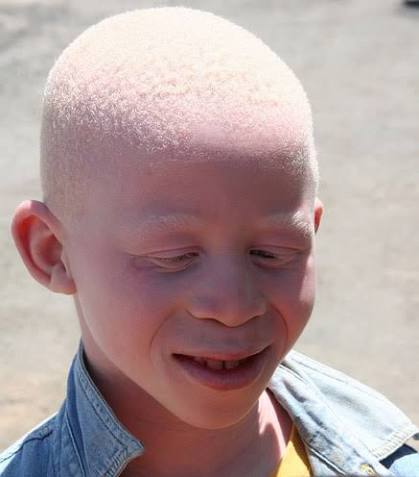
Definition: Albinism refers to a group of inherited disorders characterized by reduced or absent melanin production in the skin, hair, and eyes. It results from mutations in genes involved in melanin biosynthesis or melanosome transport. Albinism affects approximately 1 in 17,000 to 20,000 individuals worldwide, with higher prevalence (e.g., 1 in 1,000) in some African communities.
2.1 Etiology and Genetics
Melanin is synthesized from the amino acid tyrosine via the enzyme tyrosinase in melanocytes. Albinism results from mutations in genes encoding tyrosinase or proteins involved in melanosome formation and transport.
- Oculocutaneous albinism (OCA): Affects skin, hair, and eyes. Caused by autosomal recessive mutations in genes including TYR (OCA1), OCA2 (OCA2), TYRP1 (OCA3), and SLC45A2 (OCA4).
- Ocular albinism (OA): Primarily affects the eyes with minimal skin involvement. Caused by X-linked recessive mutations in the GPR143 gene.
- Hermansky-Pudlak syndrome and Chediak-Higashi syndrome: Rare syndromic forms associated with bleeding disorders and immunodeficiency.
2.2 Types of Oculocutaneous Albinism
- OCA1 (Tyrosinase-negative): Complete absence of tyrosinase activity. Affected individuals have white hair, very pale skin, and pink-blue irides. No melanin is produced.
- OCA1B (Tyrosinase-positive): Reduced tyrosinase activity. Some melanin may develop with age; hair may darken slightly; skin may tan minimally.
- OCA2 (P gene mutation): Most common type globally (especially in sub-Saharan Africa). Yellow, blond, or light brown hair; cream to light brown skin; blue to hazel irides. Some melanin production occurs.
- OCA3 (TYRP1 mutation): Rufous/red albinism. Red-bronze skin, ginger-red hair, hazel or brown irides. Predominantly seen in African and African-descendant populations.
- OCA4 (SLC45A2 mutation): Similar phenotype to OCA2. Most common in East Asian populations.
2.3 Clinical Features
- Skin: Very pale, creamy white, or light brown skin that does not tan and burns easily with sun exposure. In OCA1, skin remains white throughout life.
- Hair: White, yellow, blond, or light brown hair depending on the type. Eyelashes and eyebrows are similarly affected.
- Eyes: Iris translucency (red reflex visible on transillumination), nystagmus, strabismus, photophobia, reduced visual acuity (typically 20/60 to 20/400), and foveal hypoplasia.
- Visual Impairment: Results from abnormal routing of optic nerve fibers at the chiasm (decreased decussation) and foveal underdevelopment.
2.4 Complications
- Photodamage: Severe sunburn, actinic keratoses, and premature photoaging due to lack of UV protection.
- Skin cancer: Markedly increased risk of squamous cell carcinoma (SCC), basal cell carcinoma (BCC), and melanoma. In tropical Africa, SCC is the leading cause of death in albinism.
- Visual disability: Significant impairment affecting education, employment, and quality of life.
- Psychosocial impact: Stigma, discrimination, and social exclusion in many communities.
2.5 Management
- Sun protection: Strict avoidance of direct sun exposure; use of broad-spectrum sunscreen (SPF 50+), protective clothing, wide-brimmed hats, and UV-protective sunglasses.
- Dermatological surveillance: Regular skin examinations (every 6–12 months) for early detection of premalignant and malignant lesions.
- Ophthalmological care: Corrective lenses, low-vision aids, tinted lenses for photophobia, and management of strabismus.
- Genetic counseling: Family education regarding autosomal recessive inheritance patterns and recurrence risks (25% for each pregnancy in carrier couples).
In sub-Saharan Africa, individuals with albinism face extreme vulnerability to skin cancer due to high UV exposure and limited access to sun protection. Early detection and surgical excision of skin cancers are life-saving interventions.
3. BURNS
Definition: A burn is an injury to the skin and underlying tissues caused by thermal, chemical, electrical, or radiation energy. Burns are a significant global health burden, particularly in low- and middle-income countries.
3.1 Etiology and Classification
By Cause:
- Thermal burns: Caused by contact with flames, hot liquids (scalds), hot surfaces, or steam. Most common type.
- Chemical burns: Caused by acids, alkalis, or organic compounds. Alkalis penetrate deeper and cause more extensive damage than acids.
- Electrical burns: Caused by contact with electrical current. Severity depends on voltage, current type, and duration. May cause deep tissue necrosis with minimal surface injury.
- Radiation burns: Caused by ultraviolet radiation (sunburn), ionizing radiation (radiation therapy, nuclear accidents), or microwaves.
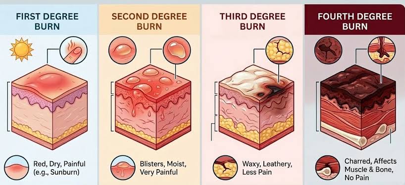
By Depth (Degree):
| Degree | Classification | Structures Involved | Clinical Features |
|---|---|---|---|
| First-degree | Superficial | Epidermis only | Erythema, pain, mild edema, no blisters. Heals in 3–6 days. |
| Second-degree | Partial-thickness | Epidermis and part of dermis | Erythema, severe pain, and fluid-filled blisters. Heals in 1–3 weeks. |
| Third-degree | Full-thickness | Entire epidermis and dermis | White, waxy, leathery, or charred. Pain is absent due to destruction of nerve endings. Requires skin grafting. |
| Fourth-degree | Deep full-thickness | Extends to fascia, muscle, and bone | Charred and insensate. Associated with high mortality; requires amputation in many cases. |
3.2 Pathophysiology
Burn injury triggers a complex local and systemic inflammatory response:
- Local response: Direct thermal damage causes protein denaturation and cell death. Surrounding tissue exhibits three concentric zones: coagulation (irreversible necrosis), stasis (potentially salvageable with adequate resuscitation), and hyperemia (inflammatory response with increased blood flow).
- Systemic response: Large burns (>20% TBSA) trigger a massive inflammatory cascade, releasing cytokines (TNF-α, IL-1, IL-6). This causes increased capillary permeability, leading to fluid shift from intravascular to interstitial spaces (burn shock).
- Hypermetabolic state: Characterized by increased oxygen consumption, protein catabolism, and energy expenditure persisting for months.
3.3 Assessment
- Extent of burns: Estimated using the Rule of Nines (adults: head 9%, each arm 9%, anterior trunk 18%, posterior trunk 18%, each leg 18%, perineum 1%) or the Lund-Browder chart (more accurate for children).
- Depth assessment: Based on appearance, pain sensation, and blanching.
- Airway assessment: Suspect inhalation injury in burns involving the face, singed nasal hairs, hoarseness, carbonaceous sputum, or exposure to fire in enclosed spaces.
- Circumferential burns: May cause compartment syndrome requiring escharotomy.
3.4 Management
Immediate First Aid:
- Stop the burning process: Remove from source, extinguish flames, remove hot clothing.
- Cool the burn: Apply cool (not ice-cold) running water for 20 minutes as soon as possible.
- Cover the burn: Use clean, non-adherent dressing (cling film, sterile gauze).
- Do NOT apply ice, butter, oils, or toothpaste to burns.
Hospital Management:
- Fluid resuscitation: The Parkland formula guides initial fluid administration for burns >15% TBSA (adults) or >10% TBSA (children): 4 mL × weight (kg) × %TBSA burn, given as half in first 8 hours and half in next 16 hours. Use Ringer's lactate.
- Wound care: Clean with mild antiseptic; debride necrotic tissue; apply topical antimicrobial agents (silver sulfadiazine, mafenide acetate).
- Surgical management: Escharotomy for circumferential burns; excision and grafting for deep partial-thickness and full-thickness burns.
- Partial-thickness burns >10% TBSA.
- Full-thickness burns.
- Burns involving face, hands, feet, genitalia, or major joints.
- Electrical or chemical burns.
- Inhalation injury.
4. SKIN TUMORS
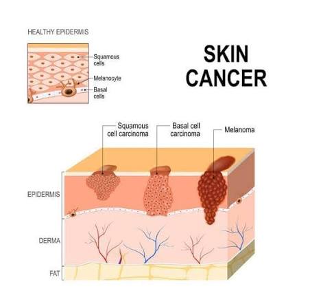
Skin tumors are neoplastic growths arising from epidermal, dermal, or subcutaneous tissues. Malignant skin cancers are the most common human cancers, with basal cell carcinoma (BCC), squamous cell carcinoma (SCC), and melanoma being the principal types.
4.1 Risk Factors
- Ultraviolet (UV) radiation: Chronic sun exposure, tanning beds, and living at high altitude.
- Fair skin: Fitzpatrick skin types I and II.
- Immunosuppression: Organ transplant recipients have 65-100-fold increased risk of SCC.
- Chronic wounds and scars: Marjolin ulcer (SCC arising in chronic burns or scars).
- Chemical exposure: Arsenic, coal tar, creosote.
4.2 Basal Cell Carcinoma (BCC)
- Epidemiology: Most common skin cancer (70–80% of all skin cancers). Slow-growing, locally invasive, rarely metastasizes.
- Pathology: Arises from basaloid cells of the epidermis. Histology shows nests of basaloid cells with peripheral palisading.
- Clinical features: Nodular pearly papule with telangiectasia and rolled border; superficial (erythematous scaly plaque).
- Management: Surgical excision with clear margins, Mohs micrographic surgery, curettage and electrodesiccation.
4.3 Squamous Cell Carcinoma (SCC)
- Epidemiology: Second most common skin cancer (20% of skin cancers). Greater metastatic potential than BCC.
- Pathology: Arises from keratinocytes of the epidermis. Histology shows atypical keratinocytes, keratin pearls, and invasive growth.
- Clinical features: Firm, hyperkeratotic nodule or plaque, often on sun-exposed areas. May ulcerate and bleed.
- Precursor lesions: Actinic keratosis (rough, scaly patches) and Bowen disease (SCC in situ).
4.4 Melanoma
- Epidemiology: Most aggressive skin cancer, responsible for 75% of skin cancer deaths. Incidence is increasing globally.
- Pathology: Arises from melanocytes. Histology shows atypical melanocytes with nuclear pleomorphism and pagetoid spread.
- ABCDE Rule for Detection:
— Asymmetry
— Border irregularity
— Color variation
— Diameter >6 mm
— Evolving (changing) - Staging: Based on Breslow thickness (depth of invasion in mm), ulceration status, and lymph node involvement.
- Management: Wide local excision, Sentinel lymph node biopsy (SLNB), and adjuvant immunotherapy (pembrolizumab).
5. SUMMARY TABLE
| Disorder | Etiology | Key Clinical Features | Management | Complications |
|---|---|---|---|---|
| Albinism | Autosomal recessive; TYR, OCA2 mutations | Hypopigmented skin/hair/eyes, nystagmus | Sun protection, skin surveillance, ophthalmic care | Skin cancer (SCC, BCC), visual loss |
| Burns | Thermal, chemical, electrical | Erythema, blisters, charring; pain (partial) or painless | Fluid resuscitation, wound care, topical antibiotics | Infection/sepsis, shock, scarring |
| BCC | UV radiation, fair skin | Pearly papule, rolled border, telangiectasia | Surgical excision, Mohs surgery | Local invasion; rare metastasis |
| SCC | UV radiation, immunosuppression, scars | Firm hyperkeratotic nodule; may ulcerate | Surgical excision, Mohs surgery, radiotherapy | Metastasis (5–10%); higher in scars |
| Melanoma | UV radiation, fair skin, atypical moles | ABCDE features; asymmetry, irregular border, color | Wide excision, SLNB, immunotherapy | Metastasis; high mortality if advanced |
