Disorders of the Respiratory System
Comprehensive clinical notes covering the etiology, pathophysiology, clinical presentation, and management of major respiratory conditions: Asthma, Pneumonia, and Emphysema.
1. Asthma
Asthma is a chronic inflammatory disorder of the airways characterized by reversible airflow obstruction, bronchial hyperresponsiveness, and airway remodeling. It affects approximately 262 million people worldwide and is a leading cause of morbidity in children and young adults.
1.1 Definition and Epidemiology
- Nature: Chronic condition with variable and recurring symptoms and reversible airflow limitation.
- Prevalence: 5–10% globally; notably higher in developed countries and urban areas.
- Onset: Frequently in childhood; may persist or remit and relapse later in life.
1.2 Etiology and Risk Factors
- Atopic (allergic) asthma: Most common form; associated with IgE-mediated hypersensitivity to allergens such as dust mites, pollen, pet dander, and mold.
- Non-atopic asthma: Triggered by infections, irritants, exercise, cold air, or stress; involves no allergen sensitization.
- Occupational asthma: Caused by workplace exposures to isocyanates, flour, wood dust, or chemicals.
- Risk Factors: Family history of atopy, childhood respiratory infections, obesity, smoking, air pollution, and low birth weight.
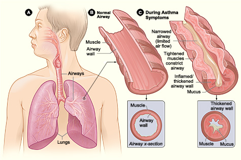
1.3 Pathophysiology
- Acute phase: Allergen exposure triggers IgE-mediated mast cell degranulation, releasing histamine, leukotrienes, prostaglandins, and cytokines.
- Bronchoconstriction: Smooth muscle contraction narrows airways within minutes.
- Mucus hypersecretion: Goblet cell hyperplasia and submucosal gland hypertrophy produce thick, tenacious mucus plugs.
- Airway edema: Increased vascular permeability causes mucosal swelling.
- Chronic inflammation: Eosinophils, T-helper 2 (Th2) cells, mast cells, and neutrophils infiltrate the airway wall.
- Airway remodeling: Subepithelial fibrosis, smooth muscle hypertrophy, and basement membrane thickening cause irreversible airflow limitation in long-standing disease.
1.4 Clinical Features
Classic triad: Wheeze, cough, and dyspnea.
- Symptoms: Variable, often worse at night or early morning; triggered by exercise, allergens, or cold air.
- Physical examination: Prolonged expiratory phase, bilateral wheezing, use of accessory muscles, tachypnea, and tachycardia.
Severe Acute Asthma
Signs of a life-threatening attack include a Silent chest (absence of wheeze due to minimal airflow), cyanosis, altered consciousness, exhaustion, and bradycardia (a pre-arrest sign).
1.5 Diagnosis
- History: Pattern of symptoms, specific triggers, and family history.
- Spirometry: Reduced FEV₁ and FEV₁/FVC ratio (<0.70). Diagnosis is supported by significant reversibility (increase in FEV₁ >12% and >200 mL after bronchodilator).
1.7 Management
Stepwise Approach (GINA Guidelines):
- Step 1: SABA as needed.
- Step 2: Low-dose Inhaled Corticosteroid (ICS) daily + SABA.
- Step 3: Low-dose ICS/LABA combination or medium-dose ICS.
- Step 4: Medium-dose ICS/LABA.
- Step 5: High-dose ICS/LABA + add-on therapy (LAMA, biologics).
Status Asthmaticus (Acute Severe Asthma)
Requires urgent hospitalization. Management includes:
— Continuous nebulized salbutamol + ipratropium bromide.
— Systemic corticosteroids (Oral prednisolone or IV hydrocortisone).
— Oxygen to maintain SpO₂ 94–98%.
— IV Magnesium sulfate for bronchodilator effect.
— Mechanical ventilation if refractory.
2. Pneumonia
Pneumonia is an acute infection of the lung parenchyma, including the alveolar spaces and interstitial tissue. It is a major cause of mortality worldwide, particularly in children under 5 and the elderly.
2.1 Classification
| Category | Definition / Key Feature |
|---|---|
| CAP | Community-acquired; acquired outside healthcare facilities. |
| HAP | Hospital-acquired; occurs >48 hours after admission. |
| VAP | Ventilator-associated; occurs >48 hours after intubation. |
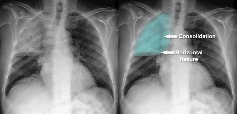
| Lobar | Consolidation of an entire lobe (typically S. pneumoniae). |
| Interstitial | Inflammation confined to alveolar walls (typical of viruses/Mycoplasma). |
2.3 Etiology
| Pathogen | Characteristics | Risk Factors |
|---|---|---|
| S. pneumoniae | Most common cause of CAP; Lobar consolidation. | Alcoholism, COPD, asplenia. |
| H. influenzae | Common cause of bronchopneumonia. | COPD, smoking, elderly. |
| Staph. aureus | Necrotizing pneumonia; Abscesses. | Post-influenza infection, IV drug use. |
| K. pneumoniae | Currant jelly sputum; Bulging fissure sign. | Alcoholism, Diabetes. |
| M. pneumoniae | Atypical; interstitial pattern. | Young adults, schools/military. |
| Legionella | Atypical; Hyponatremia; GI symptoms. | AC systems, hotels, smokers. |
2.7 Management
- General measures: Oxygen to maintain SpO₂ >94% (88–92% in COPD); hydration; analgesia.
- Antibiotic Therapy (Empirical):
— Low risk CAP: Amoxicillin 500 mg TDS for 5 days.
— Moderate CAP: Amoxicillin-clavulanate + macrolide.
— ICU CAP: Ceftriaxone + azithromycin.
Untreated pneumonia can lead to Pleural effusion, Empyema, Lung abscess, Septicemia, and ARDS.
3. Emphysema
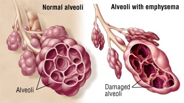
Emphysema is a form of COPD characterized by the permanent enlargement of airspaces distal to the terminal bronchioles, accompanied by destruction of alveolar walls without significant fibrosis.
3.2 Etiology and Risk Factors
- Cigarette smoking: Accounts for 80–90% of cases.
- Alpha-1 antitrypsin (AAT) deficiency: Genetic cause; AAT inhibits neutrophil elastase. Homozygous ZZ genotype causes early-onset panacinar emphysema.
- Air pollution: Indoor biomass fuel exposure.
3.3 Pathophysiology
- Protease-antiprotease imbalance: Neutrophil/macrophage proteases (elastase) destroy alveolar walls.
- Alveolar wall destruction: Loss of elastic fibers and capillary beds; airspaces enlarge and coalesce.
- Airway collapse: Loss of elastic recoil causes airways to collapse during expiration, leading to dynamic hyperinflation.
- V/Q mismatch: Reduced surface area causes hypoxemia.
3.4 Types of Emphysema
| Type | Location | Associations |
|---|---|---|
| Centriacinar | Central part of acinus; Upper lobes. | Cigarette smoking. |
| Panacinar | Entire acinus; Lower lobes. | AAT deficiency. |
| Paraseptal | Distal acinus near pleura. | Spontaneous pneumothorax; Subpleural bullae. |
3.6 Diagnosis
- Clinical hallmark: Progressive dyspnea, initially on exertion.
- Spirometry: Irreversible obstruction (FEV₁/FVC <0.70).
- Chest X-ray: Hyperinflation, flattened diaphragms, increased retrosternal airspace, and a small heart shadow.
- CT Chest: Gold standard for detecting low-attenuation areas and bullae.
Oxygen therapy (LTOT): Indicated if PaO₂ ≤55 mmHg. Must be used >15 hours/day to improve survival.
Surgical options: Bullectomy (removal of large bullae) or Lung Volume Reduction Surgery (LVRS).
4. Key Points Summary
- Asthma: Reversible airway obstruction; managed with ICS and SABA.
- Pneumonia: Infection of parenchyma; CURB-65 guides assessment and antibiotic choice.
- Emphysema: Alveolar wall destruction and loss of recoil; smoking is the primary cause.
- Red Flag: The Silent Chest in an asthma attack is a medical emergency indicating near-total absence of airflow.
