Gas Exchange and Transport
Comprehensive notes of respiratory physiology, the dynamics of the blood-air barrier, hemoglobin kinetics, and clinical correlations associated with hypoxemia and acid-base disturbances.
1. Introduction
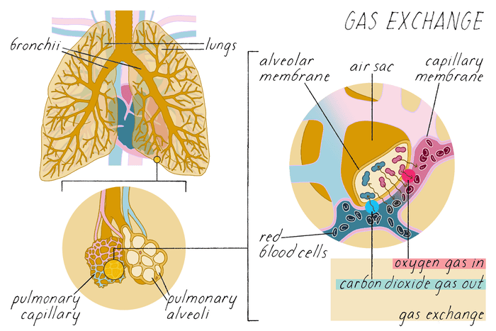
Gas exchange is the physiological process by which oxygen is transferred from alveolar air to pulmonary capillary blood, and carbon dioxide is transferred from blood to alveolar air for elimination. The subsequent transport of these gases in the blood ensures the continuous delivery of oxygen to tissues for aerobic metabolism and the removal of carbon dioxide, a metabolic byproduct. These processes are critically dependent on partial pressure gradients, the structural integrity of the blood-air barrier, and the biochemical properties of hemoglobin.
2. Partial Pressures of Respiratory Gases
The partial pressure of a gas is the pressure it would exert if it alone occupied the total volume. Gas exchange occurs strictly down partial pressure gradients via passive diffusion.
| Location | PO₂ (mmHg) | PCO₂ (mmHg) |
|---|---|---|
| Atmospheric air (dry) | 159 | 0.3 |
| Alveolar air | 100 | 40 |
| Arterial blood | 95–100 | 40 |
| Venous blood | 40 | 46 |
| Tissues (rest) | ~40 | ~46 |
| Expired air | 120 | 27 |
- Humidification: Alveolar PO₂ is lower than atmospheric PO₂ because inspired air is humidified in the upper airways; water vapor dilutes the other gases.
- O₂ Gradient: The pressure gradient for O₂ is 60 mmHg (Alveolar 100 → Venous blood 40), providing a powerful drive for O₂ uptake.
- CO₂ Gradient: The pressure gradient for CO₂ is only 6 mmHg (Venous blood 46 → Alveolar 40). Despite this small gradient, CO₂ diffuses rapidly due to its high solubility.
3. External Respiration (Pulmonary Gas Exchange)
3.1 Diffusion Across the Blood-Air Barrier
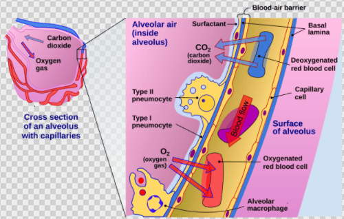
Oxygen diffuses from alveolar air into pulmonary capillary blood, while carbon dioxide diffuses in the opposite direction. Diffusion is extremely efficient due to:
- Barrier Thickness: The membrane is ultra-thin (0.2–0.5 µm).
- Surface Area: The total alveolar surface area is massive (70–100 m²).
- Equilibration Time: Gas equilibration occurs within 0.25 seconds. Since blood spends ~0.75 seconds in the pulmonary capillaries, there is a large safety margin.
3.2 Factors Affecting External Respiration
- Thickness: Increased in pulmonary fibrosis, edema, or pneumonia, which slows diffusion.
- Surface Area: Decreased in emphysema (alveolar destruction), pneumonectomy, or atelectasis.
- Pressure Gradients: Reduced at high altitudes (low inspired PO₂) or in hypoventilation.
- Diffusion Coefficient: CO₂ is ~20× more soluble than O₂, allowing it to diffuse just as quickly despite its smaller pressure gradient.
3.3 Ventilation-Perfusion (V/Q) Ratio
Optimal gas exchange requires matching of alveolar ventilation (V) to pulmonary perfusion (Q). Normal V/Q ratio is ~0.8 (4 L/min ventilation ÷ 5 L/min blood flow).
V/Q Mismatch
- High V/Q (Dead Space): Alveoli are ventilated but unperfused. Classic example: Pulmonary Embolism.
- Low V/Q (Shunt): Alveoli are perfused but unventilated. Seen in atelectasis, airway obstruction, or consolidation.
- Hypoxic Vasoconstriction: A local compensatory mechanism where alveolar hypoxia causes constriction of local pulmonary arterioles to divert blood to better-ventilated regions.
4. Oxygen Transport in Blood
Oxygen is transported in the blood in two forms: dissolved in plasma (1.5%) and bound to hemoglobin (98.5%).
4.1 Hemoglobin Structure and Function
Hemoglobin (Hb) is a tetrameric protein (two alpha and two beta chains). Each chain contains a heme group with an iron atom (Fe²⁺) that binds one O₂ molecule. Thus, one Hb molecule can bind four O₂ molecules. 1 gram of Hb can carry 1.34 mL of O₂.
4.2 Oxyhemoglobin Dissociation Curve
The relationship between PO₂ and Hb saturation is sigmoid (S-shaped) due to cooperative binding.
- P₅₀: The PO₂ at which Hb is 50% saturated; normally ~26–27 mmHg.
- Plateau (PO₂ 60–100 mmHg): Hb remains highly saturated (>90%), providing a buffer against moderate hypoxemia.
- Steep Portion (PO₂ 10–40 mmHg): Small changes in PO₂ cause large changes in saturation, facilitating O₂ unloading in tissues.
4.3 Factors Shifting the Oxyhemoglobin Curve
| Factor | Right Shift (↓ Affinity) | Left Shift (↑ Affinity) |
|---|---|---|
| Temperature | Increased (fever, exercise) | Decreased (hypothermia) |
| pH (Bohr effect) | Decreased (acidosis) | Increased (alkalosis) |
| PCO₂ | Increased | Decreased |
| 2,3-DPG | Increased (chronic hypoxia, anemia) | Decreased (stored blood) |
| CO | — | Increased (CO poisoning) |
The Bohr effect states that increased H⁺ (acidosis) and CO₂ decrease Hb affinity for O₂. This is physiologically vital as it promotes O₂ release in metabolically active tissues where CO₂ and acid levels are highest.
5. Carbon Dioxide Transport
CO₂ is transported in three forms: dissolved (7%), as bicarbonate (70%), and bound to hemoglobin as carbaminohemoglobin (23%).
5.2 Bicarbonate (HCO₃⁻) Formation
This is the most important form of CO₂ transport. The process involves several steps:
- CO₂ enters the RBC and combines with H₂O to form carbonic acid (H₂CO₃), catalyzed by carbonic anhydrase.
- H₂CO₃ dissociates into H⁺ and HCO₃⁻.
- Chloride Shift (Hamburger Phenomenon): HCO₃⁻ diffuses out of the RBC into plasma in exchange for Cl⁻ to maintain electrical neutrality.
- H⁺ binds to deoxygenated Hb (which acts as a buffer), preventing significant pH changes.
The Haldane effect states that deoxygenated hemoglobin binds CO₂ more readily than oxygenated hemoglobin. This facilitates CO₂ loading in the tissues and CO₂ unloading in the lungs as Hb becomes oxygenated.
6. Internal Respiration (Tissue Gas Exchange)
In systemic tissues, O₂ diffuses from capillary blood (PO₂ 95–100 mmHg) into tissues (PO₂ ~40 mmHg). Simultaneously, CO₂ diffuses from tissues (PCO₂ ~46 mmHg) into capillary blood (PCO₂ 40 mmHg).
Factors Affecting Delivery: Oxygen delivery (DO₂) depends on cardiac output and arterial O₂ content. Capillary density increases in exercise to shorten diffusion distance, while edema increases it, impairing exchange.
7. Clinical Correlations
Defined as reduced arterial PO₂ (<80 mmHg) or O₂ saturation (<92%).
- Hypoxic hypoxia: Low inspired O₂ or impaired pulmonary gas exchange.
- Anemic hypoxia: Reduced Hb (anemia, hemorrhage, CO poisoning).
- Stagnant hypoxia: Reduced tissue perfusion (shock, heart failure).
- Histotoxic hypoxia: Impaired cellular O₂ utilization (e.g., cyanide poisoning).
CO binds to Hb with 240× greater affinity than O₂, forming carboxyhemoglobin (COHb). This shifts the dissociation curve to the left, preventing O₂ unloading in tissues. Features include cherry-red skin, headache, and confusion. Treatment is 100% O₂ or hyperbaric oxygen.
Bluish discoloration of skin due to increased deoxygenated Hb (>5 g/dL).
- Central cyanosis: Due to arterial hypoxemia (cardiac or pulmonary disease). Visible in tongue/lips.
- Peripheral cyanosis: Due to reduced peripheral blood flow (cold, shock). Visible in extremities.
Metabolic Acidosis: Stimulates peripheral and central chemoreceptors, leading to increased ventilation (Kussmaul respirations) to blow off CO₂ and raise pH. This respiratory compensation is rapid but limited.
8. Key Points Summary
- Gas exchange depends on partial pressure gradients, surface area, and barrier thickness.
- The V/Q ratio mismatch is a primary cause of hypoxemia in clinical practice.
- O₂ is primarily transported bound to Hb; the curve is sigmoid and shifted by temperature, pH, PCO₂, and 2,3-DPG.
- CO₂ is primarily transported as bicarbonate via the carbonic anhydrase reaction and the chloride shift.
- The Bohr effect aids O₂ unloading at tissues, while the Haldane effect aids CO₂ loading at tissues and unloading at lungs.
