Organs of the Lower Respiratory System
Comprehensive anatomical study covering the structural organization, internal architecture, neurovascular supply, and clinical correlations of the trachea, bronchial tree, and lungs.
1. Introduction
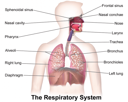
The lower respiratory tract comprises the trachea, bronchi, bronchioles, and lungs. These structures function primarily to conduct air to the alveoli, where gas exchange occurs. The lower respiratory tract is lined by respiratory epithelium and supported by cartilaginous, muscular, and elastic tissues that maintain airway patency while providing the necessary flexibility for respiration and body movement.
2. The Trachea
The trachea (windpipe) is a fibrocartilaginous tube measuring approximately 10–11 cm in length and 2 cm in diameter. it extends from the inferior border of the cricoid cartilage (C6) to the carina at the level of the sternal angle (T4–T5).
2.1 Structure
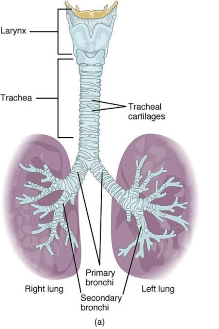
- Cartilaginous Support: Composed of 16–20 C-shaped hyaline cartilage rings that prevent airway collapse during inspiration.
- Trachealis Muscle: The posterior wall is membranous and contains the trachealis muscle (smooth muscle). This arrangement allows the esophagus to expand during swallowing.
- Histology: Lined by ciliated pseudostratified columnar epithelium with goblet cells. Submucosal glands secrete mucus that traps inhaled particles.
- Mucociliary Escalator: Ciliary action moves trapped particles upward toward the pharynx for swallowing or expectoration.
2.2 Anatomical Relations
| Relation | Structures |
|---|---|
| Anterior | Isthmus of thyroid gland, inferior thyroid veins, sternohyoid and sternothyroid muscles, manubrium sterni. |
| Posterior | Esophagus and recurrent laryngeal nerves. |
| Lateral | Lobes of the thyroid gland, carotid sheaths. |
2.3 Blood Supply and Innervation
- Arterial Supply: Inferior thyroid arteries.
- Venous Drainage: Brachiocephalic veins via inferior thyroid veins.
- Lymphatic Drainage: Pretracheal and paratracheal nodes.
- Innervation:
— Parasympathetic: Vagus nerve (causes bronchoconstriction and increased mucus secretion).
— Sympathetic: Sympathetic trunks (causes bronchodilation and reduced secretion).
3. The Bronchial Tree
The bronchial tree is the branching system of airways that distributes air from the trachea to the alveoli.
3.1 Main (Primary) Bronchi
The trachea bifurcates at the carina into right and left main bronchi. Both are supported by C-shaped cartilage rings and lined by respiratory epithelium.
- Right main bronchus: Wider, shorter (~2.5 cm), and more vertical than the left. Foreign bodies more commonly enter the right bronchus due to this verticality.
- Left main bronchus: Longer (~5 cm), narrower, and more horizontal. It passes beneath the aortic arch and anterior to the esophagus.
3.2 Lobar (Secondary) Bronchi
- Right lung: Three lobar bronchi (superior, middle, lower) corresponding to the three lobes.
- Left lung: Two lobar bronchi (upper and lower) corresponding to the two lobes.
3.3 Segmental (Tertiary) Bronchi
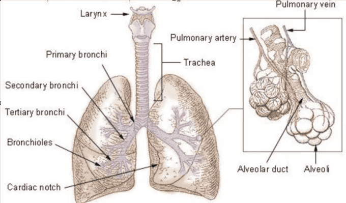
Each lobar bronchus divides into segmental bronchi. There are typically 10 on the right and 8–10 on the left. Each segmental bronchus supplies a bronchopulmonary segment.
A bronchopulmonary segment is a functionally independent unit with its own artery, vein, and lymphatic drainage. These segments are important surgically because resection of a segment does not compromise adjacent segments.
3.4 Bronchioles
- Conducting bronchioles: Diameter <1 mm; they lack cartilage and glands. Lined by ciliated cuboidal epithelium.
- Terminal bronchioles: The most distal part of the conducting zone; each supplies a pulmonary acinus.
- Respiratory bronchioles: The first part of the respiratory zone; they have scattered alveoli budding directly from their walls.
4. The Lungs
The lungs are the paired, cone-shaped organs of respiration located in the thoracic cavity on either side of the mediastinum.
4.1 Gross Anatomy
- Right lung: Larger and heavier; has three lobes (superior, middle, inferior) separated by horizontal and oblique fissures.
- Left lung: Smaller due to the cardiac notch; has two lobes (superior and inferior) separated by the oblique fissure. It contains the lingula (homologous to the right middle lobe).
- Apex: Projects into the root of the neck, 2–4 cm above the clavicle.
- Base: Concave, resting on the diaphragm.
- Hilum: Medial surface depression where bronchi, pulmonary vessels, bronchial vessels, lymphatics, and nerves enter and exit.
- Surfaces: Costal surface (convex, related to ribs) and Mediastinal surface (related to mediastinal structures).
4.2 Bronchopulmonary Segments
| Lobe | Right Lung Segments | Left Lung Segments |
|---|---|---|
| Upper Lobe | Apical, Posterior, Anterior | Apical-posterior, Anterior, Superior lingular, Inferior lingular |
| Middle Lobe | Lateral, Medial | — |
| Lower Lobe | Superior, Medial basal, Anterior basal, Lateral basal, Posterior basal | Superior, Anteromedial basal, Lateral basal, Posterior basal |
4.3 Pleura
- Parietal pleura: Lines the thoracic wall (costal pleura), diaphragm (diaphragmatic pleura), and mediastinum (mediastinal pleura). The cervical pleura (cupula) extends into the neck.
- Visceral pleura: Covers the lung surface, including the fissures.
- Pleural cavity: Potential space between parietal and visceral pleurae; contains a thin film of serous fluid (pleural fluid) that reduces friction during breathing.
- Pleural recesses: Potential spaces not occupied by lungs during quiet breathing, including the costodiaphragmatic recess (largest) and costomediastinal recess.
5. The Respiratory Zone
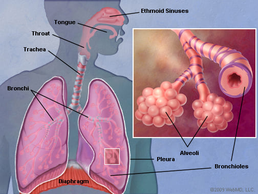
The respiratory zone is where gas exchange occurs. It includes respiratory bronchioles, alveolar ducts, alveolar sacs, and alveoli.
5.1 Alveoli
Approximately 300–500 million alveoli exist in adult lungs, providing a total surface area of 70–100 m². Each alveolus is a thin-walled sac (0.2–0.5 µm thick) lined by two types of pneumocytes:
- Type I pneumocytes: Squamous epithelial cells covering 95% of alveolar surface; the primary site of gas exchange.
- Type II pneumocytes: Cuboidal cells that produce surfactant (dipalmitoylphosphatidylcholine), which reduces surface tension and prevents alveolar collapse.
- Alveolar macrophages (dust cells): Phagocytose inhaled particles and pathogens.
5.2 Blood-Air Barrier
The barrier between alveolar air and capillary blood consists of: Type I pneumocyte, fused basement membrane, and capillary endothelial cell. The total thickness is 0.2–0.5 µm, allowing for rapid gas diffusion.
5.3 Alveolar-Capillary Network
- Each alveolus is surrounded by a dense capillary network.
- Pulmonary arteries carry deoxygenated blood to the alveolar capillaries; pulmonary veins return oxygenated blood to the left atrium.
- Bronchial arteries (systemic): Supply oxygenated blood to the lung tissue and drain partially into pulmonary veins.
6. Clinical Correlations
- Tracheal Stenosis: Narrowing of the tracheal lumen due to trauma, prolonged intubation, or granulomatous disease. Presents with stridor, dyspnea, and cough.
- Atelectasis: Collapse of lung tissue due to airway obstruction, compression, or surfactant deficiency. Types: Obstructive (resorption), Compressive (pleural effusion), Adhesive (ARDS).
- Pneumothorax: Air in the pleural cavity causing lung collapse. Can be spontaneous (rupture of bleb), traumatic (rib fracture), or iatrogenic. Tension pneumothorax is life-threatening and requires immediate needle decompression.
- Pleural Effusion: Accumulation of fluid in the pleural space. Divided into Transudative (heart failure, cirrhosis) or Exudative (pneumonia, malignancy, TB). Diagnosed using Light criteria via thoracentesis.
7. Key Points Summary
- The lower respiratory tract includes the trachea, bronchi, bronchioles, lungs, and pleura.
- The trachea has C-shaped cartilage rings and a posterior trachealis muscle; it bifurcates at the carina (T4/T5).
- The right main bronchus is wider and more vertical, making it the common site for foreign body aspiration.
- The lungs have three lobes on the right and two on the left; each lobe is divided into bronchopulmonary segments.
- The pleura consists of parietal and visceral layers with a potential pleural cavity containing lubricating fluid.
- Gas exchange occurs across the blood-air barrier in the alveoli, lined by Type I and Type II pneumocytes.
