Disorders of the Lymphatic System
Complete clinical notes covering the classification, pathophysiology, and management of lymphadenitis and Hodgkin Lymphoma.
1. Lymphadenitis
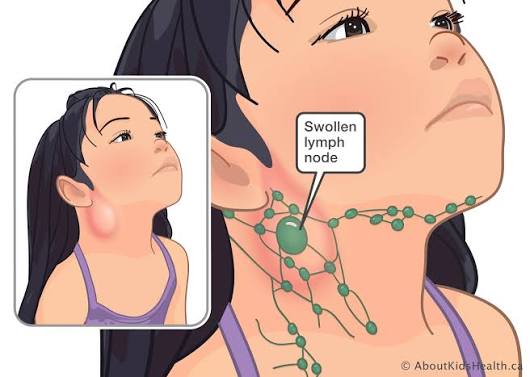
Lymphadenitis is the inflammation of the lymph nodes, usually secondary to infection, immune activation, or malignancy in the drainage territory. It represents a localized immune response and is one of the most common clinical presentations in primary care and general medicine.
1.1 Definition and Classification
- Acute lymphadenitis: Rapid onset; tender, warm, enlarged nodes; typically bacterial.
- Chronic lymphadenitis: Persistent enlargement (>4 weeks); often granulomatous or neoplastic.
- Suppurative lymphadenitis: Progression to abscess formation with central necrosis and pus.
- Generalized lymphadenitis: Involvement of multiple non-contiguous node groups; suggests systemic disease.
- Localized lymphadenitis: Confined to a single regional group; indicates local infection or malignancy.
1.2 Causes and Risk Factors
| Category | Common Causes | Key Features |
|---|---|---|
| Bacterial | Staphylococcus aureus, Streptococcus pyogenes, Bartonella henselae (cat-scratch), Mycobacterium tuberculosis | Acute, tender, erythematous; TB: matted, caseating, cold abscess |
| Viral | EBV (infectious mononucleosis), CMV, HIV, cytomegalovirus | Bilateral, symmetrical, non-suppurative; often with pharyngitis or rash |
| Fungal | Histoplasma, Coccidioides, Sporothrix | Endemic regions; chronic or granulomatous presentation |
| Parasitic | Toxoplasma gondii, filariasis, leishmaniasis | Travel history; eosinophilia may be present |
| Autoimmune | SLE, rheumatoid arthritis, sarcoidosis | Generalized; associated with other systemic features |
| Malignant | Metastatic carcinoma, lymphoma, leukemia | Hard, fixed, non-tender; progressive enlargement; systemic B symptoms |
1.3 Pathophysiology
- Infection or antigenic stimulus in the drainage area triggers immune cell recruitment to the regional node.
- Hyperplasia of germinal centers (B-cell proliferation) and paracortical expansion (T-cell response).
- Increased blood flow and vascular permeability cause redness, warmth, and swelling.
- Pressure on the capsule and surrounding tissues produces pain.
- In bacterial infections, neutrophil infiltration may lead to suppuration and abscess formation.
- In granulomatous infections (TB, fungi), epithelioid macrophages and giant cells form caseating granulomas.
1.4 Clinical Features
- Local signs: Enlarged, tender lymph node(s); overlying skin may be erythematous and warm.
- Systemic signs: Fever, malaise, anorexia; severity correlates with the underlying infection.
| Condition | Distinguishing Features |
|---|---|
| Reactive hyperplasia | Recent infection; tender, mobile, bilateral; resolves within weeks |
| Lymphoma | Painless, progressive, rubbery; may have B symptoms |
| TB | Chronic, matted, cold abscess; exposure history; positive IGRA/TST |
| Metastatic carcinoma | Hard, fixed, non-tender; known primary malignancy |
| Kikuchi-Fujimoto disease | Young women; cervical nodes; self-limiting; histology diagnostic |
| Castleman disease | Localized or multicentric; hypervascular on imaging; biopsy diagnostic |
1.7 Management
- Supportive care: Rest, hydration, analgesia (paracetamol or NSAIDs), warm compresses.
- Antibiotics: Empirical therapy targeting likely pathogens.
— Community-acquired: amoxicillin-clavulanate or cephalexin for 7–10 days.
— MRSA risk: clindamycin or trimethoprim-sulfamethoxazole.
— Cat-scratch disease: azithromycin if severe; usually self-limiting.
— TB lymphadenitis: standard anti-TB regimen (RIPE: rifampicin, isoniazid, pyrazinamide, ethambutol for 2 months, then rifampicin + isoniazid for 4 months). - Surgical drainage: Incision and drainage for fluctuant abscesses; needle aspiration may suffice for small collections.
- Treatment of underlying cause: Antivirals where indicated, antiretroviral therapy for HIV, immunosuppressants for autoimmune disease.
- Follow-up: Reassess at 2–4 weeks; persistent or enlarging nodes require further investigation.
- Abscess formation and spontaneous drainage.
- Bacteremia and sepsis.
- Chronic sinus tract formation (especially TB).
- Post-inflammatory fibrosis and persistent node enlargement.
- Misdiagnosis of underlying malignancy.
2. Hodgkin's Disease (Hodgkin Lymphoma)
Hodgkin lymphoma (HL) is a malignant neoplasm of the lymphatic system characterized by the presence of Reed-Sternberg cells in a background of inflammatory cells. It accounts for approximately 10% of all lymphomas and has a bimodal age distribution with peaks in young adulthood and after age 55.
2.1 Epidemiology and 2.2 Etiology
- Incidence: 2–3 per 100,000 annually in developed countries. Slight male predominance (M:F ratio 1.3:1).
- Infectious: Epstein-Barr virus (EBV) implicated in 40–50% of cases; HIV increases risk 10-fold.
- Genetic: Familial clustering suggests genetic susceptibility; HLA associations identified.
- Immunodeficiency: Congenital immunodeficiency, post-transplant immunosuppression.
- Environmental: No strong environmental carcinogen identified; smoking may be a minor risk factor.
2.3 Pathophysiology
- Originates from germinal center B cells that have undergone failed apoptosis.
- The neoplastic cell is the Reed-Sternberg (RS) cell or its variant, the Hodgkin cell.
- RS cells are large, binucleated or multinucleated cells with prominent eosinophilic nucleoli ('owl-eye' appearance).
- RS cells constitute <1% of the tumor mass; the majority is a reactive inflammatory infiltrate.
- RS cells express CD30 and CD15; they are usually negative for CD20 and CD45 (LCA).
- Cytokine production (IL-5, IL-10, TGF-beta) by RS cells contributes to the inflammatory background and systemic symptoms.
2.4 Classification (WHO 2016)
| Subtype | Frequency | Characteristics |
|---|---|---|
| Nodular sclerosis (NSHL) | 70% of cases | Most common in young adults; collagen bands dividing nodules; lacunar RS cell variant; good prognosis. |
| Mixed cellularity (MCHL) | 20–25% | Older adults and HIV-positive patients; numerous RS cells; EBV association common. |
| Lymphocyte-rich (LRHL) | 5% | Few RS cells; abundant small lymphocytes; excellent prognosis. |
| Lymphocyte-depleted (LDHL) | <5% | Rare; elderly or HIV-positive; numerous RS cells, few lymphocytes; aggressive. |
| Nodular lymphocyte-predominant (NLPHL) | Distinct entity | Popcorn cells (LP cells); CD20+, CD30-; indolent; male predominance. |
2.6 Staging (Ann Arbor System)
| Stage | Description |
|---|---|
| I | Single lymph node region or single extralymphatic site (IE). |
| II | Two or more lymph node regions on the same side of the diaphragm. |
| III | Lymph node regions on both sides of the diaphragm; may include spleen (IIIS) or localized extralymphatic site (IIIE). |
| IV | Diffuse or disseminated involvement of one or more extralymphatic organs with or without lymph node involvement. |
- A: No systemic symptoms.
- B: Presence of B symptoms (fever, night sweats, weight loss).
- E: Involvement of a single extranodal site contiguous with a known nodal site.
- X: Bulky disease (mediastinal mass >1/3 thoracic diameter or nodal mass >10 cm).
- S: Spleen involvement.
2.7 Investigations
- Laboratory: Full blood count (anemia, leukocytosis, eosinophilia, or thrombocytosis); ESR and LDH (elevated levels correlate with disease burden); Liver and renal function tests; Serum albumin (low albumin is a poor prognostic factor); HIV and hepatitis B/C serology.
- Imaging: CT neck/chest/abdomen/pelvis (defines nodal and organ involvement); PET-CT (Gold standard for initial staging and response assessment).
- Biopsy: Excisional lymph node biopsy is mandatory; fine needle aspiration is insufficient for diagnosis. Immunohistochemistry: CD30+, CD15+, PAX5+ (weak), CD20 variable, CD45 negative.
- Bone Marrow Biopsy: Required in selected cases (stage IV, cytopenias, or if PET-CT is unavailable).
2.8 Differential Diagnosis
| Condition | Distinguishing Features |
|---|---|
| Non-Hodgkin lymphoma | More common; diverse histology; CD30-/CD15-; no RS cells. |
| Infectious mononucleosis | Acute onset; pharyngitis; atypical lymphocytes; positive Monospot; self-limiting. |
| Tuberculosis | Chronic; matted nodes; caseating granulomas; positive AFB stain/culture. |
| Sarcoidosis | Bilateral hilar lymphadenopathy; non-caseating granulomas; ACE elevated. |
| Metastatic carcinoma | Known primary; older age; cytokeratin positive on IHC. |
| Castleman disease | Hypervascular nodes; hyaline vascular or plasma cell variant; biopsy diagnostic. |
2.9 Treatment and 2.10 Prognosis
Advanced Stage (Stages III-IV) typically requires 6–8 cycles of ABVD chemotherapy. Escalated BEACOPP is used for high-risk patients.
- A: Doxorubicin (Adriamycin) — cardiotoxicity risk.
- B: Bleomycin — pulmonary fibrosis risk.
- V: Vinblastine — neurotoxicity, myelosuppression.
- D: Dacarbazine — nausea, myelosuppression.
Prognosis: Overall 5-year survival exceeds 85%. International Prognostic Score (IPS) for advanced HL includes seven factors: Male sex, age >45 years, stage IV, albumin <40 g/L, hemoglobin <105 g/L, leukocytosis >15,000/µL, lymphocytopenia <600/µL or <8% of WBC. Each factor reduces freedom from progression by approximately 7–8%.
- Secondary malignancies: Breast cancer (after chest radiotherapy), lung cancer, acute myeloid leukemia, non-Hodgkin lymphoma.
- Cardiovascular: Coronary artery disease, valvular disease, cardiomyopathy (doxorubicin, mediastinal radiation).
- Pulmonary: Pulmonary fibrosis (bleomycin), radiation pneumonitis.
- Endocrine: Hypothyroidism (neck radiation), infertility (alkylating agents).
3. Key Points Summary
- Lymphadenitis is inflammation of lymph nodes, most commonly due to infection; acute cases are tender and warm, while chronic or malignant nodes are firm and non-tender.
- Diagnosis of lymphadenitis requires history, examination, targeted laboratory tests, and imaging; biopsy is indicated for persistent or suspicious nodes.
- Management of lymphadenitis is directed at the underlying cause: antibiotics for bacterial, anti-TB therapy for tuberculosis, and drainage for abscesses.
- Hodgkin lymphoma is characterized by Reed-Sternberg cells and has a bimodal age distribution.
- Clinical presentation includes painless lymphadenopathy and B symptoms (fever, night sweats, weight loss).
- Staging uses the Ann Arbor system; PET-CT is the gold standard for staging and response assessment.
- ABVD chemotherapy, with or without radiotherapy, is the standard curative treatment; prognosis is excellent with modern therapy.
- Long-term follow-up is essential to monitor for secondary malignancies, cardiovascular disease, and endocrine dysfunction.
