Structure of the Lymphatic System
A comprehensive note covering the lymphatic tissues, vessels, and organs.
1. Introduction and Overview
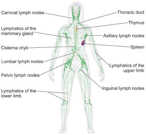
The lymphatic system is a network of tissues, vessels, and organs that works in close partnership with the cardiovascular system. It performs essential roles in fluid homeostasis, immune surveillance, and lipid absorption. Unlike the closed circulatory system, the lymphatic system is an open, one-way transport network that returns excess interstitial fluid to the bloodstream.
Definition: The lymphatic system comprises lymphatic vessels, lymphoid tissue, lymph nodes, and lymphoid organs that collect and transport lymph—a clear fluid derived from interstitial fluid—back to the venous circulation.
Key Components:
- Lymphatic vessels: Including capillaries, collecting vessels, trunks, and ducts.
- Lymph nodes: Distributed along vessel pathways for filtration.
- Lymphoid organs: Classified as primary (bone marrow, thymus) or secondary (spleen, tonsils, MALT).
- Lymph: The actual fluid transported within the system.
Understanding lymphatic anatomy is fundamental for interpreting lymphadenopathy, planning cancer staging, assessing lymphedema, and performing safe surgical dissections.
2. Lymphatic Vessels
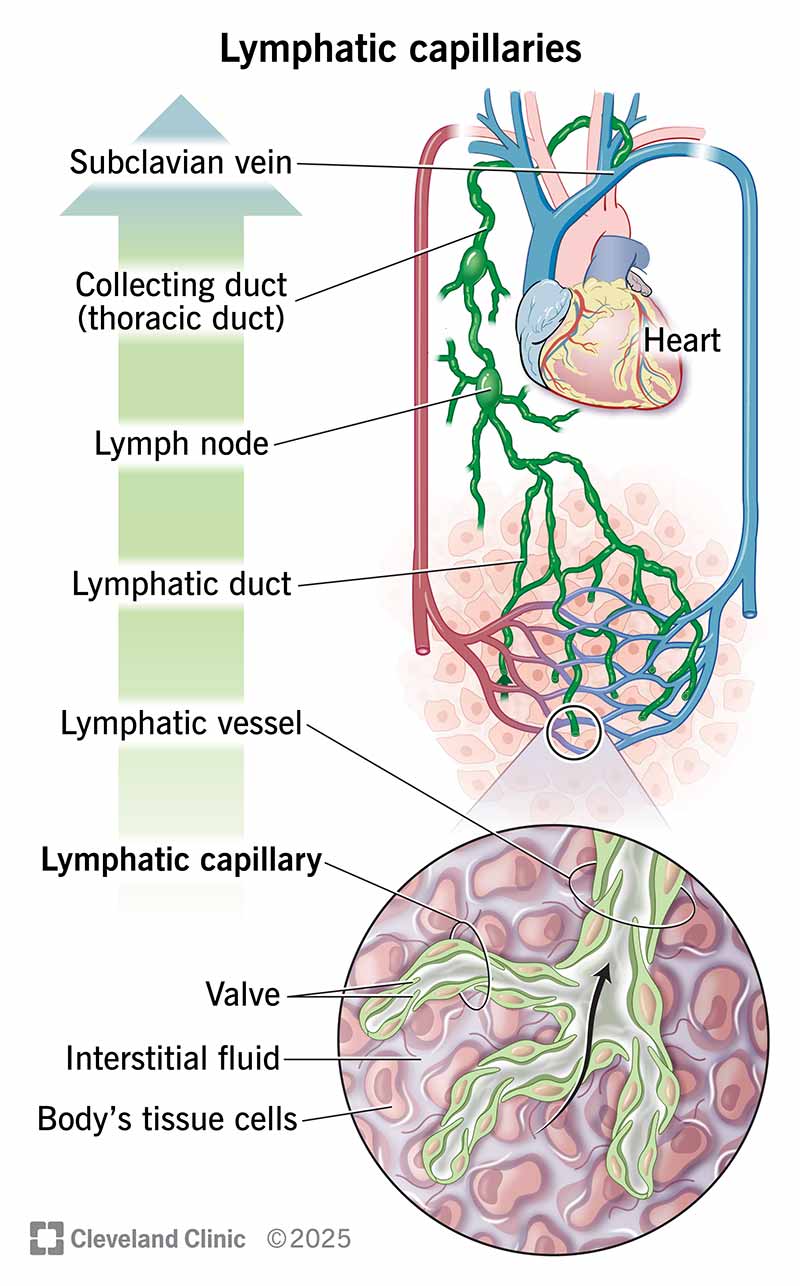
Lymphatic vessels form a hierarchical network that begins as blind-ended capillaries in tissue spaces and progressively converges into larger ducts that drain into the venous system.
2.1 Lymphatic Capillaries
- Structure: Blind-ended, thin-walled vessels composed of a single layer of overlapping endothelial cells.
- Mechanism: Endothelial cells are anchored to surrounding connective tissue by fine filaments; increased interstitial pressure opens the junctions to allow fluid entry.
- Distribution: Present in most tissues except avascular structures (cartilage, epidermis, cornea) and the central nervous system.
- Specialized Capillaries: Found in the small intestine, these are called lacteals and are responsible for absorbing dietary lipids.
2.2 Collecting Vessels
- Formed by the union of lymphatic capillaries.
- Valves: Contain valves (formed by intimal folds) that ensure unidirectional flow toward the heart.
- Walls: Thin walls with three layers: endothelium, smooth muscle media, and fibrous adventitia.
- Location: Superficial vessels drain skin and subcutaneous tissue; deep vessels accompany arteries and veins.
2.3 Lymphatic Trunks
Formed by the convergence of collecting vessels. There are five principal trunks:
- Jugular: Drains head and neck.
- Subclavian: Drains upper limbs.
- Bronchomediastinal: Drains the thorax.
- Intestinal: Drains abdominal viscera.
- Lumbar: Drains lower limbs and pelvis.
2.4 Lymphatic Ducts
- Thoracic duct: The largest lymphatic vessel (~38–45 cm long). It originates from the cisterna chyli (at L1-L2), ascends through the posterior mediastinum, and drains into the junction of the left subclavian and internal jugular veins. It collects lymph from the entire body except the right upper quadrant.
- Right lymphatic duct: A short vessel (1–2 cm) that drains into the right venous angle. It collects lymph from the right side of the head and neck, right upper limb, and right thorax.
3. Lymph Nodes
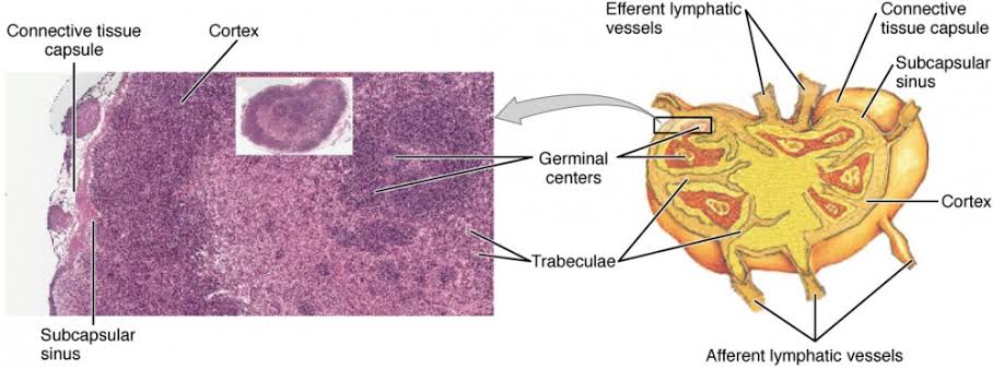
Lymph nodes are small, bean-shaped organs (2–10 mm) distributed along lymphatic vessels. They function as filtration stations and sites of immune cell activation.
3.1 Gross Structure
- Each node is enclosed by a fibrous capsule.
- Afferent vessels: Penetrate the convex surface.
- Efferent vessels: Exit at the hilum.
- An internal framework of trabeculae extends from the capsule, supporting parenchymal tissue.
3.2 Microscopic Architecture
- Cortex: Outer zone containing lymphoid follicles (B-cell zones). Primary follicles are dense; secondary follicles develop germinal centers after antigen exposure.
- Paracortex: Deep cortical region rich in T lymphocytes. Contains high endothelial venules (HEVs) that allow lymphocyte entry from blood.
- Medulla: Innermost zone with medullary cords (plasma cells, B cells, macrophages) and medullary sinuses.
- Sinuses: Subcapsular, trabecular, and medullary sinuses allow slow lymph flow, facilitating phagocytosis and antigen presentation.
3.3 Regional Lymph Node Groups
| Region | Major Groups | Drainage Area |
|---|---|---|
| Head and Neck | Cervical, submandibular, preauricular, parotid | Scalp, face, oral cavity, pharynx |
| Upper Limb | Axillary, epitrochlear, infraclavicular | Breast, upper limb, chest wall |
| Thorax | Hilar, mediastinal, paratracheal | Lungs, heart, mediastinum |
| Abdomen | Mesenteric, para-aortic, celiac | GI tract, liver, spleen, kidneys |
| Lower Limb | Inguinal, popliteal | Lower limb, perineum, external genitalia |
4. Lymphoid Organs
Lymphoid organs are classified as primary (sites of lymphocyte development) or secondary (sites of immune response activation).
4.1 Primary Lymphoid Organs
Bone Marrow:
- Site of hematopoiesis and B-lymphocyte maturation.
- Progenitor cells differentiate into mature B cells expressing surface immunoglobulins.
- Also the source of T-cell progenitors that migrate to the thymus.
Thymus:
- Bilobed organ located in the anterior superior mediastinum.
- Largest in childhood; undergoes involution after puberty, replaced by adipose tissue.
- Cortex: Densely packed with immature T lymphocytes (thymocytes).
- Medulla: Less cellular; contains Hassall corpuscles and mature T cells.
- Site of T-cell maturation and positive/negative selection (self-tolerance).
4.2 Secondary Lymphoid Organs
Spleen:
The largest lymphoid organ, located in the left upper quadrant beneath the diaphragm.
- Functions: Filtration of blood, removal of senescent erythrocytes, immune response to blood-borne pathogens.
- White pulp: Lymphoid tissue surrounding central arteries; contains periarteriolar lymphoid sheaths (T cells) and lymphoid follicles (B cells).
- Red pulp: Vascular sinusoids and splenic cords (cords of Billroth); site of blood filtration.
- Marginal zone: Interface between white and red pulp; rich in macrophages.
Tonsils:
- Aggregates of lymphoid tissue in the oropharynx: palatine, pharyngeal (adenoids), and lingual.
- Form Waldeyer ring, a first-line defense against inhaled/ingested pathogens.
- Lack a capsule and have deep crypts lined by epithelium.
Mucosa-Associated Lymphoid Tissue (MALT):
- Diffuse lymphoid tissue in mucosal surfaces (respiratory, GI, GU tracts).
- Peyer patches: Aggregated lymphoid follicles in the ileum; specialized for sampling intestinal antigens via M cells.
- Appendix: Contains abundant lymphoid tissue for gut immune surveillance.
5. Clinical Correlations
The sentinel node is the first lymph node to receive drainage from a tumor site. Identification guides cancer staging and surgical planning (e.g., in melanoma or breast cancer).
Accumulation of lymph in interstitial spaces due to impaired drainage.
- Primary: Congenital lymphatic hypoplasia.
- Secondary: Post-surgical, post-radiation, or caused by filariasis.
Enlargement of the spleen due to infections (malaria, mononucleosis), hematologic disorders, or portal hypertension. May cause hypersplenism with cytopenias.
6. Key Points Summary
- The lymphatic system is an open, one-way network returning interstitial fluid to venous circulation.
- Lymphatic capillaries are blind-ended; collecting vessels contain valves.
- The thoracic duct drains most of the body; the right lymphatic duct drains the right upper quadrant.
- Lymph nodes filter lymph and activate immune responses via B-cell follicles and T-cell paracortex.
- Primary lymphoid organs (bone marrow, thymus) produce mature lymphocytes; secondary organs (spleen, MALT, tonsils) mount immune responses.
- The spleen filters blood and has white pulp (immune) and red pulp (filtration) compartments.
