Disorders of Blood Circulation
Complete study notes on Heart Failure and Arteriosclerosis, covering definitions, classifications, pathophysiology, clinical manifestations, investigations, and the "Four Pillars" of modern management.
1. HEART FAILURE (HF)
1.1 Definition
Heart Failure (HF) is a clinical syndrome characterized by structural or functional abnormalities of the heart that result in impaired ventricular filling or ejection of blood. It is not a single disease but a constellation of signs and symptoms arising from inadequate cardiac output to meet the metabolic demands of the body, or from elevated cardiac filling pressures at rest or with exertion.
1.2 Classification
A. By Ejection Fraction (2022 AHA/ACC/HFSA Guidelines)
| Type | LVEF | Pathophysiological Description |
|---|---|---|
| HFrEF (Reduced) | ≤ 40% | Systolic dysfunction; impaired contractility. |
| HFmrEF (Mildly Reduced) | 41–49% | Heart Failure with mildly reduced Ejection Fraction; intermediate group. |
| HFpEF (Preserved) | ≥ 50% | Diastolic dysfunction; impaired relaxation and filling. |
| HFimpEF (Improved) | > 40% | Previously HFrEF with subsequent LVEF > 40% on repeated measurement. |
B. By AHA/ACC/HFSA Stages (Progressive Framework)
- Stage A: At risk for HF but without symptoms, structural heart disease, or cardiac biomarkers.
- Stage B: Structural heart disease but no prior or current signs/symptoms of HF (e.g., prior MI, LV hypertrophy, valvular disease).
- Stage C: Structural heart disease with prior or current symptoms of HF.
- Stage D: Marked HF symptoms that interfere with daily life; recurrent hospitalizations despite optimized medical therapy (GDMT).
C. By NYHA Functional Classification (Symptom Severity)
- Class I: No limitation of physical activity. Ordinary activity does not cause symptoms.
- Class II: Slight limitation. Comfortable at rest. Ordinary activity causes fatigue, palpitation, dyspnea, or chest pain.
- Class III: Marked limitation. Comfortable at rest. Less than ordinary activity causes symptoms.
- Class IV: Unable to perform any physical activity without symptoms. Symptoms present at rest.
1.3 Etiology and Risk Factors
| Category | Specific Causes |
|---|---|
| Ischemic heart disease | Myocardial infarction, chronic angina, hibernating myocardium. |
| Hypertension | Chronic pressure overload leading to LV hypertrophy and remodeling. |
| Valvular disease | Aortic stenosis/regurgitation, mitral stenosis/regurgitation. |
| Cardiomyopathies | Dilated (alcohol, viral, genetic), hypertrophic, restrictive. |
| Arrhythmias | Atrial fibrillation (tachycardia-induced cardiomyopathy). |
| Toxins/Drugs | Alcohol, cocaine, anthracyclines (doxorubicin), trastuzumab. |
| Metabolic | Diabetes, thyroid disease, obesity, Thiamine deficiency (Beriberi). |
| High-output states | Anemia, hyperthyroidism, arteriovenous fistula. |
4.1.4 Pathophysiology
A. Left-Sided Heart Failure
- Forward failure (low output): Reduced cardiac output → tissue hypoperfusion → fatigue, weakness, oliguria, altered mental status.
- Backward failure (pulmonary congestion): LV fails to pump effectively → increased left ventricular end-diastolic pressure (LVEDP) → increased left atrial pressure → pulmonary venous congestion → Pulmonary Edema.
B. Right-Sided Heart Failure
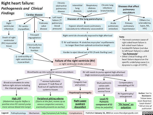
- Commonly caused by Left-sided HF, pulmonary hypertension, or chronic lung disease (Cor Pulmonale).
- Right ventricle fails to pump effectively → increased right ventricular and right atrial pressure → Systemic Venous Congestion.
- Clinical consequences: Jugular venous distension (JVD), hepatomegaly, peripheral edema (ankles, sacrum), and ascites.
C. Neurohormonal Activation
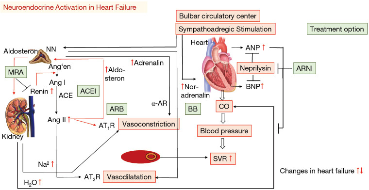
- Sympathetic Nervous System: Increased norepinephrine → increased heart rate and contractility. Chronic activation causes myocardial toxicity and arrhythmias.
- RAAS System: Decreased renal perfusion → Renin release → Angiotensin II (vasoconstriction) + Aldosterone (sodium/water retention) → Volume Overload.
- Natriuretic Peptides (BNP, NT-proBNP): Released from stretched myocardium to promote vasodilation and diuresis (compensatory).
Cardiac Remodeling
Concentric Hypertrophy: Increased wall thickness due to pressure overload (HTN, Aortic Stenosis).
Eccentric Hypertrophy: Chamber dilation due to volume overload (MI, Mitral Regurgitation).
4.1.5 Clinical Features
| Symptom/Sign | Left-Sided HF | Right-Sided HF |
|---|---|---|
| Dyspnea | Exertional, Orthopnea, PND | Less prominent |
| Cough | Dry or productive (pink frothy) | Not typical |
| Edema | Pulmonary edema (crackles) | Peripheral pitting edema |
| JVP | Normal or elevated | Elevated (key sign) |
| Heart Sounds | S3 gallop | Loud P2 (pulmonic) |
Acute Pulmonary Edema (Severe Left HF)
Presents with severe dyspnea at rest, anxiety, diaphoresis, and pink frothy sputum. This is a medical emergency requiring immediate intervention (Oxygen, Diuretics, Vasodilators).
4.1.6 Investigations
- Chest X-ray: Cardiomegaly (ratio > 50%), pulmonary congestion, Kerley B lines, and pleural effusions.
- ECG: Evidence of arrhythmias (Atrial Fibrillation), LV hypertrophy, Q waves (prior MI), or LBBB.
- Echocardiography: Gold standard for assessing LVEF, chamber size, and valvular function.
- BNP / NT-proBNP: Elevated levels (> 400 pg/mL BNP) support HF; levels < 100 pg/mL make HF unlikely.
- Troponins: Elevated in acute decompensation or myocardial stretch/necrosis.
4.1.7 Management of Heart Failure
A. Guideline-Directed Medical Therapy (GDMT) — "The Four Pillars"
For HFrEF, the standard of care includes the simultaneous or rapid sequence initiation of:
- ARNI (Sacubitril/Valsartan): Or ACEI/ARB; inhibits RAAS and neprilysin.
- Beta-blockers: (Carvedilol, Bisoprolol, Metoprolol succinate) to block sympathetic overactivation.
- MRA: (Spironolactone, Eplerenone) to block aldosterone.
- SGLT2 Inhibitors: (Dapagliflozin, Empagliflozin) to improve cardiovascular outcomes.
B. Acute Management
- Oxygen therapy if hypoxemic.
- IV loop diuretics (Furosemide) to reduce fluid volume.
- Vasodilators (Nitrates) if hypertensive.
- Inotropes (Dobutamine) if in cardiogenic shock.
4.2 ARTERIOSCLEROSIS
4.2.1 Definition
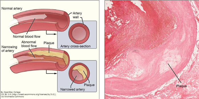
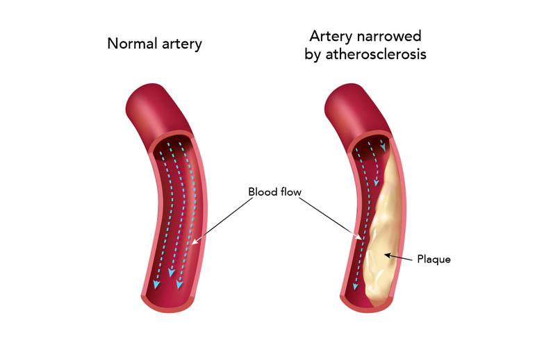
Arteriosclerosis is an umbrella term for the thickening, hardening, and loss of elasticity of arterial walls. Atherosclerosis is a specific type involving atheromatous plaque formation in the tunica intima.
4.2.2 Types of Arteriosclerosis
| Type | Affected Vessels | Key Feature |
|---|---|---|
| Atherosclerosis | Large and medium arteries | Lipid-rich plaques in tunica intima; inflammatory. |
| Monckeberg medial sclerosis | Medium-sized muscular arteries | Calcium deposits in tunica media; no luminal narrowing. |
| Arteriolosclerosis | Small arteries and arterioles | Hyaline or hyperplastic thickening; associated with HTN/Diabetes. |
4.2.3 Atherosclerosis Pathophysiology
Response-to-Injury Hypothesis:
- Endothelial injury: Caused by smoking, hypertension, or diabetes.
- Lipid accumulation: LDL enters the intima and becomes oxidized (Ox-LDL).
- Inflammatory response: Macrophages engulf Ox-LDL to become Foam Cells.
- Smooth muscle proliferation: Migration from media to intima; production of collagen.
- Plaque progression: Fibrous cap forms over a lipid core.
Plaque Morphology
| Feature | Stable Plaque | Unstable (Vulnerable) Plaque |
|---|---|---|
| Fibrous cap | Thick, well-developed | Thin or absent |
| Lipid core | Small | Large (> 40% of volume) |
| Risk | Gradual narrowing (Angina) | Rupture → Thrombosis (MI/Stroke) |
Summary & Key Clinical Pearls
- The Four Pillars of HFrEF therapy (ARNI, beta-blocker, MRA, SGLT2i) are all mortality-reducing.
- BNP/NT-proBNP are essential biomarkers for excluding heart failure.
- Atherosclerosis is a systemic disease; management of one territory (e.g., coronary) requires modification of all vascular beds.
- Statins are indicated for all patients with established atherosclerotic disease regardless of baseline LDL.
- Ankle-brachial index (ABI) < 0.9 indicates Peripheral Artery Disease (PAD).
