Types of Circulation
Complete study notes covering Pulmonary, Systemic, and Fetal circulation, including physiological mechanisms, anatomical pathways, special circulatory systems, and clinical correlations.
3.1 Introduction
The circulatory system is responsible for transporting blood throughout the body. In adults, there are two primary circuits: pulmonary circulation (blood flow between the heart and lungs) and systemic circulation (blood flow between the heart and all body tissues). During fetal development, a specialized fetal circulation exists to bypass the non-functioning lungs and liver.
3.2 Pulmonary Circulation
3.2.1 Definition and Purpose
Pulmonary circulation is the portion of the cardiovascular system that transports deoxygenated blood from the right side of the heart to the lungs and returns oxygenated blood to the left side of the heart.
Purpose:
- Gas exchange: Loading oxygen and unloading carbon dioxide in pulmonary capillaries.
- Blood volume reservoir: Pulmonary vessels contain approximately 9% of total blood volume.
- Filtration: Removal of small blood clots and air bubbles by pulmonary capillaries.
3.2.2 Pathway of Pulmonary Circulation
The precise anatomical route follows this sequence:
Right ventricle → Pulmonary valve → Pulmonary trunk → Right and left pulmonary arteries → Lobar and segmental arteries → Pulmonary capillaries (alveoli) → Pulmonary venules → Pulmonary veins (4 total: 2 from each lung) → Left atrium
- Pulmonary arteries carry deoxygenated blood (exception to the general rule).
- Pulmonary veins carry oxygenated blood (exception to the general rule).
- Pulmonary circulation is a low-pressure, low-resistance system.
- Pulmonary artery pressure: 15-30 mmHg systolic, 4-12 mmHg diastolic.
- Pulmonary vascular resistance is approximately 1/10 of systemic resistance.
3.2.3 Physiology of Pulmonary Circulation
Hypoxic vasoconstriction:
- Unique response of pulmonary vessels: alveolar hypoxia causes local vasoconstriction.
- Directs blood away from poorly ventilated alveoli toward well-ventilated regions.
- Optimizes ventilation-perfusion (V/Q) matching.
- Contrast with systemic circulation: hypoxia causes vasodilation.
Gravity effects:
- In upright position, blood flow is greater at lung bases than apices.
- Zone 1 (apex): Alveolar pressure > arterial pressure; minimal flow (potential for alveolar dead space).
- Zone 2 (mid-lung): Arterial pressure > alveolar pressure > venous pressure; intermittent flow.
- Zone 3 (base): Arterial and venous pressures > alveolar pressure; continuous flow.
3.2.4 Clinical Correlation
Pulmonary Embolism (PE)
Obstruction of pulmonary arteries by thrombus (usually from deep vein thrombosis). Symptoms include sudden dyspnea, pleuritic chest pain, tachypnea, tachycardia, and hypoxemia. Diagnosis via CT pulmonary angiography (CTPA), D-dimer, and Wells score. Management involves anticoagulation or thrombolysis in massive PE.
Pulmonary Hypertension
Mean pulmonary artery pressure > 20 mmHg (2022 ESC/ERS guidelines). Causes include left heart disease, lung disease, or idiopathic factors. Leads to right ventricular hypertrophy and eventually right heart failure (cor pulmonale).
3.3 Systemic Circulation
3.3.1 Definition and Purpose
Systemic circulation is the portion of the cardiovascular system that transports oxygenated blood from the left side of the heart to all body tissues and returns deoxygenated blood to the right side of the heart.
Purpose:
- Delivery of oxygen and nutrients to all body tissues.
- Removal of carbon dioxide and metabolic waste products.
- Transport of hormones from endocrine glands to target organs.
- Regulation of body temperature (thermoregulation via blood flow to skin).
- Maintenance of fluid and electrolyte balance.
3.3.2 Pathway of Systemic Circulation
Left ventricle → Aortic valve → Ascending aorta → Aortic arch → Descending thoracic aorta → Abdominal aorta → Common iliac arteries → Arteries of lower limbs → Arterioles → Capillary beds (all tissues) → Venules → Veins → Inferior vena cava (from below heart) + Superior vena cava (from above heart) → Right atrium
3.3.3 Major Arterial Branches
From aortic arch:
- Brachiocephalic trunk → right subclavian + right common carotid.
- Left common carotid → head and neck.
- Left subclavian → left upper limb.
From descending aorta:
- Intercostal arteries → chest wall.
- Celiac trunk → liver, stomach, spleen.
- Superior mesenteric artery → small intestine, proximal colon.
- Renal arteries → kidneys.
- Inferior mesenteric artery → distal colon, rectum.
- Common iliac arteries → pelvis and lower limbs.
3.3.4 Major Venous Drainage
- Superior vena cava: Formed by union of brachiocephalic veins. Drains head, neck, upper limbs, chest wall, and upper thoracic organs.
- Inferior vena cava: Formed by union of common iliac veins. Drains lower limbs, pelvis, abdomen, kidneys, and liver (hepatic veins).
Hepatic Portal System
A unique venous system: capillaries (GI tract) → portal vein → capillaries (liver) → hepatic veins → IVC. Carries nutrient-rich, deoxygenated blood from digestive organs to the liver for processing. This is a critical site for first-pass metabolism and detoxification.
3.3.5 Special Circulations
- Coronary circulation: Supplies heart muscle itself. Arises from ascending aorta; drains into right atrium via coronary sinus.
- Cerebral circulation: Internal carotid arteries and vertebral arteries form the Circle of Willis. Protected by the blood-brain barrier (tight junctions in continuous capillaries).
- Renal circulation: High blood flow (20-25% of cardiac output) for filtration. Features two capillary beds in series: glomerular capillaries (filtration) and peritubular capillaries (reabsorption).
- Splanchnic circulation: Blood flow to GI tract, liver, spleen, and pancreas. Highly variable; increases after meals (active hyperemia).
3.3.6 Regulation of Systemic Circulation
Short-term regulation (seconds to minutes):
- Baroreceptor reflex: carotid sinus and aortic arch receptors detect pressure changes; mediate heart rate and vasomotor tone via autonomic nervous system.
- Chemoreceptor reflex: carotid and aortic bodies detect changes in O2, CO2, and pH.
- Local autoregulation: myogenic response (Bayliss effect) and metabolic factors.
Long-term regulation (hours to days):
- Renin-angiotensin-aldosterone system (RAAS): regulates blood volume and pressure.
- Antidiuretic hormone (ADH/vasopressin): regulates water reabsorption in kidneys.
- Atrial natriuretic peptide (ANP): promotes sodium and water excretion.
3.4 Fetal Circulation
3.4.1 Overview
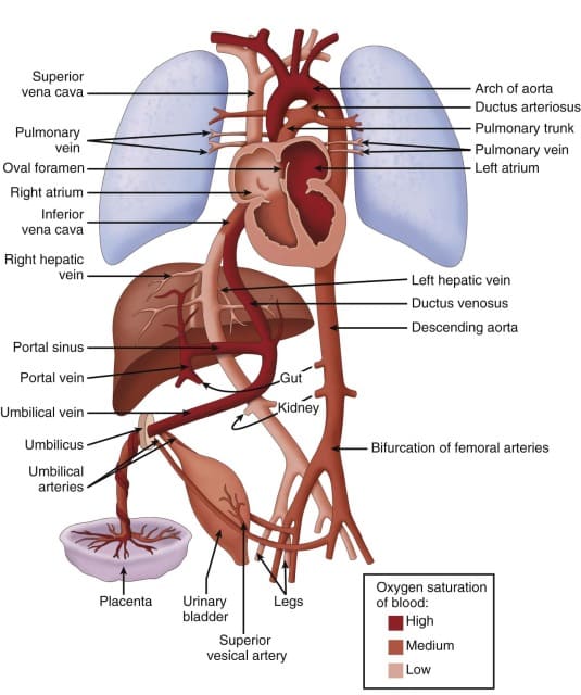
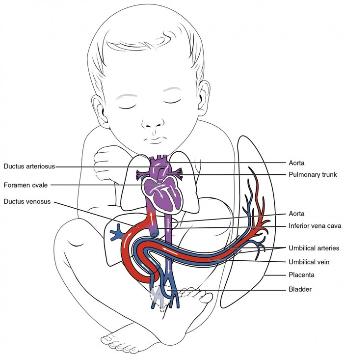
Fetal circulation is distinctly different from adult circulation because the fetus does not use its lungs for gas exchange. Instead, the placenta serves as the site of oxygenation, nutrient delivery, and waste removal. The fetal heart and blood vessels contain special shunts that bypass the lungs and liver.
3.4.2 Key Structures in Fetal Circulation
- Placenta: Organ of exchange between maternal and fetal circulations. Maternal oxygenated blood and nutrients diffuse across placental barrier into fetal blood. Fetal waste products (CO2, urea) diffuse into maternal blood. Connected to fetus via umbilical cord.
- Umbilical Cord: Contains two umbilical arteries (carry deoxygenated blood to placenta) and one umbilical vein (carries oxygenated blood from placenta to fetus). Surrounded by Wharton jelly (protective connective tissue).
3.4.3 The Three Fetal Shunts
- Ductus Venosus:
— Shunt between umbilical vein and inferior vena cava.
— Function: Bypasses the hepatic portal circulation (liver).
— Allows oxygenated blood from placenta to flow directly toward the heart.
— Approximately 50% of umbilical venous blood passes through ductus venosus. - Foramen Ovale:
— Opening in the interatrial septum between right and left atria.
— Function: Allows blood to flow from right atrium to left atrium, bypassing the lungs.
— Pressure in right atrium is higher than left atrium in fetus (due to high pulmonary vascular resistance).
— Oxygenated blood from IVC (via ductus venosus) is directed by crista dividens toward the foramen ovale.
— After birth: increased left atrial pressure causes functional closure within minutes; anatomical closure (fossa ovalis) occurs within months. - Ductus Arteriosus:
— Connection between pulmonary trunk and descending aorta (distal to left subclavian artery).
— Function: Shunts blood from pulmonary artery to aorta, bypassing the lungs.
— Most blood ejected from right ventricle passes through ductus arteriosus (high pulmonary resistance prevents significant pulmonary blood flow).
— After birth: constriction begins within 10-15 hours due to increased oxygen tension; functional closure within 1-3 days; anatomical closure (ligamentum arteriosum) within 2-3 weeks.
3.4.4 Pathway of Fetal Circulation
Oxygenated blood pathway:
Placenta → Umbilical vein → Ductus venosus (bypasses liver) → IVC → Right atrium → Foramen ovale → Left atrium → Left ventricle → Ascending aorta → Brain, coronary arteries, upper limbs (most oxygenated blood)
Deoxygenated blood pathway:
Superior vena cava → Right atrium → Right ventricle → Pulmonary trunk → Ductus arteriosus → Descending aorta → Lower body, abdominal organs → Umbilical arteries → Placenta (for reoxygenation)
Mixed blood:
- Some blood from SVC enters right ventricle and small amount goes to lungs (for growth).
- Blood from IVC mixes with SVC blood in right atrium; the more oxygenated IVC blood is preferentially shunted through foramen ovale.
3.4.5 Changes at Birth
First breath:
- Lungs expand, alveolar fluid is cleared.
- Pulmonary vascular resistance drops dramatically (80-90% reduction).
- Pulmonary blood flow increases significantly.
Umbilical cord clamping:
- Eliminates placental circulation.
- Systemic vascular resistance increases.
Closure of shunts:
| Structure | Fetal Function | Adult Remnant | Closure Time |
|---|---|---|---|
| Umbilical vein | Oxygenated blood to fetus | Ligamentum teres (round ligament of liver) | Immediate (cord clamp) |
| Ductus venosus | Bypasses liver | Ligamentum venosum | 2-3 days |
| Foramen ovale | Right-to-left atrial shunt | Fossa ovalis | Functional: minutes; Anatomical: months |
| Ductus arteriosus | Pulmonary-to-aortic shunt | Ligamentum arteriosum | Functional: 1-3 days; Anatomical: 2-3 weeks |
| Umbilical arteries | Deoxygenated blood to placenta | Medial umbilical ligaments | Immediate (cord clamp) |
3.4.6 Clinical Correlation: Patent Ductus Arteriosus (PDA)
- Failure of ductus arteriosus to close after birth.
- Incidence: approximately 1 in 2,000 full-term births; more common in premature infants.
- Pathophysiology: left-to-right shunt (aortic pressure > pulmonary pressure after birth) causes increased pulmonary blood flow, pulmonary congestion, and left heart volume overload.
- Clinical features: continuous "machinery" murmur at left upper sternal border, wide pulse pressure, bounding pulses, signs of heart failure in large shunts.
- Diagnosis: echocardiography (definitive).
- Management:
— Medical: indomethacin or ibuprofen (prostaglandin inhibitors) in premature infants.
— Surgical: ligation or catheter-based closure (coil, device) if medical therapy fails.
3.4.7 Clinical Correlation: Patent Foramen Ovale (PFO)
- Present in approximately 25-30% of adults (incomplete anatomical closure).
- Usually asymptomatic; potential route for paradoxical embolism (venous thrombus crosses to arterial circulation).
- Associated with cryptogenic stroke, migraine with aura, decompression sickness.
- Diagnosis: echocardiography with bubble study.
- Management: closure (device or surgery) in selected patients with recurrent cryptogenic stroke.
Summary — Types of Circulation
| Feature | Pulmonary | Systemic | Fetal |
|---|---|---|---|
| Purpose | Gas exchange in lungs | Deliver O2/nutrients to tissues; remove CO2/waste | Placental gas exchange; bypass non-functioning lungs and liver |
| Blood in arteries (exception) | Deoxygenated | Oxygenated | Mixed (umbilical vein = oxygenated) |
| Blood in veins (exception) | Oxygenated | Deoxygenated | Mixed (umbilical arteries = deoxygenated) |
| Pressure / Resistance | Low (15/5 mmHg) | Low | High (120/80 mmHg) | High | Low (placental resistance) |
| Special Features | Hypoxic vasoconstriction | Baroreceptor/chemoreceptor reflexes; autoregulation | Three shunts: ductus venosus, foramen ovale, ductus arteriosus |
- Pulmonary arteries carry deoxygenated blood; pulmonary veins carry oxygenated blood.
- The foramen ovale closes due to increased left atrial pressure after birth.
- The ductus arteriosus closes due to increased oxygen tension and decreased prostaglandins.
- PDA causes a left-to-right shunt with a continuous machinery murmur.
- Understanding fetal circulation is essential for managing congenital heart disease.