Structure and Functions of the Heart
Complete study notes covering macroscopic and microscopic anatomy, the cardiac cycle, neurovascular supply, and physical examination correlations.
1. Introduction
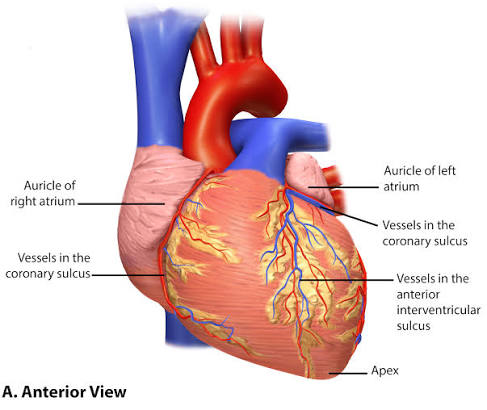
The heart is a hollow, muscular, cone-shaped organ located in the mediastinum, between the lungs. Approximately two-thirds of its mass lies to the left of the midline. It functions as a double pump maintaining continuous blood circulation through the pulmonary and systemic circuits.
Key Parameters:
- Size: Approximately 12 cm long, 9 cm wide, 6 cm thick.
- Weight: Approximately 250-350 g in adult males; slightly less in females.
- Location: Posterior to the sternum, anterior to the vertebral column at levels T5-T8.
- Apex: Directed anteriorly, inferiorly, and to the left (located at the 5th intercostal space, midclavicular line).
- Base: Directed posteriorly, superiorly, and to the right.
2. Structure of the Heart
2.1 Pericardium
The heart is enclosed within the pericardium, a double-walled sac consisting of two main layers:
- Fibrous pericardium: A tough, outer layer of dense connective tissue that prevents overfilling and anchors the heart to surrounding structures.
- Serous pericardium:
- Parietal layer: Lines the inner surface of the fibrous pericardium.
- Visceral layer (epicardium): Covers the external surface of the heart.
- Pericardial cavity: The potential space between the parietal and visceral layers containing 10-20 mL of serous fluid to reduce friction.
2.2 Heart Wall
The heart wall consists of three distinct layers:
| Layer | Name | Characteristics |
|---|---|---|
| Outer | Epicardium | Visceral layer of serous pericardium; contains coronary vessels, nerves, and adipose tissue. |
| Middle | Myocardium | Cardiac muscle tissue; the thickest layer; responsible for the pumping action/contraction. |
| Inner | Endocardium | Endothelium-lined; smooth surface that reduces friction and prevents blood clot formation; covers valves. |
2.3 Chambers of the Heart
The heart has four chambers: two atria (upper) and two ventricles (lower).
Right Atrium:
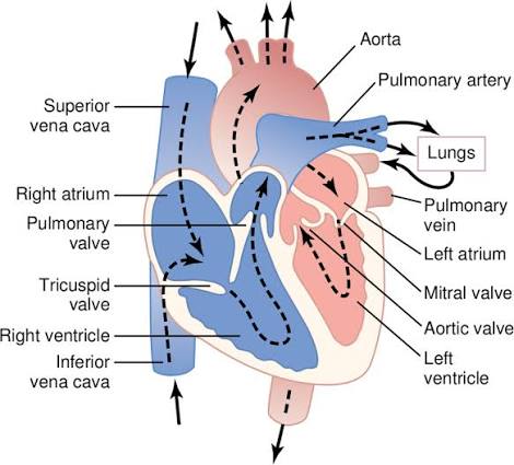
- Receives deoxygenated blood from systemic circulation via the superior vena cava (head, neck, limbs), inferior vena cava (abdomen, pelvis, lower limbs), and coronary sinus (heart wall).
- Contains the fossa ovalis (remnant of fetal foramen ovale).
- Houses the sinoatrial (SA) node — the natural pacemaker.
Right Ventricle:
- Receives blood from the right atrium via the tricuspid valve.
- Pumps blood through the pulmonary valve into the pulmonary trunk.
- Wall is thinner than the left ventricle as it pumps against low pulmonary resistance (15-30 mmHg).
- Contains the moderator band (septomarginal trabecula) carrying the right bundle branch.
Left Atrium:
- Receives oxygenated blood from the lungs via four pulmonary veins (two from each lung).
- Smaller but thicker-walled than the right atrium.
Left Ventricle:
- Receives oxygenated blood from the left atrium via the mitral (bicuspid) valve.
- Pumps blood through the aortic valve into the ascending aorta for systemic circulation.
- Wall is 2-3 times thicker than the right ventricle to pump against high systemic pressure (80-120 mmHg).
- Forms the apex of the heart.
3. Valves and Blood Flow
3.1 Atrioventricular (AV) Valves
- Tricuspid Valve (Right AV): Three cusps (anterior, posterior, septal). Prevents backflow to RA during ventricular systole.
- Mitral (Bicuspid) Valve (Left AV): Two cusps (anterior, posterior). Most commonly affected by rheumatic heart disease and mitral valve prolapse.
- Both are anchored by chordae tendineae to papillary muscles of the ventricles.
3.2 Semilunar Valves
- Pulmonary Valve: Three semilunar cusps. Located between RV and pulmonary trunk.
- Aortic Valve: Three semilunar cusps (right, left, non-coronary). Right and left coronary cusps give rise to the coronary arteries.
AV valves close when ventricular pressure exceeds atrial pressure during systole. Chordae tendineae prevent the cusps from everting into the atria. Semilunar valves close when arterial pressure exceeds ventricular pressure during diastole.
3.3 Fibrous Skeleton of the Heart
A dense connective tissue framework that:
- Provides attachment for valves and cardiac muscle fibers.
- Electrically insulates atria from ventricles.
- The AV bundle (Bundle of His) is the only normal electrical connection between atria and ventricles.
4. Coronary Circulation
4.1 Arterial Supply
- Right Coronary Artery (RCA): Arises from the right aortic sinus. Supplies the RA, most of the RV, SA node (in 60%), AV node (in 80%), and posterior 1/3 of the septum.
- Left Coronary Artery (LCA): Arises from the left aortic sinus. Divides into:
- Left Anterior Descending (LAD): Supplies anterior 2/3 of septum, apex; known as the "widow maker" when occluded.
- Circumflex artery: Supplies LA and lateral/posterior walls of the LV.
Dominance is determined by which artery gives rise to the posterior interventricular artery (PDA).
— Right-dominant (85%): PDA from RCA.
— Left-dominant (10%): PDA from Circumflex.
— Co-dominant (5%): PDA from both.
4.2 Venous Drainage
- Great cardiac vein: Accompanies LAD; drains into coronary sinus.
- Middle cardiac vein: Accompanies posterior interventricular artery.
- Coronary sinus: Drains into the right atrium (main venous return).
5. Functions and Physiology
5.1 The Cardiac Cycle
Sequence of events in one heartbeat (~0.8 seconds at 75 bpm):
- Atrial Systole (0.1 sec): SA node fires; atria contract, forcing 20-30% of blood into ventricles ("atrial kick").
- Isovolumetric Ventricular Contraction (0.05 sec): Ventricular pressure rises; AV valves close (S1 sound). All valves closed.
- Ventricular Ejection (0.25 sec): Pressure exceeds arterial pressure; semilunar valves open. SV is ~70 mL.
- Isovolumetric Ventricular Relaxation (0.08 sec): Semilunar valves close (S2 sound). All valves closed.
- Ventricular Filling (0.4 sec): AV valves open; blood flows passively from atria.
5.2 Heart Sounds
| Sound | Timing | Cause | Clinical Significance |
|---|---|---|---|
| S1 (Lubb) | Beginning of systole | Closure of AV valves | Loud in mitral stenosis; soft in mitral regurgitation. |
| S2 (Dubb) | End of systole | Closure of Semilunar valves | Splitting: normal (A2 then P2 on inspiration). |
| S3 | Early diastole | Rapid ventricular filling | Normal in children; pathological in heart failure. |
| S4 | Late diastole | Atrial contraction against stiff ventricle | Indicates decreased compliance (HTN, AS). |
5.3 Cardiac Output and Stroke Volume
Cardiac Output (CO): Volume of blood pumped per minute (SV x HR). Normal: 4.0-8.0 L/min.
Ejection Fraction (EF): Percentage of end-diastolic volume ejected. Normal: 55-70%.
Factors Affecting Stroke Volume:
- Preload: Frank-Starling mechanism (increased venous return stretches muscle, increasing force).
- Contractility: Force of contraction (increased by Sympathetics, Digoxin).
- Afterload: Systemic vascular resistance (pressure the heart must pump against).
6. Conduction System
- Sinoatrial (SA) Node: Pace-maker; generates impulses at 60-100 bpm.
- Atrioventricular (AV) Node: Located in interatrial septum; delays impulse (0.1 sec) to allow ventricular filling.
- AV Bundle (His): Passes through fibrous skeleton.
- Bundle Branches: Conduct impulses to respective ventricles.
- Purkinje Fibers: Rapidly conduct impulses to ventricular myocardium (20-40 bpm escape rhythm).
- P wave: Atrial depolarization.
- PR interval: AV node delay (0.12-0.20 sec).
- QRS complex: Ventricular depolarization (0.08-0.10 sec).
- T wave: Ventricular repolarization.
7. Clinical Correlation: Common Cardiac Examinations
Bedside Cardiac Assessment
Inspection: Look for Jugular Venous Pressure (JVP), peripheral edema, and cyanosis.
Palpation: Apex beat should be at 5th ICS, MCL. Heaves indicate ventricular hypertrophy.
Percussion: Dullness over the heart; useful in diagnosing pericardial effusion.
Auscultation: Identify S1-S4 and murmurs (Grades I-VI). A scratchy sound indicates a pericardial friction rub (pericarditis).
