Muscle Disorders: Myositis, Strain, and Paralysis
A comprehensive clinical resource for the Muscular System (Sub-topic 2.3), detailing the pathophysiology, classification, diagnosis, and management of inflammatory, mechanical, and neurological muscle conditions.
1. INTRODUCTION
Muscle disorders represent a significant category of clinical conditions encountered in general medical practice. These disorders may result from inflammation, mechanical injury, or neurological dysfunction. This chapter examines three specific entities: myositis (inflammation of muscle), muscle strain (mechanical injury to fibers), and paralysis (loss of function due to neurological impairment).
2. MYOSITIS
Myositis is defined as the inflammation of skeletal muscle. It may be acute or chronic, localized or generalized, occurring as an isolated condition or as part of a systemic disease. Involvement may include muscle fibers, connective tissue, or intramuscular blood vessels.
2.1 Classification and Types
- Infectious Myositis: Caused by bacterial, viral, fungal, or parasitic organisms.
- Bacterial myositis (pyomyositis): Common in tropical regions and immunocompromised states; Staphylococcus aureus is the most common pathogen.
- Viral myositis: Caused by influenza, coxsackievirus, EBV, or HIV.
- Parasitic myositis: Includes trichinellosis (Trichinella spiralis) and toxoplasmosis.
- Idiopathic Inflammatory Myopathies (IIM): Autoimmune disorders including:
- Dermatomyositis: Features proximal muscle weakness and characteristic skin rashes (heliotrope rash on eyelids, Gottron papules over knuckles). Associated with malignancy and interstitial lung disease.
- Polymyositis: Progressive, symmetric proximal weakness without skin involvement.
- Inclusion body myositis: Most common in older adults; causes asymmetric weakness of finger flexors and quadriceps. Often refractory to treatment.
- Drug-induced Myositis: Triggered by statins (cholesterol-lowering), colchicine, chloroquine, and zidovudine. Ranges from myalgia to severe rhabdomyolysis.
2.2 Pathophysiology
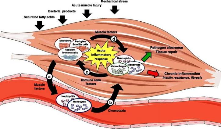
The mechanism varies by etiology. In infectious forms, organisms invade tissue directly, triggering an acute response with neutrophils and macrophages releasing proteolytic enzymes. In autoimmune forms, autoreactive T cells target muscle antigens leading to fiber necrosis. In dermatomyositis, a complement-mediated microangiopathy causes ischemic damage to the fibers.
2.3 Clinical Features
The hallmark is proximal muscle weakness (shoulders and hips). Patients report difficulty climbing stairs, rising from chairs, or lifting objects overhead.
- Muscle pain (myalgia): Severe in infectious; mild/absent in idiopathic.
- Muscle tenderness: Palpation reveals tenderness and potential swelling.
- Systemic: Fatigue, fever (infectious), and dysphagia (pharyngeal involvement).
- Dermatological: Heliotrope rash and shawl sign (dermatomyositis).
2.4 Diagnosis and Management
- Blood tests: Creatine Kinase (CK) is the most sensitive marker for damage. Autoantibodies (anti-Jo-1, anti-Mi-2) support IIM diagnosis.
- Imaging: MRI (T2-weighted/STIR) is the modality of choice for identifying inflammation.
- Muscle Biopsy: The gold standard; shows inflammatory infiltration and fiber necrosis.
Autoimmune Myositis: First-line treatment is high-dose corticosteroids (prednisone 1 mg/kg/day). Steroid-sparing agents (methotrexate, azathioprine) are added for resistant cases.
Drug-induced: Immediate discontinuation of the offending agent. Severe cases require aggressive hydration to prevent acute kidney injury.
3. MUSCLE STRAIN
A muscle strain (pulled muscle) is an injury to a muscle or its tendons caused by overstretching or excessive force. Strains involve muscles/tendons, whereas sprains involve ligaments.
3.1 Etiology and Vulnerability
Occurs during sudden acceleration or deceleration. Common mechanisms include direct trauma, muscle fatigue, and inadequate warm-up. The hamstrings (biceps femoris) are most vulnerable as they cross two joints (hip and knee).
3.2 Classification by Severity
| Grade | Severity | Pathology | Healing Time |
|---|---|---|---|
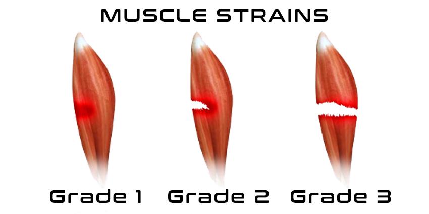
| Grade I | Mild | Few fibers torn; minimal swelling; no strength loss. | 1–2 weeks |
| Grade II | Moderate | Moderate fibers torn; intact muscle; noticeable weakness/bruising. | 3–6 weeks |
| Grade III | Severe | Complete rupture of muscle or tendon; palpable defect; complete loss of function. | 3–6 months |
3.4 Management: The PRICE Protocol
In the acute phase (first 48 hours):
- P — Protection: Use splints or crutches.
- R — Rest: Avoid activities that cause pain (relative rest).
- I — Ice: Apply cold packs for 15–20 mins every 2–3 hours.
- C — Compression: Elastic bandages to minimize swelling.
- E — Elevation: Raise limb above heart level to reduce edema.
Surgical management is reserved for Grade III strains with complete rupture, particularly involving the Achilles tendon or pectoralis major in athletes.
4. PARALYSIS
Paralysis is the complete or partial loss of muscle function resulting from damage anywhere along the motor pathway, from the motor cortex to the neuromuscular junction (NMJ).
4.1 Classification
- By Distribution:
— Monoplegia: One limb.
— Hemiplegia: One side of the body.
— Paraplegia: Both lower limbs.
— Quadriplegia: All four limbs. - By Muscle Tone:
— Flaccid: Loss of tone (hypotonia), absent reflexes, muscle atrophy. Caused by LMN lesions.
— Spastic: Increased tone (hypertonia), hyperreflexia, clonus, Babinski sign. Caused by UMN lesions.
4.2 Causes and Clinical Features
Upper Motor Neuron (UMN) Lesions: Originate in the cortex. Causes: Stroke, MS, Cerebral Palsy. Features: Spasticity, weakness in extensors (upper) and flexors (lower).
Lower Motor Neuron (LMN) Lesions: Originate in the anterior horn. Causes: Polio, GBS, Nerve trauma. Features: Fasciculations and severe neurogenic atrophy.
Facial Paralysis
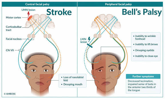
In Bell's palsy (LMN), the entire half of the face is affected (cannot wrinkle forehead). In a Stroke (UMN), the forehead is spared because the frontalis muscle receives bilateral cortical innervation.
4.6 Summary Comparison Table: UMN vs. LMN
| Feature | Upper Motor Neuron (UMN) | Lower Motor Neuron (LMN) |
|---|---|---|
| Muscle Tone | Increased (Spasticity) | Decreased (Flaccidity) |
| Reflexes | Hyperreflexia | Hyporeflexia or absent |
| Babinski Sign | Present (Upgoing toe) | Absent |
| Atrophy | Mild (Disuse) | Severe (Neurogenic) |
| Fasciculations | Absent | Present |
Cauda Equina Syndrome
Compression of nerve roots below L1–L2. Red flags: saddle anesthesia, bladder/bowel dysfunction. Decompression within 48 hours is required to improve outcomes.
5. KEY POINTS SUMMARY
- Myositis: CK is the most sensitive lab marker; MRI is the choice for imaging.
- Muscle Strain: Grade III is a complete rupture requiring potential surgery; hamstrings are most frequently injured.
- Paralysis: UMN lesions result in spasticity; LMN lesions result in flaccidity and fasciculations.
- Bell's Palsy: An LMN facial nerve lesion affecting the entire ipsilateral half of the face.
