Foundational Concepts of Limb Anatomy
Complete medical notes covering Bones, Joints, Muscles, Vessels, Nerves, and Surface Anatomy of the Upper and Lower Limbs.
1. INTRODUCTION TO LIMB ANATOMY
The limbs are paired appendages that enable locomotion, manipulation of objects, and interaction with the environment. Each limb consists of a girdle (pectoral or pelvic), a proximal segment, an intermediate segment, and a distal segment containing multiple digits. The upper limb is specialized for prehension and fine motor control, while the lower limb is adapted for weight-bearing and locomotion.
1.1 Comparative Organization of Upper and Lower Limbs
The upper and lower limbs share a common fundamental pattern of organization. This similarity reflects their common evolutionary origin.
| Feature | Upper Limb | Lower Limb |
|---|---|---|
| Girdle | Pectoral (shoulder) girdle: clavicle, scapula | Pelvic girdle: hip bone (ilium, ischium, pubis) |
| Proximal bone | Humerus | Femur |
| Intermediate bones | Radius and ulna | Tibia and fibula |
| Proximal carpal/tarsal bones | Scaphoid, lunate, triquetrum, pisiform, trapezium, trapezoid, capitate, hamate | Talus, calcaneus, navicular, cuboid, cuneiforms (3) |
| Distal metacarpals/metatarsals | 5 metacarpals | 5 metatarsals |
| Digits | 5 digits (thumb has 2 phalanges; fingers have 3) | 5 digits (hallux has 2 phalanges; toes have 3) |
| Primary function | Prehension, manipulation, fine motor control | Weight-bearing, locomotion, stability |
| Mobility vs stability | High mobility, low stability | High stability, lower mobility |
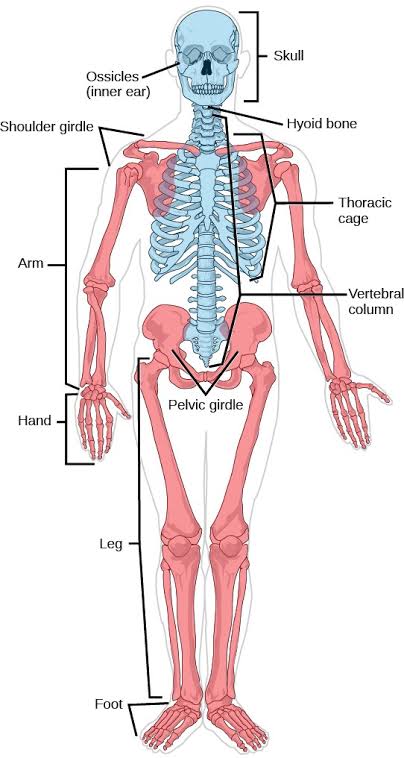
2. BONES OF THE LIMBS
2.1 Upper Limb Bones
- Clavicle: An S-shaped long bone. It articulates medially with the manubrium (sternoclavicular joint) and laterally with the acromion (acromioclavicular joint). It is the only bony attachment of the upper limb to the axial skeleton. Fractures commonly occur at the junction of the middle and lateral thirds.
- Scapula: A flat, triangular bone on the posterior thoracic wall. It has three borders (superior, medial, lateral), three angles (superior, inferior, lateral), and two surfaces (costal and posterior). The glenoid cavity articulates with the head of the humerus. The spine of the scapula divides the posterior surface into supraspinous and infraspinous fossae. The acromion and coracoid process are important muscle attachment and articulation sites.
- Humerus: The longest bone of the upper limb. The head articulates with the glenoid cavity. The anatomical neck separates the head from the tubercles; the surgical neck is the constricted region below the tubercles and is a common fracture site. The greater tubercle receives insertions of supraspinatus, infraspinatus, and teres minor. The lesser tubercle receives the subscapularis. The deltoid tuberosity is the insertion of the deltoid. The medial and lateral epicondyles are palpable landmarks. The olecranon fossa posteriorly and coronoid fossa anteriorly receive the ulna during elbow movement. The trochlea articulates with the ulna; the capitulum articulates with the radius.
- Radius: The lateral bone of the forearm. Its head articulates with the capitulum of the humerus and the radial notch of the ulna. The radial tuberosity is the insertion of biceps brachii. The styloid process is palpable on the lateral aspect of the wrist.
- Ulna: The medial bone of the forearm. Its olecranon forms the point of the elbow. The trochlear notch articulates with the trochlea of the humerus. The coronoid process and radial notch are additional articular surfaces.
- Carpal Bones: Arranged in two rows. Proximal row (lateral to medial): scaphoid, lunate, triquetrum, pisiform. Distal row (lateral to medial): trapezium, trapezoid, capitate, hamate. The scaphoid is the most frequently fractured; the lunate is commonly dislocated.
Some Lovers Try Positions That They Cannot Handle: Scaphoid, Lunate, Triquetrum, Pisiform, Trapezium, Trapezoid, Capitate, Hamate.
2.2 Lower Limb Bones
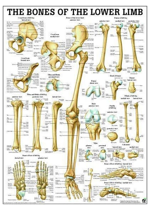
- Hip bone (os coxae): Formed by the fusion of three bones: ilium, ischium, and pubis. The acetabulum is the deep socket for the head of the femur. The obturator foramen is the large opening formed by the pubis and ischium. The iliac crest is palpable from ASIS to PSIS. The ischial tuberosity is the weight-bearing point in sitting.
- Femur: The longest and strongest bone in the body. The head articulates with the acetabulum. The fovea capitis is the pit for the ligament of the head of the femur. The neck connects the head to the shaft and is a common fracture site in elderly patients. The greater and lesser trochanters are muscle attachment sites. The linea aspera is the rough ridge on the posterior shaft. The medial and lateral condyles articulate with the tibia; the patellar surface articulates with the patella.
- Patella: The largest sesamoid bone, embedded in the quadriceps tendon. It protects the knee joint and improves the mechanical advantage of the quadriceps.
- Tibia: Large, medial weight-bearing bone. The tibial tuberosity is the insertion of the patellar ligament. The anterior border (shin) is subcutaneous. The medial malleolus forms the medial prominence of the ankle.
- Fibula: Slender, lateral bone. The lateral malleolus forms the lateral prominence of the ankle and extends more distally than the medial malleolus.
- Tarsal bones: include the talus, calcaneus (heel bone, largest), navicular, cuboid, and three cuneiforms.
3. JOINTS OF THE LIMBS
Most limb joints are synovial, characterized by a joint cavity, articular cartilage, synovial membrane, and joint capsule.
3.1 Shoulder Joint (Glenohumeral)
A ball-and-socket synovial joint; the most mobile joint in the body. Stability is provided by the rotator cuff muscles (SITS): supraspinatus, infraspinatus, teres minor, and subscapularis. Also supported by the glenoid labrum and coracohumeral ligament.
3.2 Elbow Joint
A hinge (ginglymus) joint involving three bones and three articulations: humeroulnar, humeroradial, and proximal radioulnar. The annular ligament encircles the radial head.
3.4 Hip Joint
A ball-and-socket joint. The iliofemoral ligament (Y ligament of Bigelow) is the strongest in the body and prevents hyperextension. The pubofemoral ligament limits abduction, and the ischiofemoral ligament limits internal rotation.
3.5 Knee Joint
The largest and most complex joint. It consists of three articulations: lateral tibiofemoral, medial tibiofemoral, and patellofemoral. Stabilized by:
- MCL and LCL: Medial and lateral collateral ligaments.
- ACL and PCL: Anterior and posterior cruciate ligaments.
- Menisci: Medial and lateral fibrocartilaginous discs.
3.6 Ankle Joint (Talocrural)
A hinge joint between the distal tibia, fibula, and talus. The deltoid ligament (medial) is very strong. Lateral ligaments (anterior talofibular, posterior talofibular, calcaneofibular) are weaker and more commonly sprained.
4. MUSCLE COMPARTMENTS OF THE LIMBS
Organized into compartments separated by deep fascia and intermuscular septa. Each contains muscles with similar functions and innervation.
4.1 Upper Limb Muscle Compartments
- Arm: Anterior (coracobrachialis, biceps brachii, brachialis) — musculocutaneous nerve; Posterior (triceps brachii) — radial nerve.
- Forearm: Anterior (flexor-pronator) — primarily median nerve (except FCU and medial half of FDP, which are ulnar nerve); Posterior (extensor-supinator) — radial nerve.
- Hand: Thenar (median nerve, except adductor pollicis); Hypothenar (ulnar nerve); Central (lumbricals/interossei — dual innervation).
4.2 Lower Limb Muscle Compartments
- Thigh: Anterior (quadriceps, sartorius) — femoral nerve; Medial (adductors, gracilis, pectineus) — primarily obturator nerve; Posterior (hamstrings) — sciatic nerve (tibial division).
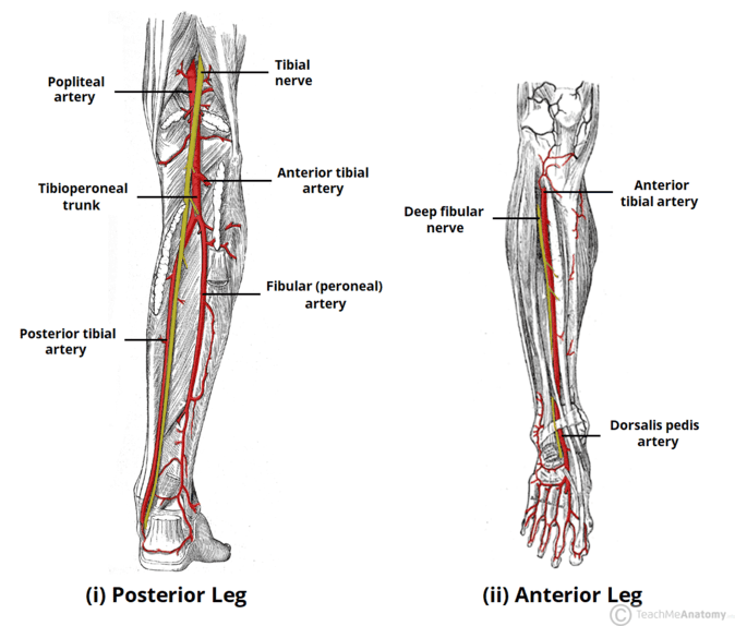
- Leg: Anterior (tibialis anterior, extensors) — deep fibular nerve; Lateral (fibularis longus/brevis) — superficial fibular nerve; Posterior (gastrocnemius, soleus, flexors) — tibial nerve.
5. BLOOD SUPPLY OF THE LIMBS
5.1 Upper Limb Arteries
Subclavian Artery → Axillary Artery (at 1st rib) → Brachial Artery (at teres major). The brachial artery bifurcates in the cubital fossa into the Radial Artery (lateral) and Ulnar Artery (medial).
5.2 Lower Limb Arteries
External Iliac Artery → Femoral Artery (at inguinal ligament) → Popliteal Artery (at adductor hiatus). The popliteal artery bifurcates into the Anterior Tibial Artery (continues as Dorsalis Pedis) and the Posterior Tibial Artery (branches into medial/lateral plantar arteries).
5.3 Venous Drainage
- Upper Limb: Cephalic vein (lateral) and Basilic vein (medial) are superficial. The median cubital vein connects them in the cubital fossa.
- Lower Limb: Great saphenous vein (medial, drains to femoral vein) and Small saphenous vein (lateral, drains to popliteal vein).
6. NERVE SUPPLY OF THE LIMBS
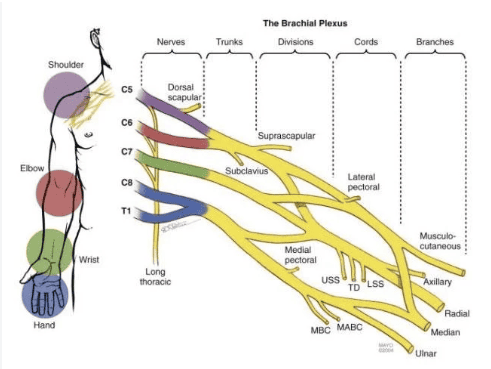
6.1 Brachial Plexus (C5-T1)
Organized into Roots, Trunks, Divisions, Cords, and Terminal Branches.
- Musculocutaneous: Lateral cord; anterior arm muscles.
- Median: Lateral and medial cords; most anterior forearm and lateral hand.
- Ulnar: Medial cord; medial forearm and intrinsic hand muscles.
- Axillary: Posterior cord; deltoid and teres minor.
- Radial: Posterior cord; posterior arm and forearm muscles.
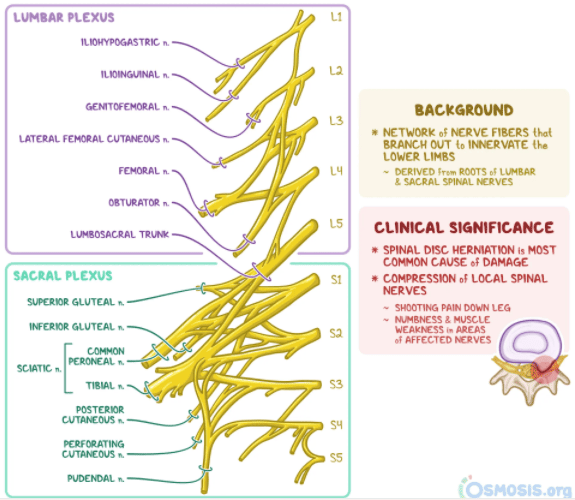
6.2 Lumbosacral Plexus (L1-S4)
- Lumbar Plexus (L1-L4): Iliohypogastric, Ilioinguinal, Genitofemoral, Lateral femoral cutaneous, Femoral nerve (anterior thigh), and Obturator nerve (medial thigh).
- Sacral Plexus (L4-S4): Superior/Inferior gluteal, Sciatic nerve (largest nerve), Posterior femoral cutaneous, and Pudendal nerve.
6.3 Summary of Major Nerves and Functions
| Nerve | Origin | Motor Function | Sensory Distribution | Clinical Significance |
|---|---|---|---|---|
| Median | Lat/Med cords | Wrist/Digit flexion | Lat. palm; digits 1-3.5 | Carpal tunnel; thenar wasting |
| Ulnar | Medial cord | Intrinsic hand; FCU | Medial palm; digits 4-5 | Claw hand (ulnar paradox) |
| Radial | Post. cord | Elbow/Wrist/Digit extension | Post. arm/forearm; lateral dorsum hand | Wrist drop (Saturday night palsy) |
| Femoral | Lumbar plexus | Knee extension; Hip flexion | Ant. thigh; medial leg (saphenous) | Femoral nerve palsy |
| Sciatic | Sacral plexus | Knee flexion; all below knee | Skin post. thigh/leg | Sciatica; foot drop (fibular division) |
7. LYMPHATIC DRAINAGE OF THE LIMBS
- Upper Limb: superficial vessels drain to axillary lymph nodes (5 groups: anterior, posterior, lateral, central, apical). The apical group drains to the subclavian lymph trunk.
- Lower Limb: superficial vessels drain to superficial inguinal lymph nodes. Lymph from lateral heel/foot drains to popliteal nodes, then deep inguinal nodes.
8. SURFACE ANATOMY OF THE LIMBS
- Upper Limb: Landmarks include the clavicle, acromion, medial/lateral epicondyles, olecranon, and the anatomical snuffbox (containing the scaphoid and radial artery). The cubital fossa contains the biceps tendon, brachial artery, and median nerve.
- Lower Limb: Landmarks include the ASIS, PSIS, greater trochanter, patella, medial/lateral malleoli, and calcaneus. The femoral triangle (NAVEL: Nerve, Artery, Vein, Empty space, Lymphatics) and popliteal fossa are critical clinical regions.
9. CLINICAL RELEVANCE
9.1 Fractures
- Colles fracture: Distal radius fracture with dorsal displacement ('dinner fork' deformity).
- Humeral surgical neck: Common in elderly; risk to axillary nerve.
- Tibial shaft: High risk of compartment syndrome due to tight fascial envelope.
9.2 Nerve Injuries
- Erb palsy (C5-C6): 'Waiter's tip' posture.
- Radial nerve (Spiral groove): Causes wrist drop.
- Common fibular nerve (Fibular neck): Causes foot drop.
Compartment Syndrome
Caused by increased pressure within a muscle compartment. Recognized by the '6 Ps': Pain out of proportion, Pain on passive stretch, Pallor, Pulselessness, Paresthesia, and Paralysis. Requires emergency fasciotomy.
10. SUMMARY TABLE: FOUNDATIONAL CONCEPTS
| System | Upper Limb | Lower Limb | Clinical Pearl |
|---|---|---|---|
| Main joints | Glenohumeral, elbow, wrist | Hip, knee, ankle | Shoulder dislocation; ankle sprain |
| Main arteries | Axillary, brachial, radial, ulnar | Femoral, popliteal, tibial | Femoral artery puncture site |
| Lymph nodes | Axillary (5 groups) | Inguinal, popliteal | Sentinel node biopsy (breast cancer) |
11. KEY POINTS
- The brachial plexus (C5-T1) supplies the upper limb; the lumbosacral plexus (L1-S4) supplies the lower limb.
- The shoulder is the most mobile joint; the hip is the most stable large joint.
- The great saphenous vein is the longest in the body and is used for bypass grafting.
- Nerve injuries produce characteristic postures: Wrist drop (Radial), Claw hand (Ulnar), Foot drop (Common Fibular).
