Histology of Glands
Microscopic anatomy of exocrine, endocrine, and mixed glands. A detailed study of functional organization, modes of secretion, and cellular architecture essential for medical clinical practice.
1. Introduction to Glands
A gland is an organized group of cells specialized to synthesize and secrete substances for use elsewhere in the body or for elimination. Glands are derived from epithelial tissue. During embryonic development, epithelial cells proliferate and invaginate into the underlying connective tissue, differentiating into secretory units and, in some cases, duct systems.
The secretory products of glands include enzymes, hormones, mucus, sweat, and sebum, all of which are essential for maintaining homeostasis, digestion, and protection.
2. Classification of Glands
Glands are primarily classified based on the presence or absence of a duct system and their mode of secretion.
2.1 Classification by Presence of Ducts
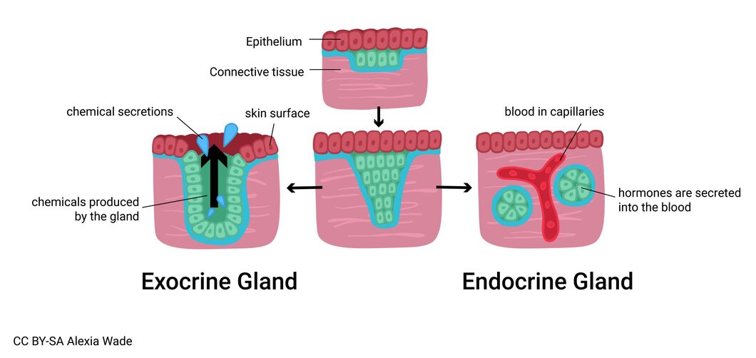
- Exocrine glands: Possess ducts that transport secretions directly onto an epithelial surface. Examples include salivary glands and sweat glands.
- Endocrine glands: Ductless glands that release hormones directly into the bloodstream or interstitial fluid. They are characterized by rich capillary networks.
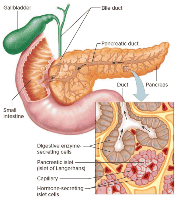
- Mixed glands: Contain both exocrine and endocrine components. The pancreas is the quintessential example.
2.2 Comparison of Exocrine and Endocrine Glands
| Feature | Exocrine Glands | Endocrine Glands |
|---|---|---|
| Ducts | Present | Absent |
| Secretion route | Onto epithelial surfaces/into ducts | Directly into blood or lymph |
| Secretory products | Enzymes, mucus, sweat, oil, wax | Hormones |
| Distance of action | Local (near site of secretion) | Distant (target organs via blood) |
| Blood supply | Moderate | Extremely rich capillary network |
| Cell arrangement | Acini, tubules, or alveoli | Cords, clusters, or follicles |
3. Exocrine Glands
Exocrine glands constitute the majority of glands in the body. They consist of a secretory portion (where the product is made) and a duct system (which delivers and may modify the product).
3.1 Structural Classification
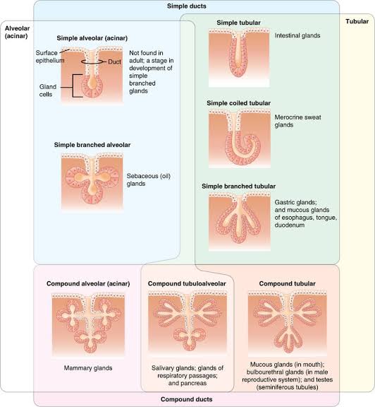
3.1.1 Based on Duct System
- Simple glands: Possess an unbranched duct.
— Simple tubular: Intestinal glands.
— Simple coiled tubular: Merocrine sweat glands.
— Simple branched tubular: Gastric glands.
— Simple branched alveolar: Sebaceous glands. - Compound glands: Possess a branched duct system.
— Compound tubular: Mucous glands of the mouth.
— Compound alveolar (acinar): Mammary glands.
— Compound tubuloalveolar: Salivary glands and pancreas.
3.2 Functional Classification (Modes of Secretion)
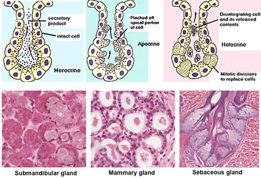
Glands are classified by the mechanism used to release their products:
- Merocrine (Eccrine) Secretion: The product is released via exocytosis. The cell remains completely intact. This is the most common mode (e.g., Salivary glands, Pancreas).
- Apocrine Secretion: The apical portion of the cytoplasm is pinched off and released with the product. The cell repairs itself afterward (e.g., Apocrine sweat glands).
- Holocrine Secretion: The entire cell disintegrates to become the secretion. Cells are replaced by mitotic division of the basal layer (e.g., Sebaceous glands).
| Mode | Mechanism | Cell Fate | Examples |
|---|---|---|---|
| Merocrine | Exocytosis of vesicles | Cell remains intact | Salivary, pancreatic acini |
| Apocrine | Apical cytoplasm pinched off | Cell repairs/continues | Mammary gland, axillary sweat |
| Holocrine | Entire cell disintegrates | Cell dies and is shed | Sebaceous glands |
3.4 Histological Types of Secretory Cells
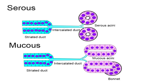
- Serous Cells: Produce watery, protein-rich secretions (enzymes). Pyramidal shape with round basal nuclei. Cytoplasm is basophilic (RER) at the base and eosinophilic (zymogen granules) at the apex.
- Mucous Cells: Produce viscous mucinogen. Columnar shape with flattened nuclei pressed against the basal membrane. Cytoplasm appears pale/vacuolated in H&E.
- Mixed (Seromucous) Cells: Contain both types. Often show serous demilunes (crescent caps of serous cells over mucous acini). Example: Submandibular gland.
4. Endocrine Glands
Ductless glands characterized by a lack of a surface connection and a very high density of fenestrated capillaries. Secretions (hormones) diffuse into the blood for systemic distribution.
4.1 Major Endocrine Histology
- Thyroid Gland: Unique follicular arrangement. Spherical follicles filled with colloid (thyroglobulin). Follicular cells produce T3/T4; Parafollicular (C cells) produce Calcitonin.
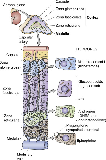
- Adrenal Gland:
— Cortex (3 Zones): Glomerulosa (Aldosterone), Fasciculata (Cortisol), Reticularis (Androgens).
— Medulla: Contains Chromaffin cells secreting Epinephrine and Norepinephrine. - Pituitary (Hypophysis): Anterior (Adenohypophysis) with 5 cell types; Posterior (Neurohypophysis) containing axons and pituicytes.
- Pancreatic Islets (Langerhans): Scattered clusters. Beta cells (Insulin), Alpha cells (Glucagon), Delta cells (Somatostatin).
5. Mixed Glands (Quintessential Example: Pancreas)
The pancreas is approximately 99% exocrine (serous acini producing digestive enzymes) and 1% endocrine (islets of Langerhans).
6. Clinical Relevance
Cystic Fibrosis (CF)
An autosomal recessive disorder caused by mutations in the CFTR gene. It results in defective chloride transport, leading to thick, viscous secretions that obstruct ducts in the pancreas, sweat glands, and lungs, causing pancreatic insufficiency and malabsorption.
Iodine Deficiency & Goitre
Enlargement of the thyroid (Goitre) is frequently due to Iodine deficiency, which remains a significant public health concern in several regions of Uganda. Without iodine, follicular cells cannot synthesize T3/T4, leading to compensatory hypertrophy.
- Sialadenitis: Inflammation of salivary glands (often parotid) due to mumps or sialoliths (stones).
- Addison Disease: Primary adrenal insufficiency due to destruction of the cortex (low cortisol/aldosterone).
- Pituitary Adenomas: Benign tumors that can cause hormone hypersecretion or bitemporal hemianopia by compressing the optic chiasm.
7. Summary Table: Major Glands of the Body
| Gland | Type | Mode/Mechanism | Clinical Significance |
|---|---|---|---|
| Parotid | Exocrine | Merocrine | Mumps, Sjögren syndrome |
| Thyroid | Endocrine | Follicular/Blood | Goitre, Hypothyroidism |
| Adrenal Cortex | Endocrine | Cord/Blood | Addison, Cushing syndrome |
| Adrenal Medulla | Endocrine | Cluster/Blood | Pheochromocytoma |
| Sebaceous | Exocrine | Holocrine | Acne, cysts |
| Mammary | Exocrine | Apocrine/Merocrine | Mastitis, cancer |
8. Key Points Summary
- Glands are organized epithelial cells classified as exocrine (ducts) or endocrine (ductless).
- The three release modes are merocrine (most common), apocrine (apical loss), and holocrine (cell death).
- The pancreas is a vital mixed gland; its endocrine dysfunction leads to Diabetes Mellitus.
- Adrenal cortex zones are ordered: Glomerulosa, Fasciculata, Reticularis (Salt, Sugar, Sex).
