Physiology of Muscle Contraction
A comprehensive study of the physiological processes that convert chemical energy into mechanical force, covering the neuromuscular junction, molecular mechanisms of the sliding filament theory, excitation-contraction coupling, and metabolic regulation.
1. Introduction
Muscle contraction is a complex physiological process that converts chemical energy into mechanical force. Understanding these mechanisms is essential for diagnosing and treating neuromuscular disorders, prescribing exercise programs, and managing patients with muscle-related conditions. This study covers the neuromuscular junction (NMJ), the sliding filament theory, excitation-contraction coupling, energy metabolism, and the distinct types of muscle contraction.
2. The Neuromuscular Junction (NMJ)
The NMJ is the specialized synapse between a motor neuron and a skeletal muscle fiber. It is the primary site where the nervous system communicates with the muscular system to initiate contraction.
2.1 Structure of the NMJ
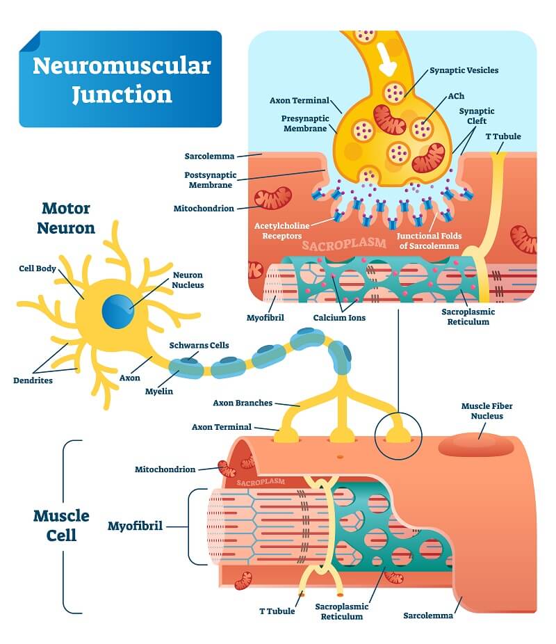
- Presynaptic Terminal: The motor neuron axon terminal containing numerous synaptic vesicles filled with acetylcholine (ACh). Voltage-gated calcium channels are embedded in this membrane.
- Synaptic Cleft: A narrow extracellular space (approximately 50 nm wide) separating the membranes. It contains the enzyme acetylcholinesterase (AChE), which hydrolyzes ACh to terminate the signal.
- Postsynaptic Membrane (Motor End Plate): The specialized region of the sarcolemma containing nicotinic acetylcholine receptors (nAChR). These are ligand-gated ion channels concentrated in junctional folds to increase surface area. Each nAChR consists of five subunits (two alpha, one beta, one gamma/delta, one epsilon).
2.2 Events at the NMJ (Step-by-Step)
- Step 1 — Arrival of action potential: An action potential travels down the motor neuron axon and reaches the presynaptic terminal, opening voltage-gated calcium ($Ca^{2+}$) channels.
- Step 2 — Calcium influx and vesicle fusion: Calcium ions enter the terminal and bind to vesicle proteins, triggering exocytosis of ACh into the synaptic cleft.
- Step 3 — ACh binding: ACh diffuses across the cleft and binds to nicotinic receptors. Binding of two ACh molecules to the alpha subunits opens the channel.
- Step 4 — Depolarization: The open channel allows sodium ($Na^+$) influx and potassium ($K^+$) efflux. This produces an end-plate potential (EPP), which is always suprathreshold under normal conditions and triggers a muscle action potential.
- Step 5 — Muscle fiber action potential: The potential spreads across the sarcolemma and into the T-tubules.
- Step 6 — ACh hydrolysis: AChE breaks down ACh into acetate and choline, which is transported back for resynthesis.
- The NMJ is the synapse between a motor neuron and a muscle fiber.
- ACh is the primary neurotransmitter.
- AChE ensures the fiber is not continuously stimulated.
- The EPP is always suprathreshold in healthy tissue.
3. Sarcomere Structure
The sarcomere is the basic functional unit of skeletal muscle contraction, defined as the region between two adjacent Z-discs.
3.1 Thin Filaments (Actin)
Thin filaments consist of F-actin (polymerized globular G-actin). In the resting state, myosin-binding sites are blocked by tropomyosin. Troponin is a complex of three subunits attached at intervals:
- Troponin T: Binds to tropomyosin.
- Troponin I: Inhibits the actin-myosin interaction.
- Troponin C: Binds calcium ions.
3.2 Thick Filaments (Myosin)
Composed of myosin II. Each molecule has a long tail and two globular heads. Each head contains an ATP-binding site and an actin-binding site.
3.3 Bands and Zones
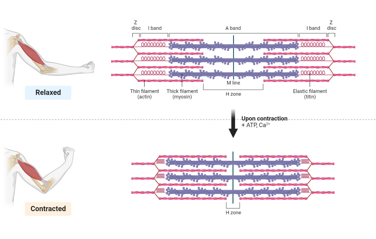
- A band: Dark band; contains the entire length of thick filaments (including overlap with thin).
- I band: Light band; contains thin filaments only; bisected by the Z-disc.
- H zone: Central region of A band with thick filaments only. Narrows during contraction.
- M line: Protein line in the center of the H zone that holds thick filaments in place.
- Z-disc: Anchors thin filaments; composed primarily of alpha-actinin.
4. The Sliding Filament Theory
Proposed by Huxley and Hanson (1954), it states that muscle shortening results from filaments sliding past one another without changing their individual lengths. Z-discs move closer, I bands and H zones narrow, and the A band remains constant.
4.1 The Cross-Bridge Cycle
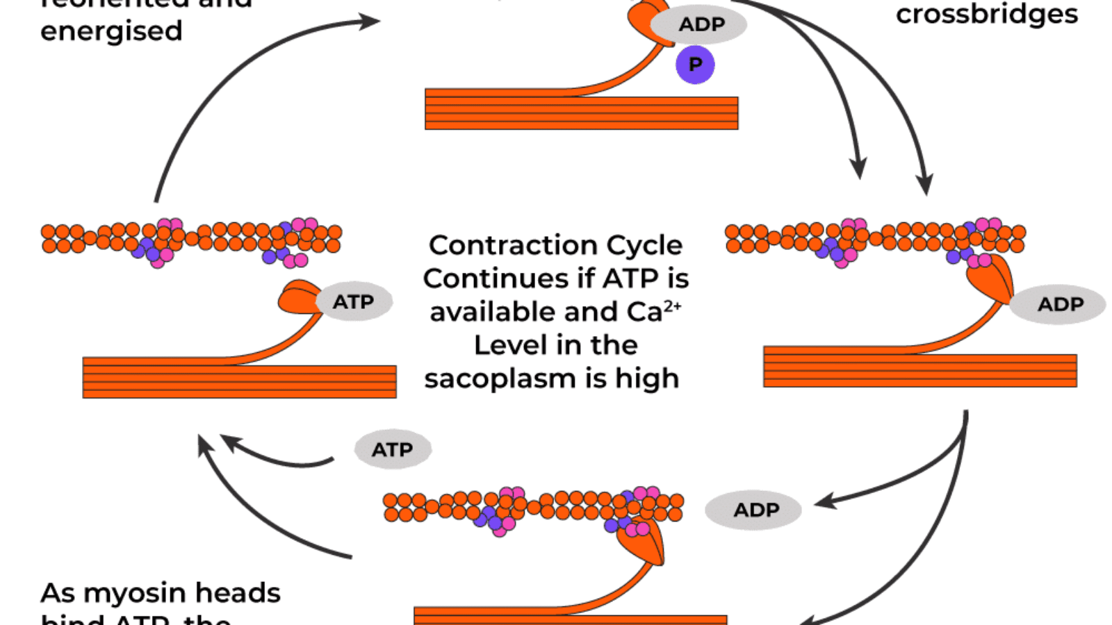
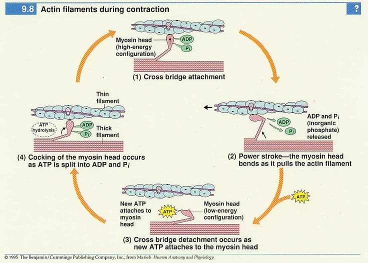
- Step 1 — Cross-bridge formation: Calcium binds to troponin C, tropomyosin shifts, and the myosin head (bound to ADP and Pi) binds to actin.
- Step 2 — Power stroke: Release of inorganic phosphate (Pi) triggers the head to pivot, pulling the thin filament ~10 nm toward the M line. ADP is then released.
- Step 3 — Cross-bridge detachment: A new ATP molecule binds to the myosin head, causing it to detach from actin.
- Step 4 — Reactivation: Myosin hydrolyzes ATP into ADP and Pi, returning the head to its high-energy "cocked" conformation.
5. Excitation-Contraction Coupling
The process by which an electrical action potential triggers mechanical contraction via calcium release from the sarcoplasmic reticulum (SR).
5.1 Steps of Coupling
- Step 1 — Action potential propagation: The potential spreads across the sarcolemma into T-tubules.
- Step 2 — DHP receptors: T-tubule dihydropyridine (DHP) receptors detect the voltage change. In skeletal muscle, they are mechanically coupled to ryanodine receptors (RyR) on the SR.
- Step 3 — Calcium release: RyR channels open, and calcium flows from the SR into the cytosol. Cytosolic concentration rises from $10^{-7}$ M to $10^{-5}$ M.
- Step 4 — Calcium-troponin binding: Calcium binds to troponin C, pulling tropomyosin away from the active sites.
- Step 5 — Muscle contraction: Cross-bridge cycling proceeds.
- Step 6 — Reuptake: SERCA (Calcium ATPase) pumps calcium back into the SR to allow relaxation.
6. Energy Sources for Muscle Contraction
| Energy System | Substrate | ATP Yield | Duration | Oxygen? |
|---|---|---|---|---|
| Phosphocreatine | Phosphocreatine | 1 ATP per PCr | 10-15 seconds | No |
| Anaerobic Glycolysis | Glucose/Glycogen | 2 ATP per glucose | 1-2 minutes | No |
| Aerobic Respiration | Glucose, fatty acids | 30-32 ATP per glucose | Hours | Yes |
7. Types of Muscle Contraction
- Isotonic Contraction: Muscle generates tension while changing length.
- Concentric: Muscle shortens while generating tension (e.g., lifting a weight).
- Eccentric: Muscle lengthens while generating tension (e.g., lowering a weight slowly).
- Isometric Contraction: Muscle generates tension without changing length (e.g., holding a heavy object still).
- Isokinetic Contraction: Muscle contracts at a constant velocity against variable resistance.
8. Clinical Relevance
An autoimmune disorder with antibodies against nAChR at the NMJ. It results in muscle weakness that improves with rest. Diagnosis involves the edrophonium test.
A pharmacogenetic disorder triggered by anesthetics, causing excessive calcium release from the SR. Treated with dantrolene (a RyR antagonist).
Produced by Clostridium botulinum; the toxin cleaves SNARE proteins, preventing ACh release from presynaptic terminals, leading to flaccid paralysis.
Breakdown of muscle fibers releasing myoglobin into the blood. Myoglobinuria can lead to acute kidney injury. Often caused by trauma or extreme exertion.
Add a Comment