Elimination of Urine (Micturition)
Comprehensive anatomical and physiological overview of the micturition process, neural control mechanisms, and clinical pathophysiology of the lower urinary tract.
1. Definition
Elimination of urine, also called micturition or voiding, is the process by which urine is expelled from the urinary bladder through the urethra to the exterior of the body. It is the final step in urine handling, following filtration by the kidneys and transport through the ureters.
2. Anatomy of the Lower Urinary Tract
The Urinary Bladder
The bladder is a hollow, muscular, distensible organ located in the anterior pelvis. In adults, it typically holds 400–600 mL of urine. The physiological desire to void typically begins when the volume reaches 200–300 mL.
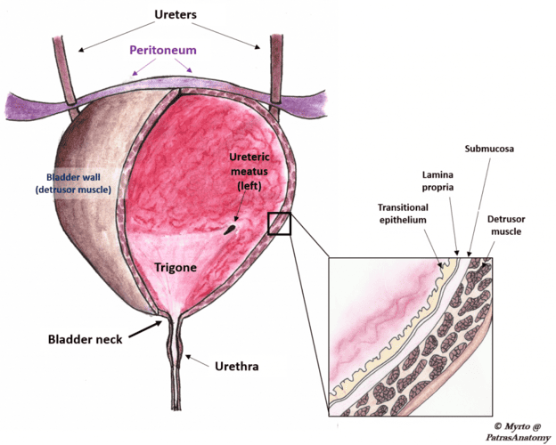
Layers of the bladder wall (inner to outer):
- Mucosa: Transitional epithelium (urothelium) that stretches without damage; forms folds called rugae when the bladder is empty.
- Submucosa: Connective tissue layer containing blood vessels and nerves.
- Muscularis (Detrusor Muscle): Smooth muscle arranged in three interlacing layers; its contraction is responsible for expelling urine.
- Adventitia/Serosa: The outermost protective covering.
Key Anatomical Regions:
- Apex: The anterior tip, connected to the umbilicus by the median umbilical ligament.
- Body: The main central portion of the bladder.
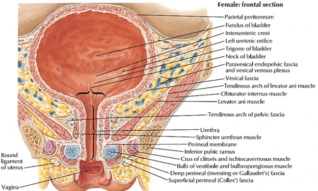
- Fundus (Base): The posterior surface containing the trigone—a smooth, triangular area bounded by the two ureteric orifices and the internal urethral orifice.
- Neck: The inferior narrowing where the bladder joins the urethra.
The trigone is highly sensitive to stretch. Because it is embryologically distinct from the rest of the bladder (mesodermal origin), it lacks rugae and remains smooth regardless of the bladder's distension state.
The Urethra
- Female urethra: Approximately 3–4 cm long. Its short length increases susceptibility to urinary tract infections (UTIs) as bacteria can ascend more easily.
- Male urethra: Approximately 18–20 cm long, divided into three main portions: prostatic, membranous, and spongy (penile).
The Urethral Sphincters
| Sphincter | Location | Muscle Type | Control | Function |
|---|---|---|---|---|
| Internal urethral sphincter | Bladder neck | Smooth muscle | Involuntary (autonomic) | Closes during filling; relaxes during voiding. |
| External urethral sphincter | Deep perineal pouch / pelvic floor | Skeletal muscle | Voluntary (somatic) | Consciously prevents or permits urination. |
3. Physiology of Micturition
Micturition involves two alternating phases controlled by a combination of reflex (involuntary) and voluntary mechanisms.
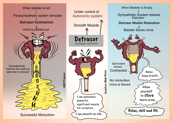
Phase 1: Storage (Filling)
- Urine flows continuously from the kidneys via the ureters into the bladder.
- The detrusor muscle remains relaxed to allow for high compliance.
- Both sphincters remain contracted to maintain continence.
- Sympathetic (Hypogastric) and somatic (Pudendal) nerves are active.
- Parasympathetic activity is inhibited.
- Stretch receptors in the bladder wall send signals to the spinal cord. At low volumes, these are suppressed by higher brain centres.
Phase 2: Voiding (Emptying)
Triggered consciously when the setting is appropriate through the following sequence:
- Voluntary relaxation of the external urethral sphincter (cortical inhibition of the pudendal nerve).
- Sympathetic inhibition → internal sphincter relaxes.
- Parasympathetic activation (pelvic splanchnic nerves, S2–S4) → detrusor muscle contracts strongly.
- Intravesical pressure rises; the bladder neck opens.
- Urine flows out through the urethra.
- Sensory feedback from the urethra reinforces detrusor contraction until the bladder is completely empty.
4. Neural Control
Afferent (Sensory) Pathways
Stretch receptors in the bladder wall and trigone detect distension. Signals travel via the pelvic splanchnic nerves to the sacral spinal cord (S2–S4), then ascend to the pontine micturition centre (PMC) and the cerebral cortex.
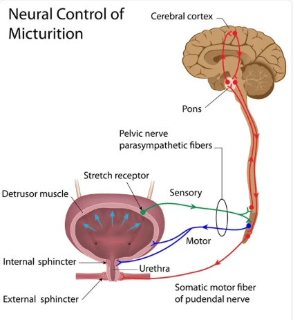
Efferent (Motor) Pathways
| Pathway | Nerve | Spinal Origin | Neurotransmitter | Effect |
|---|---|---|---|---|
| Parasympathetic | Pelvic splanchnic nerves | S2–S4 | Acetylcholine (M3 receptors) | Detrusor contraction; internal sphincter relaxation. |
| Sympathetic | Hypogastric nerve | T11–L2 | Noradrenaline (α1 and β3 receptors) | Detrusor relaxation (β3); internal sphincter contraction (α1). |
| Somatic | Pudendal nerve | S2–S4 | Acetylcholine (nicotinic) | External sphincter contraction (voluntary control). |
Higher Control Centres
- Sacral Micturition Centre (S2–S4): Contains the basic reflex arc. In infants, this produces involuntary voiding.
- Pontine Micturition Centre (PMC): Coordinates the switch between storage and voiding; ensures detrusor contraction and sphincter relaxation occur together.
- Cerebral Cortex: Provides conscious, voluntary control. Allows for the delay of voiding or initiation at will.
5. Pathophysiology
Urinary Retention
Inability to empty the bladder. Causes include:
- Obstructive: Benign prostatic hyperplasia (BPH), urethral stricture, bladder stones.
- Neurogenic: Spinal cord injury, cauda equina syndrome, diabetic neuropathy.
- Pharmacological: Anticholinergics, opioids, tricyclic antidepressants.
- Postoperative: Occurs frequently after pelvic or spinal surgery.
Urinary Incontinence
The involuntary leakage of urine, classified by mechanism:
| Type | Mechanism | Key Features |
|---|---|---|
| Stress | Weak pelvic floor / sphincter | Leakage with cough, sneeze, or exertion. |
| Urge | Detrusor overactivity | Sudden urgency, large-volume loss. |
| Overflow | Chronic retention with passive overflow | Dribbling, palpable bladder, weak stream. |
| Functional | Physical/cognitive barriers | Inability to reach the toilet in time despite normal tract function. |
Special Pathological Conditions
- Neurogenic Bladder (Spastic): Lesion above sacral cord → detrusor overactivity, small capacity, and reflux risk.
- Neurogenic Bladder (Flaccid): Lesion at/below sacral cord → detrusor areflexia, large capacity, and high residual urine.
- Vesicoureteral Reflux: Backflow of urine to ureters due to failure of the ureterovesical valvular mechanism. Predisposes to pyelonephritis.
6. Clinical Relevance
- Acute retention in elderly males: BPH is the leading cause; requires urgent catheterisation.
- UTI: Extremely common, especially in females; diagnosed by urinalysis and culture.
- Obstetric fistula: Resulting from prolonged obstructed labour; causes continuous incontinence and social stigmatization.
- Spinal cord injury: Requires intermittent catheterisation to prevent secondary renal damage.
Patient Assessment
- History: Voiding pattern, hematuria, and neurological symptoms.
- Examination: Abdominal palpation for bladder distension, digital rectal examination (males).
- Investigations: Urine culture, bladder scan, renal ultrasound, and urodynamics.
7. Summary: Storage Phase vs. Voiding Phase
| Feature | Storage Phase | Voiding Phase |
|---|---|---|
| Detrusor muscle | Relaxed | Contracted |
| Internal sphincter | Closed | Open |
| External sphincter | Closed (voluntary) | Open (voluntary relaxation) |
| Parasympathetic | Inhibited | Active |
| Sympathetic | Active | Inhibited |
| Intravesical pressure | Low and stable | Elevated |
9. Key Points
- Micturition is a reflex act modulated by voluntary cortical control.
- Parasympathetic nerves (S2–S4, pelvic nerve) promote voiding.
- Sympathetic nerves (T11–L2, hypogastric nerve) promote storage.
- The pontine micturition centre coordinates the synergy between detrusor contraction and sphincter relaxation.
- In Uganda, BPH, UTIs, and Obstetric fistula are primary drivers of lower urinary tract clinical visits.
