Structure & Function of the Upper Urinary Tract
Comprehensive coverage of kidney and ureter anatomy, nephron physiology, renal vasculature, and detailed clinical correlations.
1. OVERVIEW
The upper urinary tract comprises the kidneys and ureters — the organs responsible for filtering blood, forming urine, and conveying it to the bladder. The kidneys are paired retroperitoneal organs that perform excretion, electrolyte regulation, acid-base balance, blood pressure control, and endocrine functions. The ureters are muscular conduits that propel urine from the renal pelvis to the urinary bladder via peristalsis.
2. THE KIDNEYS — GROSS ANATOMY
Location & Position
- Position: Paired retroperitoneal organs located between the T12 and L3 vertebrae.
- Orientation: The left kidney sits slightly more superior than the right (due to the liver).
- Shape: Bean-shaped (reniform); medial concavity = hilum; lateral convexity.
- Size: ~4–5 inches (10–12 cm) long; ~150 g in adults (size of a fist).
- Covering: Enclosed by renal fascia (Gerota's fascia) — a dense elastic connective tissue sheath; surrounded by perirenal fat.
External Features
| Feature | Description |
|---|---|
| Renal Capsule | Three layers of connective tissue/fat covering the kidney; protects and stabilizes the organ. |
| Hilum | Medial indentation where renal artery, renal vein, and ureter enter/exit. |
| Renal Pelvis | Funnel-shaped structure collecting urine; continuous with the ureter. |
| Adrenal Gland | Sits atop each kidney; produces cortisol and other hormones. |
Renal Hilum Contents
- Anterior to posterior: Renal vein → Renal artery → Ureter.
Very Angry Unicorn (Vein, Artery, Ureter).
The renal sinus is the cavity within the hilum containing fat, vessels, and the collecting system.
3. THE KIDNEYS — INTERNAL STRUCTURE
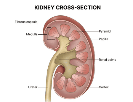
The kidney is divided into two primary regions: the renal parenchyma and the collecting system (pyelocalyceal system).
A. Renal Parenchyma
Renal Cortex (Outer Layer)
- Contains most nephron components: renal corpuscles, proximal & distal convoluted tubules, cortical collecting ducts.
- Renal Mantle: Peripheral cortical tissue covering the base of each renal pyramid.
- Renal Columns (of Bertin): Extensions of cortical tissue projecting inward between pyramids toward the renal sinus.
Renal Medulla (Inner Layer)
- Organized into renal pyramids (~9 per kidney).
- Contains medullary rays (descending/ascending limbs of Loop of Henle + medullary collecting ducts).
- Each pyramid + overlying cortex = renal lobe (anatomical unit).
- Renal Papilla: Apex of each pyramid; urine drains through the area cribrosa into minor calyces.
B. Renal Collecting System
| Structure | Description |
|---|---|
| Minor Calyces | 7–9 per kidney; each encloses a renal papilla. May be simple (1 papilla) or compound (2–3 papillae). |
| Major Calyces | Formed by convergence of 2–3 minor calyces; 2–3 per kidney. |
| Renal Pelvis | Funnel-shaped structure formed by union of major calyces; marks transition to the proximal ureter. |
4. THE NEPHRON — FUNCTIONAL UNIT
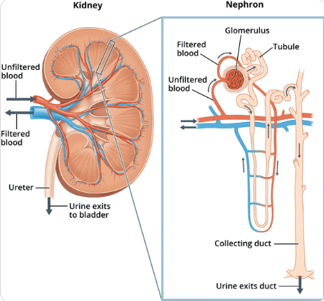
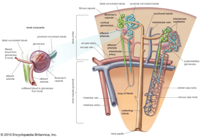
The nephron is the structural and functional unit of the kidney. Each adult kidney contains 1 to 1.5 million nephrons (occasionally >2.5 million).
Types of Nephrons
| Type | Location | Loop of Henle | Function |
|---|---|---|---|
| Cortical (Superficial) | Near cortical surface | Short | Bulk filtration; ~80% of nephrons |
| Juxtamedullary | Near corticomedullary junction | Long (deep into medulla) | Concentrating urine; countercurrent multiplication |
A. Renal Corpuscle (Filtration Unit)
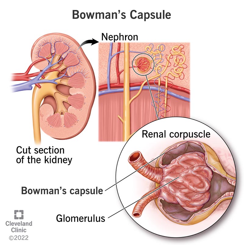
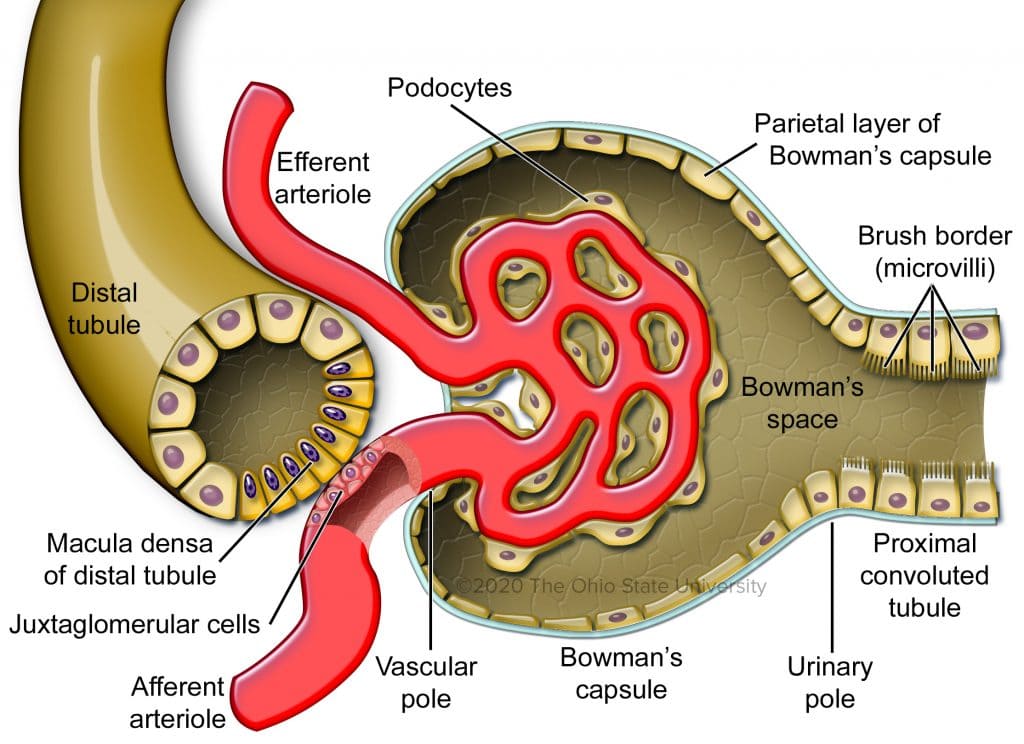
The renal corpuscle consists of the glomerulus + Bowman's capsule.
The Glomerulus
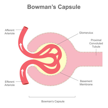
- A tuft of fenestrated capillaries supplied by the afferent arteriole and drained by the efferent arteriole.
- Filtration Barrier (3 layers):
- Fenestrated Endothelium: Pores 70–100 nm; provides size selectivity.
- Glomerular Basement Membrane (GBM): Composed of type IV collagen, laminin, fibronectin, proteoglycans; provides charge selectivity (repels negatively charged proteins).
- Podocytes (Visceral Epithelium): Specialized cells with interdigitating foot processes forming slit diaphragms — the final filtration barrier.
Bowman's Capsule
- Double-walled cup surrounding the glomerulus.
- Parietal layer: Simple squamous epithelium.
- Visceral layer: Podocytes.
- Bowman's (Capsular) Space: Collects the ultrafiltrate.
- Vascular pole: Where afferent/efferent arterioles enter/exit.
- Urinary pole: Where filtrate exits into the PCT.
B. Proximal Convoluted Tubule (PCT)
- Location: Renal cortex; adjacent to glomerulus.
- Epithelium: Simple cuboidal with brush border (dense microvilli) → massively increases surface area.
- Key Features: Abundant mitochondria for active transport; numerous vesicles for transcellular transport.
- Function: Reabsorbs 60–80% of the ultrafiltrate:
— ~65% of Na⁺, ~60% of water
— Nearly 100% of glucose and amino acids
— ~90% of bicarbonate
— Secretes organic acids and bases
C. Loop of Henle
A U-shaped structure that penetrates the renal medulla, creating the countercurrent multiplier system essential for urine concentration.
| Segment | Permeability | Key Transport |
|---|---|---|
| Thin Descending Limb | Highly permeable to H₂O; impermeable to Na⁺/Cl⁻ | Water exits by osmosis; filtrate concentrates to ~1200 mOsm/L |
| Thin Ascending Limb | Impermeable to H₂O; permeable to Na⁺/Cl⁻ | Passive salt reabsorption |
| Thick Ascending Limb (TAL) | Impermeable to H₂O; highly permeable to ions | Active Na⁺/K⁺/2Cl⁻ cotransport (NKCC2); dilutes urine |
D. Distal Convoluted Tubule (DCT)
- Location: Renal cortex; near its glomerulus of origin.
- Epithelium: Simple cuboidal; fewer microvilli than PCT.
- Key Features: Highest mitochondrial density in the nephron; extensive basolateral amplification.
- Function:
— Fine-tuning of Na⁺, K⁺, Ca²⁺, and pH.
— Aldosterone-sensitive Na⁺ reabsorption (ENaC channels).
— PTH-sensitive Ca²⁺ reabsorption.
— Variable water permeability (regulated by ADH indirectly).
E. Collecting Duct System
- Connecting Tubule (CNT): Transition zone; contains intercalated cells (H⁺/HCO₃⁻ regulation).
- Collecting Duct (CD): Lined by principal cells (Na⁺/water reabsorption) and intercalated cells (acid-base).
- Function: Final urine modification; ADH-dependent water reabsorption via aquaporin-2 channels.
- Multiple nephrons drain into a single collecting duct → medullary collecting duct → papillary duct → area cribrosa → calyx.
F. Juxtaglomerular Apparatus (JGA)
The JGA regulates glomerular filtration rate (GFR) via tubuloglomerular feedback.
| Component | Description |
|---|---|
| Macula Densa | Specialized tall cells of the distal TAL; sense NaCl concentration in tubular fluid. |
| Juxtaglomerular (Granular) Cells | Modified smooth muscle of afferent arteriole; synthesize and release renin. |
| Extraglomerular Mesangial Cells | Connect macula densa to granular cells; structural support. |
5. BLOOD SUPPLY & VASCULATURE
Renal Arterial Tree
Abdominal Aorta → Renal Artery → Segmental Arteries → Interlobar Arteries → Arcuate Arteries → Interlobular Arteries → Afferent Arterioles → Glomerulus → Efferent Arterioles → Peritubular Capillaries / Vasa Recta → Interlobular Veins → Arcuate Veins → Interlobar Veins → Renal Vein → IVC.
Unique Features
- Efferent arterioles form a second capillary bed (unusual in the body):
— Peritubular capillaries: Surround PCT and DCT in cortex.
— Vasa recta: Long, hairpin capillaries accompanying juxtamedullary nephrons' loops of Henle; maintain medullary osmotic gradient (countercurrent exchange).
6. KIDNEY FUNCTIONS SUMMARY
| Function | Mechanism | Clinical Relevance |
|---|---|---|
| Filtration & Excretion | Glomerular filtration of urea, creatinine, uric acid | Glomerular diseases → azotemia |
| Electrolyte Balance | Na⁺, K⁺, Ca²⁺, Mg²⁺, PO₄³⁻ reabsorption/secretion | Hypo-/hyperkalemia, hypernatremia |
| Acid-Base Balance | H⁺ secretion; HCO₃⁻ reabsorption; NH₃ buffer | Metabolic acidosis/alkalosis |
| Blood Pressure Regulation | RAAS (renin → Ang I → Ang II → aldosterone); Na⁺/water retention | Renovascular hypertension |
| Erythropoiesis | Erythropoietin (EPO) production by peritubular cells | Renal anemia in CKD |
| Calcium/Phosphate Metabolism | Calcitriol (1,25-OH Vitamin D) activation | Renal osteodystrophy |
| Gluconeogenesis | Glucose synthesis during fasting | Minimal in humans vs. other species |
7. THE URETERS — GROSS ANATOMY
General Characteristics
- Length: ~25–30 cm (adult).
- Diameter: Widest at renal pelvis (~10 mm); narrowest at ureterovesical junction (~1–3 mm).
- Shape: S-shaped, collapsible muscular tubes.
- Course: Retroperitoneal; descend on psoas major, cross the pelvic brim, enter bladder posterolaterally.
Three Anatomical Constrictions (Narrowings)
These are common sites for kidney stone impaction:
- Ureteropelvic Junction (UPJ) — where renal pelvis narrows into ureter.
- Pelvic Brim — where ureter crosses the iliac vessels.
- Ureterovesical Junction (UVJ) — where ureter enters the bladder wall (intramural portion).
Relations
| Structure | Right Ureter | Left Ureter |
|---|---|---|
| Anterior | Duodenum (2nd part), right colic vessels | Left colic vessels, sigmoid colon |
| Posterior | Psoas major, genitofemoral nerve | Psoas major, genitofemoral nerve |
| Medial | IVC | Aorta |
8. THE URETERS — HISTOLOGY
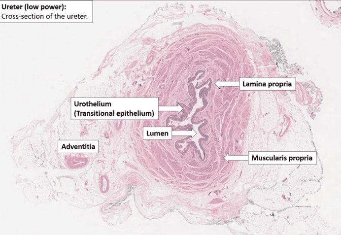
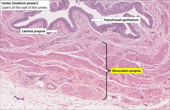
The ureter wall consists of three layers (from lumen outward):
1. Mucosa
- Urothelium (Transitional Epithelium): 4–6 cell layers; dome-shaped superficial cells that flatten when stretched. Accommodates changes in urine volume.
- Lamina Propria: Loose connective tissue; contains collagen, elastic fibers, and lymphocytes.
2. Muscularis Propria
- Upper 2/3: Inner circular + outer longitudinal smooth muscle layers.
- Lower 1/3: Additional inner longitudinal layer (three layers total) near the bladder.
- Function: Generates peristaltic waves to propel urine.
3. Adventitia
- Outer connective tissue layer with blood vessels, nerves, and lymphatics; anchors ureter to surrounding structures.
9. URETER FUNCTION & PERISTALSIS
Urine Transport
- Urine is propelled by peristaltic contractions initiated by pacemaker cells in the proximal renal pelvis.
- Contractions occur every 10–15 seconds, moving urine in small boluses.
- The ureterovesical junction acts as a physiological one-way valve:
— The intramural ureter courses obliquely through the bladder wall.
— Bladder contraction compresses the intramural ureter, preventing vesicoureteral reflux (VUR).
Neurovascular Supply of the Ureter
| Division | Source | Function |
|---|---|---|
| Sympathetic | Renal plexus, intermesenteric plexus (T11–L2) | Vasoconstriction; may modulate peristalsis |
| Parasympathetic | Pelvic splanchnic nerves (S2–S4) | Modulates peristalsis |
| Sensory | T11–L2 (visceral afferents) | Referred pain to ipsilateral groin/flank (ureteric colic) |
Arterial Supply:
— Upper: Renal, gonadal, aortic branches.
— Middle: Common iliac artery branches.
— Lower: Superior/inferior vesical arteries.
Lymphatic Drainage:
— Upper: Lumbar (para-aortic) nodes.
— Lower: Common iliac, external iliac, and internal iliac nodes.
10. CLINICAL CORRELATIONS
| Condition | Pathophysiology | Key Feature |
|---|---|---|
| Hydronephrosis | Obstruction (stone, tumor, stricture) → urine backs up → renal pelvis dilation | Flank pain, decreased GFR |
| Ureteric Colic | Stone impaction at a ureteric narrowing → severe colicky flank pain radiating to groin | Hematuria, nausea, restlessness |
| Vesicoureteral Reflux (VUR) | Failure of UVJ valve mechanism → urine refluxes to ureters/kidneys | Recurrent UTIs, pyelonephritis, renal scarring |
| Renal Cell Carcinoma (RCC) | Malignancy of proximal tubule cells; invades renal vein | Hematuria, flank mass, paraneoplastic syndromes |
| Glomerulonephritis | Inflammation of glomerular filtration barrier | Hematuria, proteinuria, hypertension, edema |
| Acute Kidney Injury (AKI) | Sudden drop in GFR from pre-renal, intrinsic, or post-renal causes | Elevated creatinine/BUN, oliguria |
| Chronic Kidney Disease (CKD) | Progressive nephron loss over ≥3 months | Anemia (↓EPO), bone disease (↓calcitriol), metabolic acidosis |
