Functions of Joints
Joints act like biomechanical and biological systems that enable movement, maintain stability, nourish tissues, sense position, and protect vital structures.
4.1 Mechanical Functions
A. Mobility
Joints allow the skeleton to move in multiple planes — sagittal, frontal, and transverse. The range of motion (ROM) is determined by the specific articular geometry and individual anatomical variations.
Types of Movement by Joint Classification:
| Joint Type | Movement Allowed | Examples |
|---|---|---|
| Uniaxial | Movement in one plane | Hinge (elbow), Pivot (atlantoaxial) |
| Biaxial | Movement in two planes | Condyloid (knuckles), Saddle (thumb) |
| Multiaxial | Movement in three planes + rotation | Ball-and-socket (shoulder, hip) |
Ball-and-socket joints provide the greatest range of motion, allowing for flexion/extension, abduction/adduction, rotation, and circumduction.
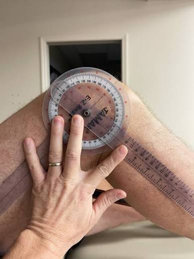
Goniometry
Measuring ROM with a goniometer is the gold standard in orthopedic and physiotherapy assessment. It quantifies joint angles in degrees, enabling objective tracking of rehabilitation progress and surgical outcomes.
B. Stability
Joints must balance mobility with stability — a fundamental trade-off in biomechanics. Stability is maintained through several integrated mechanisms:
| Stabilizer | Mechanism | Example |
|---|---|---|
| Bony architecture | Deep sockets and congruent surfaces | Deep acetabulum of the hip |
| Ligaments | Passive restraint; limits excessive motion | ACL and MCL in the knee |
| Muscles/Tendons | Dynamic stabilization via active contraction | Rotator cuff muscles in the shoulder |
| Joint capsule | Fibrous enclosure maintaining negative pressure | Glenohumeral (shoulder) capsule |
| Negative intra-articular pressure | Suction effect drawing surfaces together | All major synovial joints |
| Labrum/Meniscus | Deepens the socket and improves congruency | Glenoid labrum, acetabular labrum |
The shoulder sacrifices stability for mobility; its shallow glenoid fossa allows extensive ROM but makes it the most frequently dislocated major joint. Conversely, the hip prioritizes stability for weight-bearing via a deep acetabulum, resulting in reduced mobility.
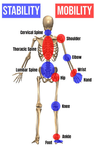
Rotator Cuff Dynamics
Shoulder dislocations account for up to 50% of all major joint dislocations. The rotator cuff muscles (supraspinatus, infraspinatus, teres minor, subscapularis) act as essential dynamic stabilizers by actively compressing the humeral head into the glenoid fossa during movement.
C. Shock Absorption
Articular cartilage and menisci distribute compressive and shear forces across joint surfaces. Synovial fluid acts as a viscous dampener, reducing friction to near-zero levels and dissipating energy.
Key Structures in Shock Absorption:
- Articular (hyaline) cartilage: Covers bone ends; matrix of type II collagen and proteoglycans. It is avascular and aneural.
- Fibrocartilage menisci: C-shaped discs that increase joint congruency and absorb 30–50% of compressive forces.
- Synovial fluid: Viscous fluid with a coefficient of friction of ~0.002–0.04.
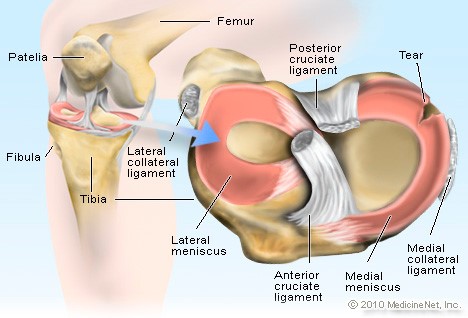
Meniscectomy Risks
Loss of shock absorption following meniscectomy leads to accelerated osteoarthritis. Partial meniscal removal can increase contact pressures on articular cartilage by 200–300%, significantly elevating degenerative risk.
D. Force Transmission
Joints transmit mechanical forces from muscles to produce movement. Weight-bearing joints transmit multiples of body weight (BW) during various activities:
| Activity | Force Relative to Body Weight |
|---|---|
| Standing | 1x BW |
| Walking | 3–4x BW |
| Running | 5–7x BW |
| Jumping/Landing | 10–12x BW |
Abnormal force transmission — due to malalignment, muscle weakness, or joint instability — is a primary driver of osteoarthritis. The knee joint, for instance, transmits forces exceeding 3,000 N during normal gait.
4.2 Nutritional & Metabolic Functions
Because articular cartilage is avascular, it relies entirely on diffusion for nutrient delivery and waste removal. Synovial fluid is the primary medium for this exchange.
Mechanism of Cartilage Nutrition:
- Synovial fluid production: Secreted by the synovial membrane as a blood plasma ultrafiltrate with added hyaluronic acid.
- Diffusion: Small solutes (glucose, oxygen) diffuse from fluid into the cartilage matrix.
- "Milking" action: Joint movement creates cyclic loading/unloading, generating a pumping mechanism that enhances fluid exchange.
- Subchondral bone contribution: Medullary cavities of underlying epiphyseal bone also contribute to nutrition in loaded joints.
Immobilization Effects
Prolonged bed rest or casting leads to cartilage degeneration. Without the "milking" action of movement, nutrient exchange diminishes, causing chondrocyte death and matrix breakdown. Early mobilization is critical post-surgery.
4.3 Proprioceptive Function
Proprioception is the body's ability to perceive its position and movement in space. Specialized mechanoreceptors provide critical sensory feedback.
Types of Joint Receptors:
| Receptor | Location | Response | Function |
|---|---|---|---|
| Type I (Ruffini) | Superficial capsule | Slow-adapting | Static joint position; sustained stretch |
| Type II (Pacinian) | Deep capsule | Rapidly-adapting | Dynamic movement; acceleration |
| Type III (Golgi) | Ligaments | High-threshold | Tension monitoring; protective inhibition |
| Type IV (Free Nerve) | Capsule/Ligaments | Nociceptive | Pain signaling |
Ascending Pathways: Information travels via the dorsal column-medial lemniscal (DCML) pathway to the somatosensory cortex and via spinocerebellar tracts to the cerebellum.
Joint injury (e.g., ACL tear) disrupts proprioception, increasing re-injury risk by 40–70%. Rehabilitation must include balance boards and perturbation exercises to restore neuromuscular control.
4.4 Protection
Joints protect underlying vital structures through various mechanical arrangements:
- Skull sutures: Rigid fibrous joints absorb impact; fontanelles allow cranial molding during birth.
- Vertebral joints: Intervertebral discs and facet joints cushion and align the vertebral canal (protecting the spinal cord).
- Rib cage joints: Provide a flexible but protective enclosure for the heart and lungs.
- Pelvic joints: Maintain the integrity of the pelvic ring to protect pelvic viscera.
Summary Table: Functions of a Joint
| Function | Key Structures | Clinical Relevance |
|---|---|---|
| Mobility | Articular surfaces, capsule, fluid | Goniometry; ROM restoration |
| Stability | Ligaments, muscles, labrum | ACL reconstruction; Rotator cuff repairs |
| Shock Absorption | Cartilage, menisci, synovial fluid | Meniscectomy -> Osteoarthritis |
| Nutrition | Synovial fluid and membrane | Immobilization -> Degeneration |
| Proprioception | Ruffini, Pacinian, Golgi receptors | Injury -> High re-injury risk |
| Protection | Sutures, discs, rib cartilage | Trauma protection; CNS safety |
