Types of Joints
Comprehensive anatomy notes covering functional and structural classifications of joints.
| Quick Reference: Mobility vs. Structure | |
|---|---|
| Functional (Mobility) | Structural (Tissue) |
| Synarthrosis — Immovable | Fibrous — Dense CT |
| Amphiarthrosis — Slightly movable | Cartilaginous — Cartilage |
| Diarthrosis — Freely movable | Synovial — Synovial cavity + fluid |
1. Introduction to Articulations
A joint (articulation) is any place where adjacent bones or bone and cartilage come together to form a connection. The 206 bones of the human body provide structural scaffolding, protect internal organs, and facilitate locomotion—but none of this is possible without joints allowing bones to articulate with one another.
Joints are classified using two complementary systems:
- Functional Classification: Based on the degree of movement permitted (synarthrosis, amphiarthrosis, and diarthrosis).
- Structural Classification: Based on the type of connective tissue binding the bones (fibrous, cartilaginous, and synovial).
When examining a patient, first determine if the joint is supposed to move (diarthrosis) or not (synarthrosis). Pathology in synarthroses presents very differently from diarthroses. For example, suture separation in infants indicates craniosynostosis—a condition fundamentally different from a synovial joint dislocation.
2. Functional Classification
Functional classification is determined by the amount of mobility found between adjacent bones. These categories form a mobility spectrum from immobile to freely movable.
A. Synarthrosis (Immovable Joint)
A synarthrosis is an immobile or nearly immobile joint. This provides for a strong union between articulating bones—critical at locations where bones protect internal organs.
Structural Basis: Fibrous or cartilaginous connections with no joint cavity.
Key Examples:
- Sutures: Fibrous joints between skull bones (e.g., sagittal, coronal, lambdoid). They protect the brain and form the face.
- Gomphosis: Specialized fibrous joint anchoring a tooth into its alveolar socket via the periodontal ligament. This is unique to the dentition.
- Synchondrosis: Cartilaginous joint where bones are joined by hyaline cartilage. The epiphyseal (growth) plate is a temporary synchondrosis. The first sternocostal joint is permanent.
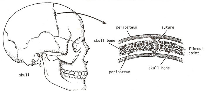
Craniosynostosis
Craniosynostosis is the premature fusion of one or more cranial sutures in infants. It can cause abnormal head shape (plagiocephaly, scaphocephaly) and increased intracranial pressure. Early surgical intervention is often required to allow normal brain growth.
B. Amphiarthrosis (Slightly Movable Joint)
An amphiarthrosis is a joint that permits limited mobility. These joints strike a balance between stability and flexibility—strong enough to maintain structural integrity, yet flexible enough to permit shock absorption.
Structural Basis: Cartilaginous (symphysis) or fibrous (syndesmosis).
Key Examples:
- Symphysis: Bones are joined by a fibrocartilage pad or disc. The pubic symphysis unites the hip bones. The intervertebral discs unite vertebrae; while each moves only slightly, they sum together for a large range of motion.
- Syndesmosis: Two parallel bones united by fibrous tissue (ligaments or interosseous membrane). Key example: the distal tibiofibular joint.
Symphysis Pubis Dysfunction (SPD) affects up to 1 in 5 pregnant women. Increased relaxin hormone causes excessive mobility, leading to pelvic pain and difficulty walking. Intervertebral disc herniation occurs when the nucleus pulposus protrudes through the annulus fibrosus, most commonly at L4-L5 and L5-S1.
C. Diarthrosis (Freely Movable Joint)
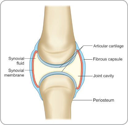
A diarthrosis is a freely mobile joint. All synovial joints are functionally classified as diarthroses. They are found predominantly in the appendicular skeleton.
Structural Basis: Feature articular cartilage, a joint cavity filled with synovial fluid, and a fibrous articular capsule.
Axes of Movement:
- Uniaxial: Movement in a single plane (one axis). Example: elbow joint (flexion/extension).
- Biaxial: Movement in two planes (two axes). Example: metacarpophalangeal (knuckle) joints.
- Multiaxial (Polyaxial/Triaxial): Movement in all three anatomical planes. Example: shoulder and hip joints.
Joint mobility is inversely related to joint strength. Synarthroses provide maximum protection but zero movement. Diarthroses allow extensive movement but are the most frequently injured due to their looser articulations.
3. Structural Classification
Based on how the bones are held together and the type of connective tissue binding the articulating surfaces.
| Structural Type | Connective Tissue | Mobility | Examples |
|---|---|---|---|
| Fibrous | Dense regular CT (collagen-rich) | Immovable / Slight | Skull sutures, syndesmoses |
| Cartilaginous | Hyaline or fibrocartilage | Immovable / Slight | Synchondroses, symphyses |
| Synovial | Articular capsule + Synovial fluid | Freely movable | Shoulder, Hip, Knee, Ankle |
A. Fibrous Joints
Adjacent bones are directly united by dense fibrous connective tissue. There is no joint cavity.
- Suture: Short fibers unite skull bones. With age, these may ossify completely (synostosis).
- Syndesmosis: Bones united by a ligament or membrane. More mobile than sutures. Example: distal tibiofibular joint.
- Gomphosis: A "peg-in-socket" joint where the tooth root is held by the periodontal ligament.
Fontanelles ("soft spots") on an infant's skull are membranous gaps between cranial bones at sutures. The anterior fontanelle closes at 12–18 months; the posterior at 2–3 months. Delayed closure may indicate hydrocephalus, hypothyroidism, or rickets.
B. Cartilaginous Joints
Bones are united by cartilage; there is no joint cavity.
- Synchondrosis: Bones joined by hyaline cartilage. Most are temporary (epiphyseal plate).
- Symphysis: Bones covered by hyaline cartilage but united by a fibrocartilage pad. These are amphiarthroses.
Synchondroses use hyaline cartilage and are typically immobile. Symphyses use fibrocartilage (containing thick collagen bundles) which provides greater resistance to pulling and bending forces and permits slight movement.
C. Synovial Joint Features
- Articular Capsule: A two-layered structure: (1) outer fibrous capsule of dense irregular CT for stability, and (2) inner synovial membrane that secretes fluid.
- Synovial Fluid: A viscous, egg-white-like fluid that lubricates the joint, nourishes avascular articular cartilage, and absorbs shock.
- Bursae & Tendon Sheaths: Fluid-filled sacs that reduce friction between adjacent structures.
Synovial Fluid Analysis
Normal fluid is clear, viscous, and pale yellow. Inflammatory arthritis (RA) produces cloudy fluid with elevated WBCs. Septic arthritis produces purulent fluid. Crystal analysis identifies needle-shaped urate (gout) or rhombic calcium pyrophosphate (pseudogout).
4. Synovial Joint Subtypes
Classified into six subtypes based on the shape of articulating surfaces and axes of movement.
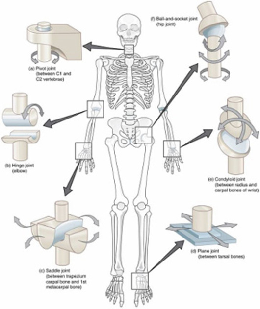
A. Plane (Gliding) Joint
- Movement: Sliding/gliding only; non-axial.
- Articular Surface: Flat or slightly curved surfaces of equal size.
- Examples: Intercarpal joints (wrist), facet (zygapophyseal) joints of the spine.
B. Hinge Joint
- Movement: Flexion/extension only; uniaxial.
- Articular Surface: Convex end of one bone fits into concave end of another.
- Examples: Elbow (humeroulnar), knee (tibiofemoral), interphalangeal joints.
C. Pivot Joint
- Movement: Rotation around one axis only.
- Articular Surface: Rounded portion of bone enclosed within a ring of bone and ligament.
- Examples: Atlantoaxial joint (C1-C2), proximal radioulnar joint (forearm rotation).
Screw-Home Mechanism
During the final 5–10° of knee extension, the tibia externally rotates (or femur internally rotates). This "locks" the knee in full extension, allowing quadriceps to relax while standing. To initiate flexion, the popliteus muscle must "unlock" the knee by internally rotating the tibia.
D. Condyloid (Ellipsoid) Joint
- Movement: Flexion/extension + Abduction/adduction; biaxial. Rotation is NOT permitted.
- Articular Surface: Oval convex surface articulates with an elliptical cavity.
- Examples: Radiocarpal (wrist) joint, knuckle (MCP) joints of fingers 2–5.
E. Saddle Joint
- Movement: Flexion/extension + Abduction/adduction; biaxial. More mobile than condyloid.
- Articular Surface: Both surfaces are saddle-shaped (concave in one direction, convex in the other).
- Examples: 1st carpometacarpal (CMC) joint of the thumb. This gives humans "opposable thumbs."
F. Ball & Socket Joint
- Movement: Multiaxial in all three planes (rotation and circumduction). Most mobile type.
- Articular Surface: Spherical head fits into a cup-like socket.
- Examples: Shoulder (glenohumeral) and Hip joints.
Bankart Lesion = tear of the anterior-inferior glenoid labrum, common in shoulder dislocation. Hill-Sachs Lesion = compression fracture of the posterolateral humeral head. These often coexist after anterior dislocation.
5. Master Summary Table
| Functional | Structural | Subtype | Axis | Key Examples |
|---|---|---|---|---|
| Synarthrosis | Fibrous | Suture | None | Skull sutures |
| Synarthrosis | Fibrous | Gomphosis | None | Tooth socket |
| Amphiarthrosis | Fibrous | Syndesmosis | Limited | Distal tibiofibular |
| Amphiarthrosis | Cartilaginous | Symphysis | Limited | Pubic symphysis, IV discs |
| Diarthrosis | Synovial | Hinge | Uniaxial | Elbow, Knee, Ankle |
| Diarthrosis | Synovial | Ball & Socket | Multiaxial | Shoulder, Hip |
6. Clinical Correlations
| Condition | Joint Type | Key Pathological Features |
|---|---|---|
| Osteoarthritis (OA) | Synovial | Cartilage degeneration, osteophytes, subchondral sclerosis. |
| Rheumatoid Arthritis | Synovial | Autoimmune synovitis, pannus formation, symmetrical. |
| Ankylosing Spondylitis | Cartilaginous | Fusion of SI joints and vertebral bodies (bamboo spine). |
| Meniscal Tear | Synovial | Traumatic or degenerative; joint line pain and effusion. |
7. Quick Reference Mnemonics
- Functional Classes (S-A-D): Synarthrosis (Immovable), Amphiarthrosis (Slight), Diarthrosis (Free).
- Structural Classes (F-C-S): Fibrous, Cartilaginous, Synovial.
- Synovial Subtypes: "PHS PCB" — Plane, Hinge, Saddle, Pivot, Condyloid, Ball & Socket.
