Functions of the Skeletal System
A Comprehensive study guide covering functions of the skeletal system.
1. Introduction to the Skeletal System
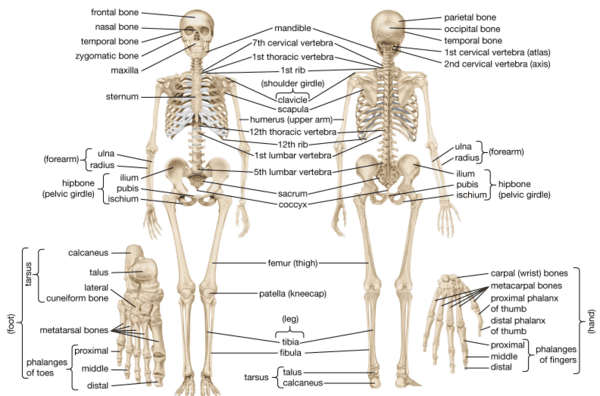
The skeletal system is one of the most important organ systems in the human body. It is composed of 206 bones in the adult human, along with cartilage, ligaments, and tendons that connect and support these bones. The word skeleton comes from the Greek word skeletos, meaning "dried up." However, bones are far from lifeless — they are living, dynamic tissues that perform many vital functions essential for life.
As a clinical medicine student, understanding the functions of the skeletal system is fundamental because bones do much more than simply hold the body together. They protect delicate organs, enable movement, produce blood cells, and regulate important minerals in the blood.
Key Terms to Remember:
- Osseous tissue: The hard, dense connective tissue that forms bones.
- Cartilage: A strong, flexible connective tissue found at joints and in other structures.
- Ligament: A band of tough tissue that connects one bone to another bone.
- Tendon: A cord of tissue that connects muscle to bone.
- Joint: The place where two or more bones meet.
- Hematopoiesis: The process of blood cell formation in the bone marrow.
2. Function 1: Support (Structural Framework)
The primary and most visible function of the skeletal system is to provide support for the entire body. Just as the steel beams of a building provide a scaffold that supports its weight, the bones and cartilage of your skeletal system compose the scaffold that supports the rest of your body.
Without the skeletal system, the human body would be a limp mass of organs, muscle, and skin. The bones provide a rigid framework that maintains the body's shape and holds the organs in their proper positions. The vertebral column (spine) supports the head and trunk, while the lower limbs support the weight of the entire body when standing.
Weakened Support
In conditions such as osteoporosis (bone thinning), the supporting function of bones is weakened. This is especially common in elderly patients and post-menopausal women. As a clinician, you will need to recognise the signs of weakened bone support, such as loss of height, stooped posture (kyphosis), and increased risk of fractures.
Examples of Support Function:
- The vertebral column supports the skull and trunk, allowing upright posture.
- The pelvic girdle supports the abdominal organs and connects the spine to the lower limbs.
- The rib cage provides a framework that supports the chest wall and assists in breathing.
- The long bones of the legs (femur, tibia) support the body's weight during standing and walking.
3. Function 2: Protection of Vital Organs
The human skeleton acts like a built-in suit of armour. Many bones are specifically shaped and positioned to protect delicate internal organs from physical injury and trauma. This protective function is critical for survival.
Key Protective Structures:
- The Skull (Cranium): The cranial bones form a hard, bony box that completely surrounds and protects the brain from non-traumatic injury. The skull also protects the eyes and the organs of hearing and balance.
- The Vertebral Column: The 33 vertebrae form a bony canal that houses and protects the spinal cord — the main pathway for nerve signals between the brain and the rest of the body. Damage to the spinal cord can result in paralysis.
- The Rib Cage: The 12 pairs of ribs, together with the sternum (breastbone) and thoracic vertebrae, form the thoracic cage. This structure protects the heart, lungs, and major blood vessels from external forces.
- The Pelvis: The pelvic bones protect the urinary bladder, reproductive organs, and parts of the large intestine.
Assessment of Protective Structures
When examining a patient who has suffered chest trauma, always consider the possibility of rib fractures and damage to the underlying heart and lungs. Similarly, head injuries require careful assessment because the skull may hide serious brain injury. Understanding which organs are protected by which bones will guide your physical examination and diagnosis.
4. Function 3: Facilitation of Movement
Bones do not move by themselves. They work together with muscles, joints, and the nervous system to produce movement. From a mechanical point of view, bones act as levers, joints serve as fulcrums (pivot points), and muscles provide the force needed to create motion.
When a muscle contracts, it pulls on the tendon attached to a bone. This pull causes the bone to move at the joint. Different types of joints allow different types of movement:
- Ball-and-socket joints (e.g., hip and shoulder) allow movement in all directions.
- Hinge joints (e.g., elbow and knee) allow bending and straightening only.
- Pivot joints (e.g., between the first and second cervical vertebrae) allow rotation.
- Gliding joints (e.g., between wrist bones) allow sliding movements.
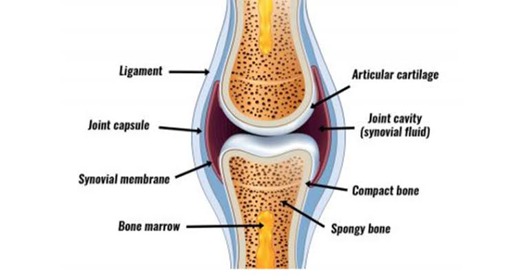
Joint diseases such as osteoarthritis (wear-and-tear of cartilage) and rheumatoid arthritis (autoimmune inflammation of joints) impair movement. Understanding normal joint anatomy helps you identify abnormal findings such as stiffness and reduced range of motion.
5. Function 4: Hematopoiesis (Blood Cell Formation)
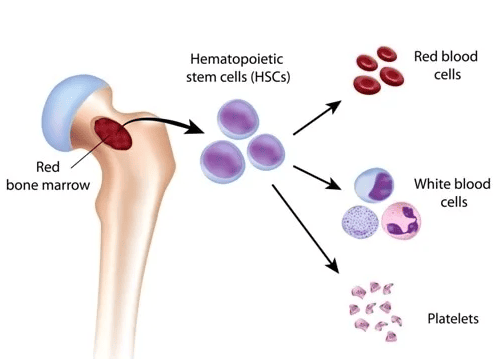
One of the most vital hidden functions of the skeletal system is hematopoiesis — the production of blood cells. This process occurs in the red bone marrow, a soft, jelly-like tissue found inside certain bones.
In the red bone marrow, special cells called hematopoietic stem cells divide and differentiate into all three types of blood cells:
- Red Blood Cells (Erythrocytes): These carry oxygen from the lungs to all body tissues and transport carbon dioxide back to the lungs. Without red blood cells, tissues cannot produce energy.
- White Blood Cells (Leukocytes): These are the soldiers of the immune system. They fight infections, destroy foreign invaders, and help the body recover from disease.
- Platelets (Thrombocytes): These small cell fragments are essential for blood clotting. When a blood vessel is injured, platelets gather at the site and form a plug to stop bleeding.
Diseases affecting the bone marrow, such as leukemia (cancer of blood-forming tissues) and aplastic anaemia (failure of the bone marrow to produce blood cells), are life-threatening conditions. Bone marrow biopsy is an important diagnostic tool used to investigate fatigue, recurrent infections, or excessive bleeding.
6. Function 5: Mineral Storage and Homeostasis
Bones serve as the body's main reservoir for important minerals, particularly calcium and phosphorus. These minerals are incorporated into the bone matrix and can be released back into the bloodstream when the body needs them.
Why is Calcium Important?
- Muscle contraction: Calcium is required for muscles to contract. Without adequate calcium, muscles cannot function.
- Nerve impulse transmission: Calcium helps nerve cells send signals to other cells, including muscle and gland cells.
- Blood clotting: Calcium is a necessary factor in the blood clotting cascade.
- Enzyme activity: Many enzymes require calcium to function properly.
How Does the Body Regulate Blood Calcium?
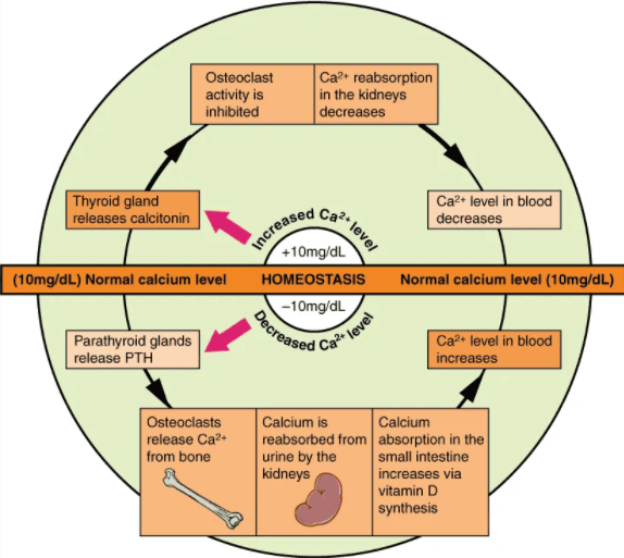
The body maintains blood calcium levels within a very narrow range (approximately 8.5–10.5 mg/dL) through a process called calcium homeostasis. Three main hormones control this process:
- Parathyroid Hormone (PTH): Released by the parathyroid glands when blood calcium is LOW. PTH stimulates osteoclasts (bone-breaking cells) to release calcium from bone into the blood.
- Calcitonin: Released by the thyroid gland when blood calcium is HIGH. Calcitonin stimulates osteoblasts (bone-building cells) to deposit calcium salts into bone, thereby lowering blood calcium levels.
- Vitamin D: Promotes calcium absorption from the diet in the small intestine.
Hypocalcaemia can cause muscle spasms and seizures. Hypercalcaemia can cause weakness and kidney stones. In Uganda, rickets (vitamin D deficiency causing soft bones) is still seen in children, particularly in urban areas with limited sunlight exposure. You should advise patients on proper nutrition and sunlight exposure.
7. Bone Structure: Compact and Spongy Bone
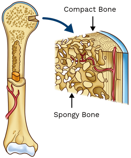
Bones are not solid throughout. They are composed of two main types of bone tissue: compact bone and spongy bone (also called cancellous or trabecular bone).
Compact Bone (Cortical Bone)
Compact bone is the hard, dense outer layer of bone. It makes up about 80% of the total bone mass in adults. It is made up of cylindrical units called osteons (Haversian systems), containing:
- Haversian canal: A central canal containing blood vessels and nerves.
- Lacunae: Small spaces containing mature bone cells called osteocytes.
- Canaliculi: Tiny channels allowing osteocytes to communicate and exchange nutrients.
- Lamellae: Concentric rings of calcified matrix.
Spongy Bone (Cancellous/Trabecular Bone)
Found at the ends of long bones (epiphyses) and inside flat bones. It has a porous, honeycomb-like structure. The trabeculae (thin bony struts) are arranged along lines of stress to provide strength with minimal weight.
8. Types of Bone Marrow
Bone marrow is the soft, spongy tissue found in the hollow interior of bones. There are two types:
Red Bone Marrow (Myeloid Tissue)
Responsible for hematopoiesis. In infants, most bones contain red marrow. In adults, it is found mainly in:
- The proximal ends of the femur and humerus.
- The sternum (breastbone).
- The ribs.
- The vertebrae (spinal bones).
- The pelvis.
- The skull bones.
Yellow Bone Marrow
Consists mainly of adipose tissue (fat cells). Found in the medullary cavity of long bones in adults. It serves as an energy source during times of starvation or prolonged exercise.
In situations of severe blood loss or chronic anaemia, yellow marrow can convert back to red marrow to increase blood cell production. This ensures the body can respond to increased demands for blood cells.
9. Clinical Relevance for Clinical Medicine Students
Common Skeletal Conditions in Ugandan Clinical Practice:
- Fractures: Common due to road traffic accidents, falls, and domestic violence. Clinicians must classify them as simple, compound, comminuted, or greenstick.
- Osteomyelitis: Bone infection, often Staphylococcus aureus. In Uganda, this may follow open fractures or spread from soft tissue infections.
- Sickle Cell Disease Bone Crisis: Common in Uganda; causes bone pain crises due to vaso-occlusion in the marrow.
- Osteoporosis: Leads to fragility fractures of the hip, wrist, and spine in post-menopausal women.
- Bone Tumours: Primary tumours like osteosarcoma or metastatic tumours from prostate, breast, or lung cancer.
10. Summary and Key Learning Points
| Function | Mechanism / Structure |
|---|---|
| Support | Rigid framework maintaining body shape; anchors muscles. |
| Protection | Protective enclosures (Skull, Vertebral Canal, Rib Cage). |
| Movement | Bones act as levers; joints act as fulcrums. |
| Hematopoiesis | Production of Erythrocytes, Leukocytes, and Platelets in red marrow. |
| Mineral Storage | Reservoir for Calcium and Phosphorus; hormonal regulation (PTH/Calcitonin). |
