The Anterior Abdominal Wall
A Comprehensive Anatomical Notes Covering Topographical Anatomy, Layered Structure, Musculature, Neurovascular Supply, Inguinal Region, and Clinical Applied Anatomy.
CHAPTER 1: Introduction and Functional Overview
1.1 Functional Significance of the Abdominal Wall
The abdominal wall is a dynamic, multilayered boundary enclosing the abdominal cavity. It represents one of the most functionally diverse anatomical regions of the human body, serving multiple critical roles that extend far beyond simple structural enclosure.
The abdominal wall functions to protect abdominal viscera, maintain or increase intra-abdominal pressure for essential physiological processes including defecation, micturition, parturition, and forced expiration, and facilitate movement and stabilization of the trunk.
The relative bony deficiency of the abdomen compared to the thorax and pelvis allows for remarkable flexibility of the trunk as well as distensibility to accommodate dynamic changes in the volume of abdominal contents. This flexibility is essential for respiration, digestion, and the accommodation of a growing fetus during pregnancy. However, this same flexibility creates potential weak points that are clinically significant, particularly in the inguinal region where hernias commonly occur.
Understanding the layered anatomy of the abdominal wall is fundamental to multiple medical and surgical disciplines, including general surgery, obstetrics and gynecology, plastic surgery, emergency medicine, and radiology. The abdominal wall is a frequent site of surgical incision, and knowledge of its layered structure, neurovascular supply, and potential anatomical variations is essential for safe and effective surgical practice.
The Abdominal Wall as a Functional Unit
The abdominal wall operates not as isolated layers but as an integrated functional unit. The three flat muscles (external oblique, internal oblique, transversus abdominis) with their orthogonal fiber orientations, the vertical rectus abdominis enclosed within the rectus sheath, and the median linea alba work in concert to provide structural integrity, mobility, and the ability to generate intra-abdominal pressure.
1.3 Overview of Abdominal Wall Structure
The anterior wall of the abdomen has nine layers. From outermost to innermost, they are:
- Skin
- Subcutaneous tissue (Superficial fascia): Divided into Camper's superficial fatty layer and Scarpa's deep membranous layer.
- Investigating (deep) fascia layering the muscles.
- External oblique muscle
- Internal oblique muscle
- Transversus abdominis muscle
- Transversalis fascia
- Extraperitoneal (preperitoneal) fat (adipose and areolar tissue).
- Parietal peritoneum.
The abdominal muscles are divided broadly into anterolateral and posterior components. The anterolateral muscles include five paired muscles: the external oblique, internal oblique, transversus abdominis, rectus abdominis, and pyramidalis. The posterior muscles include the psoas major and quadratus lumborum bilaterally.
CHAPTER 2: Topographical Anatomy and Surface Landmarks
2.1 Surface Bony Landmarks
Accurate description of abdominal anatomy and pathology requires familiarity with key surface bony landmarks that can be identified by palpation:
- Xiphoid process: The inferior-most portion of the sternum, located at the level of the T9 vertebra. It serves as the superior attachment of the linea alba and the rectus abdominis muscle.
- Costal margins (subcostal plane): The inferior borders of the rib cage, formed by the cartilages of the 7th through 10th ribs. The subcostal plane connects the lowest points of the costal margins and typically passes through the L3 vertebra.
- Iliac crest: The superior, curved border of the ilium. The highest point of the iliac crest lies at the level of the L4 vertebra and is a common landmark for lumbar puncture.
- Anterior superior iliac spine (ASIS): The anterior termination of the iliac crest. It is easily palpable and serves as the lateral attachment of the inguinal ligament.
- Pubic tubercle: A small, palpable projection on the superior aspect of the pubic bone. It serves as the medial attachment of the inguinal ligament and is an important landmark for identifying the superficial inguinal ring.
- Pubic symphysis: The midline cartilaginous joint connecting the left and right pubic bones. It serves as the inferior attachment of the rectus abdominis and linea alba.
2.2 Cutaneous Landmarks
- Umbilicus: The scar marking the site of attachment of the umbilical cord. It is typically located at the level of the L3-L4 intervertebral disc and the T10 dermatome. The umbilicus is an important surgical landmark and is the site of attachment of the falciform ligament.
- Linea alba: A visible midline tendinous strip extending from the xiphoid process to the pubic symphysis. It is formed by the interlacing fibers of the bilateral aponeuroses of the flat abdominal muscles and appears as a pale line on the skin.
- Linea semilunaris: The lateral border of the rectus abdominis muscle, visible as a curved line on the abdominal wall. It marks the transition between the rectus sheath and the aponeuroses of the flat muscles.
- Inguinal crease: The skin fold that overlies the inguinal ligament, separating the abdominal wall from the thigh.
2.3 Abdominal Quadrants
The abdomen can be divided into four quadrants using the transumbilical plane (horizontal line through the umbilicus) and the median plane (vertical line through the midline):
| Quadrant | Major Organs |
|---|---|
| Right Upper Quadrant (RUQ) | Liver, gallbladder, duodenum, head of pancreas, right kidney, right adrenal gland, hepatic flexure of colon |
| Left Upper Quadrant (LUQ) | Stomach, spleen, tail of pancreas, left kidney, left adrenal gland, splenic flexure of colon |
| Right Lower Quadrant (RLQ) | Cecum, appendix, ascending colon, right ovary/uterine tube (female), right ureter, right spermatic cord (male) |
| Left Lower Quadrant (LLQ) | Descending colon, sigmoid colon, left ovary/uterine tube (female), left ureter, left spermatic cord (male) |
2.4 Abdominal Regions (Nine-Region Scheme)
For more precise anatomical localization, the abdomen can be divided into nine regions using two vertical planes (midclavicular lines) and two horizontal planes (subcostal and intertubercular).
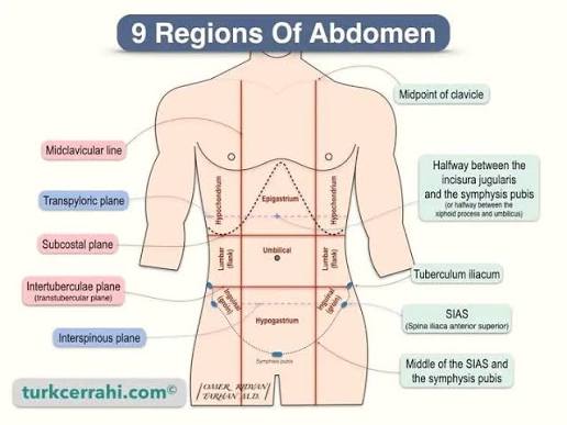
| Region | Location | Key Structures |
|---|---|---|
| Epigastric | Central upper region | Stomach (cardia and body), liver (left lobe), pancreas (body), duodenum (superior part) |
| Umbilical | Central middle region | Transverse colon, small intestine, aorta, inferior vena cava |
| Pubic (Hypogastric) | Central lower region | Urinary bladder (when full), uterus (female), rectum, sigmoid colon, small intestine |
| Right Hypochondriac | Upper right | Liver (right lobe), gallbladder, right kidney, hepatic flexure |
| Left Hypochondriac | Upper left | Spleen, stomach (fundus), left kidney, splenic flexure, tail of pancreas |
| Right Lumbar (Flank) | Middle right | Ascending colon, right kidney, duodenum (descending part) |
| Left Lumbar (Flank) | Middle left | Descending colon, left kidney, small intestine |
| Right Groin (Inguinal) | Lower right | Cecum, appendix, right ovary/uterine tube, right ureter |
| Left Groin (Inguinal) | Lower left | Sigmoid colon, left ovary/uterine tube, left ureter |
CHAPTER 3: Layers of the Anterolateral Abdominal Wall
3.1 Overview
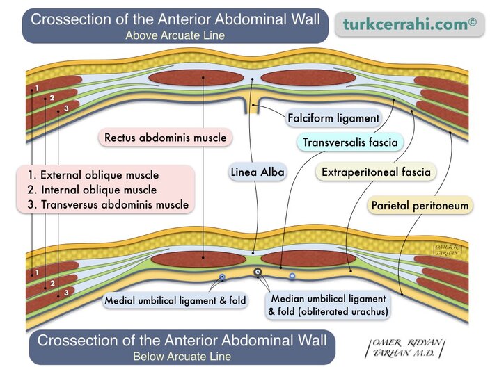
The anterolateral abdominal wall is composed of multiple layers arranged in a precise sequence from superficial to deep:
- Skin
- Superficial fatty layer (Camper's fascia)
- Deep membranous layer (Scarpa's fascia)
- Investigative (deep) fascia layering the muscles
- External oblique muscle
- Internal oblique muscle
- Transversus abdominis muscle
- Transversalis fascia
- Extraperitoneal fat
- Parietal peritoneum
3.2 Skin
The skin of the abdominal wall is relatively thin and mobile. It is innervated in a segmental pattern by the anterior rami of T7-L1 spinal nerves. The skin around the umbilicus is supplied by the T10 dermatome. The skin contains numerous sweat glands and sebaceous glands and is a common site for surgical incisions.
3.3 Superficial Fascia
The subcutaneous tissue of the anterior abdominal wall below the umbilicus separates into two distinct layers, a feature unique to this region:
3.3.1 Camper's Fascia (Superficial Fatty Layer)
Camper's fascia is the superficial fatty layer of the subcutaneous tissue. It is thick, areolar, and contains variable amounts of adipose tissue. It is continuous with the superficial fat of the rest of the body and is not structurally distinct from the subcutaneous fat of the thigh or chest. It allows for the mobility of the skin over the underlying muscular layer.
3.3.2 Scarpa's Fascia (Deep Membranous Layer)
Scarpa's fascia is the deeper membranous layer of the subcutaneous tissue. It is a thin, fibrous layer that is firmly attached to the linea alba in the midline and to the fascia lata of the thigh inferiorly. Scarpa's fascia is much thinner than Camper's fascia and contains less fat. It is continuous with:
- Colles' fascia in the perineum (inferiorly).
- The superficial fascia of the penis and scrotum (in males).
- The superficial fascia of the labia majora (in females).
- The fascia of the thigh (fascia lata) at the inguinal ligament.
Fluid Extravasation Pathways
The continuity of Scarpa's fascia has important clinical implications for fluid extravasation. Because Scarpa's fascia is firmly attached to the fascia lata below the inguinal ligament, urine or blood extravasating from the urethra (e.g., after urethral rupture) cannot pass into the thigh. Instead, it spreads into the perineum (as Colles' fascia is continuous with Scarpa's fascia) and may track superiorly into the abdominal wall. This creates a characteristic pattern of swelling and bruising that is diagnostically significant.
3.5 Transversalis Fascia
The transversalis fascia is a thin, membranous layer of connective tissue that lines the internal surface of the transversus abdominis muscle. It is continuous with the fascia lining the diaphragm superiorly, the fascia of the pelvic walls inferiorly, and the endothoracic fascia of the thorax. The transversalis fascia is an important surgical landmark, as it forms the posterior wall of the inguinal canal and the deep inguinal ring is an opening within it.
3.6 Extraperitoneal Fat
The extraperitoneal fat (also called preperitoneal fat, endoabdominal fat, or retroperitoneal fat) is a layer of loose connective tissue and adipose tissue that lies between the transversalis fascia and the parietal peritoneum. It is continuous with the retroperitoneum and provides cushioning and insulation for the abdominal organs. The amount of extraperitoneal fat varies significantly between individuals and increases with obesity.
3.7 Parietal Peritoneum
The parietal peritoneum is the innermost layer of the abdominal wall. It is a serous membrane that lines the abdominal cavity and is continuous with the visceral peritoneum that covers the abdominal organs. The peritoneal cavity is the potential space between the parietal and visceral peritoneum, normally containing only a small amount of serous fluid (approximately 50 mL) that lubricates the surfaces and reduces friction.
The parietal peritoneum is sensitive to pain, temperature, and pressure, and is innervated by the same somatic nerves that supply the overlying abdominal wall. In contrast, the visceral peritoneum is insensitive to most stimuli but is sensitive to stretch and chemical irritation.
CHAPTER 4: Musculature and Aponeuroses
4.1 Overview
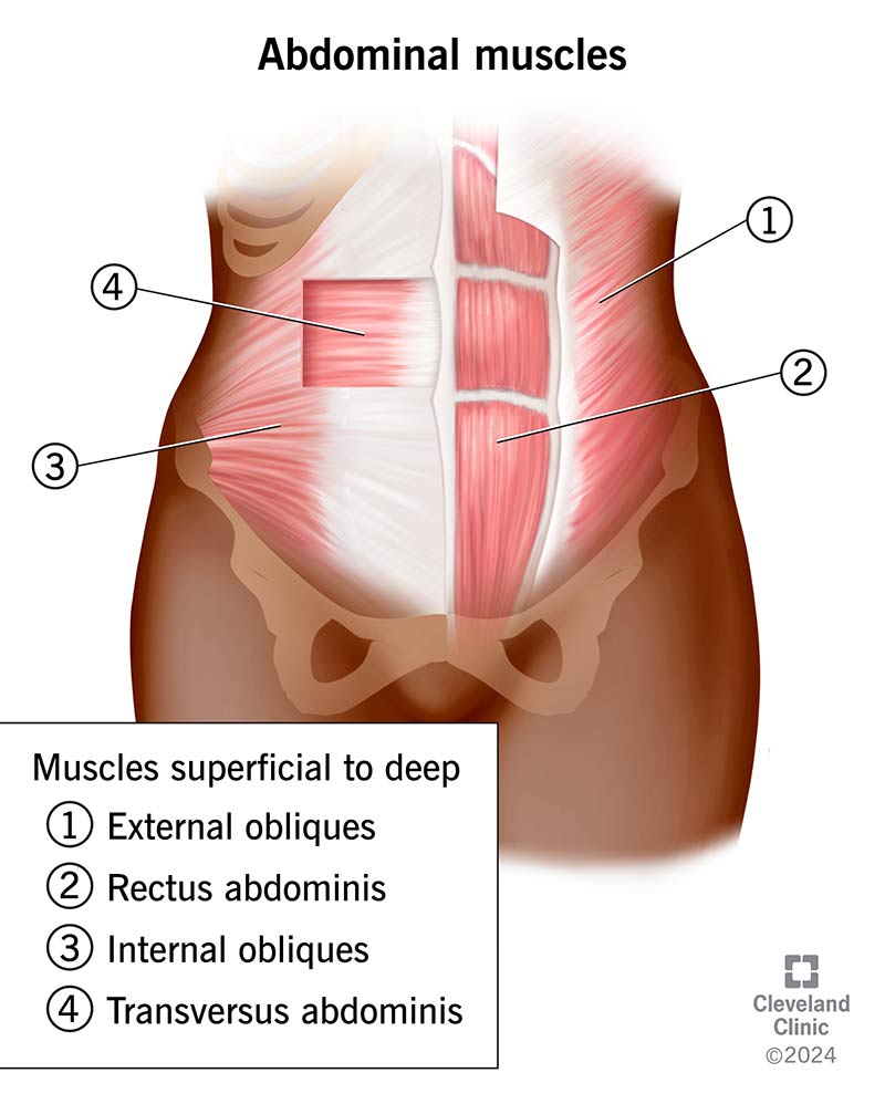
The muscles of the anterolateral abdominal wall are arranged in three layers of flat muscles laterally and two vertical muscles anteromedially. The flat muscles are characterized by their distinctive fiber orientations, which are arranged orthogonally to provide strength in multiple directions while allowing flexibility.
4.2 The Flat Muscles
4.2.1 External Oblique Muscle
The external oblique is the most superficial of the three flat muscles. Its fibers arise from the external surfaces of the fifth through twelfth ribs and run inferomedially in a characteristic "hands-in-pockets" orientation. As the muscle approaches the midclavicular line, its fibers become aponeurotic and form a broad, flat tendon that crosses the rectus abdominis to reach the linea alba in the midline.
- Origin: External surfaces of ribs 5-12.
- Insertion: Linea alba, pubic tubercle, anterior half of iliac crest.
- Innervation: Anterior rami of T7-T12 (thoracoabdominal nerves).
- Action: Bilateral contraction compresses abdomen and flexes vertebral column; unilateral contraction causes ipsilateral lateral flexion and contralateral rotation of the trunk.
The inferior border of the external oblique aponeurosis is folded posteriorly on itself to form the inguinal ligament (Poupart's ligament), which extends from the ASIS to the pubic tubercle. The external oblique aponeurosis also contains the superficial (external) inguinal ring.
4.2.2 Internal Oblique Muscle
The internal oblique lies immediately deep to the external oblique. Its fibers run superomedially, perpendicular to the external oblique, creating an orthogonal arrangement. The internal oblique originates from the lumbar fascia, iliac crest, and lateral inguinal ligament.
- Origin: Lumbar fascia, anterior two-thirds of iliac crest, lateral half of inguinal ligament.
- Insertion: Inferior borders of ribs 10-12, linea alba, pubic crest (via conjoint tendon).
- Innervation: Anterior rami of T7-T12, L1 (iliohypogastric and ilioinguinal nerves).
- Action: Bilateral contraction compresses abdomen and flexes vertebral column; unilateral contraction causes ipsilateral lateral flexion and ipsilateral rotation of the trunk.
The inferior fibers of the internal oblique arch over the spermatic cord (in males) or round ligament (in females) and, together with the transversus abdominis, form the conjoint tendon (falx inguinalis), which inserts onto the pubic crest and pectineal line. The conjoint tendon reinforces the medial part of the posterior wall of the inguinal canal.
4.2.3 Transversus Abdominis Muscle
The transversus abdominis is the deepest of the three flat muscles. Its fibers run transversely (horizontally), completing the orthogonal arrangement. The transversus abdominis is the primary muscle responsible for compressing the abdominal contents and increasing intra-abdominal pressure.
- Origin: Internal surfaces of ribs 7-12 (costal cartilages), thoracolumbar fascia, iliac crest, lateral third of inguinal ligament.
- Insertion: Linea alba, pubic crest (via conjoint tendon).
- Innervation: Anterior rami of T7-T12, L1.
- Action: Compresses abdominal contents; increases intra-abdominal pressure; stabilizes the lumbar spine.
The transversus abdominis is particularly important for core stability and is often targeted in physical therapy for low back pain. Its contraction creates a "corset" effect around the abdomen, providing support to the lumbar spine and pelvis.
4.3 The Vertical Muscles
4.3.1 Rectus Abdominis
The rectus abdominis is a long, strap-like muscle that runs vertically on either side of the midline, enclosed within the rectus sheath. It is a powerful flexor of the vertebral column and is responsible for the "six-pack" appearance in well-conditioned individuals.
- Origin: Pubic symphysis and pubic crest.
- Insertion: Xiphoid process and costal cartilages of ribs 5-7.
- Innervation: Anterior rami of T7-T12.
- Action: Flexes vertebral column (bilateral); compresses abdomen; stabilizes pelvis during walking.
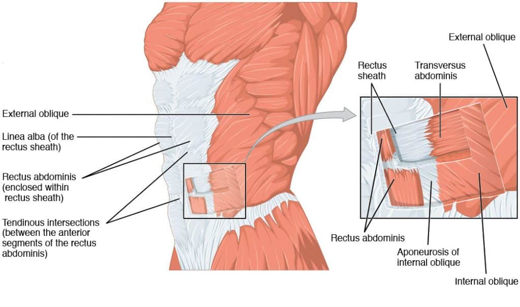
The rectus abdominis is divided by three tendinous intersections (inscriptions) into four discrete muscle bellies. These intersections are fibrous bands that firmly attach the anterior surface of the rectus abdominis to the anterior rectus sheath. They are located at the level of the umbilicus, the xiphoid process, and midway between these two points. They prevent the muscle from bulging excessively during contraction.
4.3.2 Pyramidalis
The pyramidalis is a small, triangular muscle that lies anterior to the inferior part of the rectus abdominis within the rectus sheath. It is absent in approximately 20% of individuals and is considered a vestigial muscle. It originates from the body of the pubis and inserts onto the linea alba midway between the pubis and umbilicus. It is innervated by the subcostal nerve (T12).
4.4 The Rectus Sheath
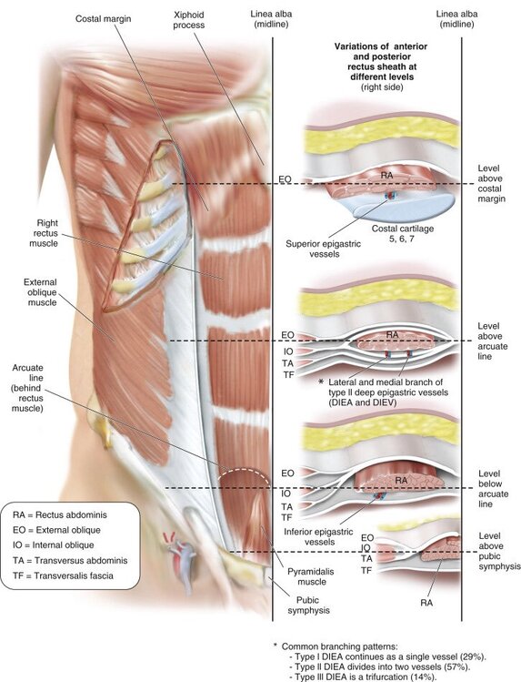
The rectus sheath is a tough, fibrous compartment that encloses the rectus abdominis and pyramidalis muscles. It is formed by the aponeuroses of the three flat muscles. The composition of the rectus sheath changes at two important levels: the costal margin and the arcuate line.
4.4.1 Above the Costal Margin
Superior to the costal margin, the anterior wall of the rectus sheath is formed by the aponeurosis of the external oblique muscle only. There is no posterior wall at this level; the rectus abdominis lies directly on the costal cartilages and the transversalis fascia.
4.4.2 Between the Costal Margin and the Arcuate Line
In the upper three-quarters of the rectus abdominis, the rectus sheath has both anterior and posterior walls:
- Anterior wall: Formed by the aponeuroses of the external oblique and the anterior lamella of the internal oblique.
- Posterior wall: Formed by the aponeuroses of the posterior lamella of the internal oblique and the transversus abdominis.
At the lateral margin of the rectus sheath, the aponeuroses of all three flat muscles meet to form the linea semilunaris.
4.4.3 Below the Arcuate Line
Inferior to the arcuate line (also called the semicircular line of Douglas), the composition of the rectus sheath changes dramatically. The arcuate line is located approximately one-third of the distance from the pubic crest to the umbilicus. Below this line:
- Anterior wall: Formed by the aponeuroses of all three flat muscles (external oblique, internal oblique, and transversus abdominis).
- Posterior wall: Absent. The rectus abdominis lies directly on the transversalis fascia and extraperitoneal fat.
This change in composition means that the rectus abdominis is not enclosed posteriorly below the arcuate line, making this region a potential site of weakness. The inferior epigastric vessels enter the rectus sheath posteriorly and ascend within the sheath.
Arcuate Line in Surgery
The arcuate line is an important surgical landmark. During laparoscopic surgery, the absence of the posterior rectus sheath below the arcuate line means that the peritoneum is separated from the rectus abdominis only by the thin transversalis fascia and extraperitoneal fat. This makes the infraumbilical region more susceptible to injury during trocar insertion. Additionally, the change in fascial architecture at the arcuate line is a common site for Spigelian hernias.
4.5 Linea Alba
The linea alba is a median fibrous band that extends from the xiphoid process to the pubic symphysis. It is formed by the interlacing fibers of the bilateral aponeuroses of all three flat muscles. The linea alba is relatively avascular and is a common site for midline surgical incisions. It is wider superiorly and narrower inferiorly. It is an important site of structural weakness, and defects can lead to epigastric hernias.
Summary of Muscle Layer Characteristics
| Muscle | Layer | Fiber Direction | Origin | Insertion | Innervation | Primary Action |
|---|---|---|---|---|---|---|
| External oblique | Superficial flat | Inferomedial ("hands-in-pockets") | Ribs 5-12 (external surfaces) | Linea alba, pubic tubercle, iliac crest | T7-T12 | Compression, flexion, contralateral rotation |
| Internal oblique | Middle flat | Superomedial (perpendicular to EO) | Lumbar fascia, iliac crest, inguinal ligament | Ribs 10-12, linea alba, pubic crest | T7-T12, L1 | Compression, flexion, ipsilateral rotation |
| Transversus abdominis | Deep flat | Transverse (horizontal) | Ribs 7-12, thoracolumbar fascia, iliac crest, inguinal ligament | Linea alba, pubic crest (via conjoint tendon) | T7-T12, L1 | Compression, increases intra-abdominal pressure |
| Rectus abdominis | Vertical (anterior) | Vertical | Pubic symphysis and crest | Xiphoid process, costal cartilages 5-7 | T7-T12 | Flexion of vertebral column, compression |
| Pyramidalis | Vertical (inferior) | Vertical | Body of pubis | Linea alba | T12 | Tenses linea alba |
CHAPTER 5: Neurovascular Supply and Lymphatics
5.1 Nerve Supply
The anterolateral abdominal wall is innervated in a segmental pattern by the anterior rami of spinal nerves T7-L1. This segmental innervation is a direct consequence of the embryological development of the body wall from somites. This pattern is clinically significant for understanding dermatomes and regional anesthesia.
5.1.1 Thoracoabdominal Nerves (T7-T12)
These are the anterior rami of the 7th-11th intercostal nerves and the subcostal nerve (T12). They exit the intercostal spaces and enter the abdominal wall, where they travel within the neurovascular plane (between transversus abdominis and internal oblique) before piercing the posterior rectus sheath. Each gives off:
- Lateral cutaneous branch: Supplies the skin of the lateral abdominal wall.
- Anterior cutaneous branch: Supplies the skin of the anterior abdominal wall after piercing the rectus abdominis and anterior sheath.
- Muscular branches: Supply the flat muscles and the rectus abdominis.
The segmental distribution follows a characteristic inferomedial slope:
- T7: Innervates skin just inferior to the xiphoid process.
- T10: Innervates skin at the level of the umbilicus.
- T12: Innervates suprapubic skin.
5.1.2 Iliohypogastric and Ilioinguinal Nerves (L1)
- Iliohypogastric nerve: Arises from L1 (with T12 contribution). It emerges from the lateral border of the psoas major, travels deep to the quadratus lumborum, and pierces the transversus abdominis superior to the iliac crest. It supplies the skin over the buttocks and ilium and the skin over the inferior rectus abdominis.
- Ilioinguinal nerve: Also arises from L1 and follows a similar course. It travels in the neurovascular plane until it reaches the superficial inguinal ring. It then enters the inguinal canal (though not through the deep ring) and exits via the superficial ring to supply the skin of the inguinal region, medial thigh, mons pubis, and external genitalia.
Ilioinguinal Nerve Injury
The ilioinguinal nerve is the nerve most at risk of damage during inguinal hernia repair. Injury can cause chronic postoperative pain, sensory loss in the groin and genitalia, and impaired motor function of the inferior abdominal muscles. Careful identification and preservation are essential.
5.2 Arterial Supply
The arterial supply ensured by three anastomosing networks:
5.2.1 Vertical Network (Superior and Inferior Epigastric Arteries)
- Superior epigastric artery: Terminal branch of the internal thoracic (mammary) artery. It descends posterior to the rectus abdominis within the sheath and anastomoses with the inferior epigastric artery at the level of the umbilicus.
- Inferior epigastric artery: Arises from the external iliac artery just proximal to the inguinal ligament. It ascends obliquely along the posterior surface of the rectus abdominis, piercing the transversalis fascia to enter the rectus sheath.
The anastomosis between the superior and inferior epigastric arteries forms an important collateral pathway between the subclavian artery (via the internal thoracic) and the external iliac artery. This is clinically significant in aortic coarctation.
5.2.2 Superficial Network
- Superficial epigastric artery: A branch of the femoral artery ascending in the superficial fascia.
- Superficial circumflex iliac artery: A branch of the femoral artery supplying skin over the iliac region.
- Superficial external pudendal artery: Supplies skin of the external genitalia and lower abdominal wall.
5.2.3 Lateral Network
- Musculophrenic artery: A branch of the internal thoracic artery.
- Posterior intercostal arteries (10th and 11th): Continue from the intercostal spaces.
- Subcostal artery: Runs below the 12th rib.
- Lumbar arteries: Four pairs arising from the posterior aspect of the aorta.
5.3 Venous Drainage
5.3.1 Superficial Venous Drainage
A network of superficial veins radiates out from the umbilicus. They drain:
- Superiorly: Into the axillary vein via the lateral thoracic vein.
- Inferiorly: Into the femoral vein via the superficial epigastric and great saphenous veins.
5.3.2 Deep Venous Drainage
Follow the arteries of the same name: Superior epigastric vein (to internal thoracic), Inferior epigastric vein (to external iliac), and Posterior intercostal/subcostal/lumbar veins (to azygos system and IVC).
5.3.3 Portosystemic Anastomosis
The thoracoepigastric vein provides a collateral pathway between the superficial systemic veins (axillary and femoral) and the portal venous system via the paraumbilical veins.
Caput Medusae
Caput medusae is the clinical sign of engorged paraumbilical veins that radiate from the umbilicus. It results from portal hypertension, most commonly due to liver cirrhosis. The paraumbilical veins (remnants of the umbilical vein) connect the portal system to the systemic superficial veins. When portal pressure rises, blood flows through these alternative pathways, causing characteristic dilated veins radiating like the snakes on the head of Medusa.
5.4 Lymphatic Drainage
The lymphatic drainage follows a characteristic pattern divided by an important watershed line at the umbilicus:
- Above the Umbilicus: Lymph drains superiorly to the axillary lymph nodes (specifically the pectoral/anterior group).
- Below the Umbilicus: Lymph drains inferiorly to the superficial inguinal lymph nodes (specifically the horizontal group).
Deep lymphatic vessels follow the arteries and drain to the external iliac, common iliac, and lumbar lymph nodes.
CHAPTER 6: The Inguinal Region (Groin)
6.1 Overview
The inguinal region is one of the most clinically significant areas. It is the site of the inguinal canal, a natural passageway representing a potential weak point and the most common site of herniation.
6.2 Inguinal Ligament
The inguinal ligament (Poupart's ligament) is not a true ligament but a structural modification formed by the folded inferior border of the external oblique aponeurosis. It extends from the ASIS to the pubic tubercle. Related structures include:
- Lacunar ligament (Gimbernat's): A crescentic extension attaching the medial end of the inguinal ligament to the pectineal line.
- Pectineal ligament (Cooper's): A strong fibrous band along the pectineal line of the superior pubic ramus.
- Reflected inguinal ligament: Fibers of the external oblique aponeurosis that reflect superomedially from the pubic tubercle.
6.3 Inguinal Canal Boundaries
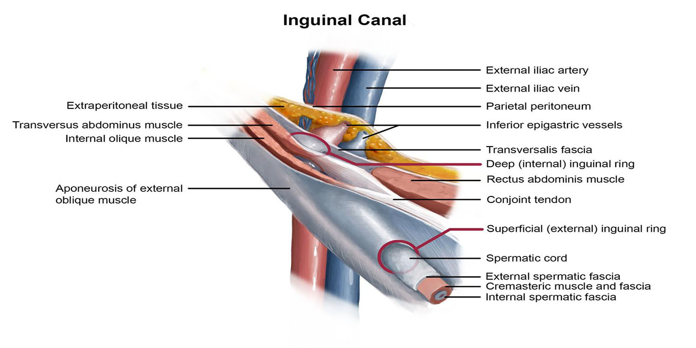
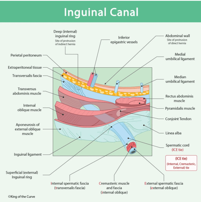
The inguinal canal is an oblique passage approximately 4 cm in length running inferomedially through the lower wall, parallel and superior to the inguinal ligament.
| Wall | Structure | Clinical Note |
|---|---|---|
| Anterior | External oblique aponeurosis (reinforced laterally by internal oblique) | Relatively strong; rarely the site of direct herniation |
| Posterior | Transversalis fascia (reinforced medially by conjoint tendon/falx inguinalis) | Weakest wall; site of direct inguinal hernias |
| Roof | Arched fibers of internal oblique and transversus abdominis muscles | Formed by muscle contraction; weakness contributes to hernia |
| Floor | Inguinal ligament and lacunar ligament | Formed by the rolled-under edge of the EO aponeurosis |
MALT (Walls of the Canal)
- Muscles form the roof.
- Aponeuroses form the anterior wall.
- Ligaments form the floor.
- Tendon/transversalis fascia forms the posterior wall.
6.4 Canal Openings
6.4.1 Deep (Internal) Inguinal Ring
An opening in the transversalis fascia located 1.5 cm superior to the midpoint of the inguinal ligament. It is lateral to the inferior epigastric vessels. It represents the internal entrance and is the site of protrusion for indirect inguinal hernias.
6.4.2 Superficial (External) Inguinal Ring
A triangular opening in the external oblique aponeurosis superolateral to the pubic tubercle. Its base is the pubic crest. It is larger in males than in females.
6.5 Canal Contents
6.5.1 In Males: The Spermatic Cord
A collection of structures passing to and from the testis. Remembered by FANO x 3:
- Fasciae (3 layers):
— External spermatic fascia: Derived from external oblique aponeurosis.
— Cremasteric muscle and fascia: Derived from internal oblique muscle/fascia.
— Internal spermatic fascia: Derived from transversalis fascia. - Arteries (3): Testicular artery (Aorta), Cremasteric artery (Inf. epigastric), Artery of the ductus deferens (Vesical artery).
- Nerves (3): Genital branch of genitofemoral (motor to cremaster), Ilioinguinal nerve (sensory), Sympathetic fibers (autonomic).
- Other structures: Vas deferens, Pampiniform venous plexus (thermoregulation), and lymphatic vessels.
6.5.2 In Females
The canal is narrower and contains the round ligament of the uterus, which extends from the uterine horn to attach to the labia majora.
6.5.3 In Both Sexes
The ilioinguinal nerve passes through part of the canal and exits via the superficial ring. Note: it does not pass through the deep ring; it pierces the internal oblique muscle and enters the canal directly.
CHAPTER 7: Clinical and Applied Anatomy
7.1 Abdominal Hernias
A hernia is the protrusion of an organ or tissue through an abnormal opening. Inguinal hernias account for approximately 75% of all abdominal wall hernias.
7.1.1 Indirect Inguinal Hernia
Herniation through the deep inguinal ring, lateral to the inferior epigastric vessels. It travels along the canal within the spermatic cord. It is congenital in origin (failure of processus vaginalis closure). It has a higher risk of strangulation due to the narrow neck at the deep ring.
7.1.2 Direct Inguinal Hernia
Abdominal contents push directly through a weakness in the posterior wall of the canal, medial to the inferior epigastric vessels, within Hesselbach's triangle. It does not pass through the deep ring. Direct hernias are acquired (weakened muscles in older males) and have a lower risk of strangulation.
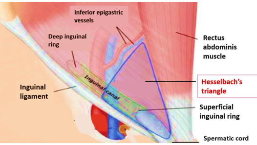
7.1.3 Hesselbach's (Inguinal) Triangle Boundaries:
- Lateral border: Inferior epigastric vessels.
- Medial border: Lateral border of the rectus abdominis muscle.
- Inferior border: Inguinal ligament.
Summary of Hernia Types
| Hernia Type | Location | Relative to Epigastric Vessels | Origin | Strangulation Risk |
|---|---|---|---|---|
| Indirect inguinal | Deep ring, inguinal canal | Lateral | Congenital | High (narrow neck) |
| Direct inguinal | Hesselbach's triangle | Medial | Acquired | Lower (wide neck) |
| Femoral | Femoral canal | Inferior to inguinal ligament | Acquired | Highest |
| Umbilical | Umbilical ring | Midline | Congenital/Acquired | Moderate |
7.3 Surgical Incisions
The choice is determined by the target, exposure needed, and desire to minimize damage.
- Midline Incision: Through the linea alba (relatively avascular). Provides rapid access with minimal blood loss. DIVIDES the linea alba, a site of weakness.
- Paramedian Incision: 2-3 cm lateral to midline. divides anterior sheath, retracts rectus laterally, then divides posterior sheath. Preserves segmental nerve supply.
- Kocher (Subcostal) Incision: Parallel to costal margin. Used for gallbladder (right) or splenic (left) surgery. involves muscle cutting.
- McBurney Incision (Gridiron): Oblique incision in RLQ over McBurney's point. Muscle-splitting approach (fibers split, not cut) preserves nerve supply. Used for appendectomy.
- Pfannenstiel Incision: Transverse incision 5 cm above pubic symphysis. Used in OB/GYN (C-section). Excellent cosmetic results with limited upper exposure.
Principles of Abdominal Incision
An ideal incision should provide adequate exposure, minimize damage to muscles/nerves/vessels, be extensible, and resist dehiscence. Muscle-splitting incisions (e.g., McBurney) are generally preferred over cutting incisions because they preserve structural integrity.
7.4 Additional Clinical Considerations
7.4.1 Rectus Sheath Hematoma
A collection of blood within the sheath, resulting from rupture of the superior or inferior epigastric vessels. Can occur after trauma, vigorous exercise ("coughing fit" hematoma), or in patients on anticoagulants. It presents as a painful, palpable abdominal mass that does not cross the midline because the linea alba separates the two sheaths.
7.4.2 Abdominal Wall Abscess
Abscesses superficial to Scarpa's fascia can spread freely, while those deep to Scarpa's fascia are contained by its attachments to the linea alba and fascia lata.
7.4.3 Diastasis Recti
A condition in which the linea alba widens, causing separation of the two rectus abdominis muscles. Common during pregnancy or in obese individuals. Unlike a hernia, it does not involve a fascial defect and the peritoneum remains intact. Treatment is usually conservative (physical therapy).
CHAPTER 8: Summary and Review - Key Anatomical Structures
| Structure | Location/Description | Clinical Relevance |
|---|---|---|
| Camper's fascia | Superficial fatty layer of subcutaneous tissue | Allows skin mobility; site of superficial infections |
| Scarpa's fascia | Deep membranous layer; continuous with Colles' | Dictates fluid extravasation pathways |
| External oblique | Superficial flat muscle; inferomedial fibers | Forms inguinal ligament; contains superficial ring |
| Internal oblique | Middle flat muscle; superomedial fibers | Forms conjoint tendon; reinforces inguinal canal |
| Transversus abdominis | Deep flat muscle; transverse fibers | Primary compressor; forms deep inguinal ring |
| Rectus abdominis | Vertical muscle in rectus sheath | Trunk flexor; site of rectus sheath hematoma |
| Arcuate line | Level where posterior rectus sheath ends | Surgical landmark; site of Spigelian hernias |
| Inguinal ligament | Folded edge of EO aponeurosis | Floor of inguinal canal; landmark for hernias |
| Deep inguinal ring | Opening in transversalis fascia; lateral to epigastric vessels | Site of indirect hernia protrusion |
| Hesselbach's triangle | Bounded by rectus, epigastric vessels, inguinal ligament | Site of direct hernia protrusion |
