The Cell: An Introduction to the Basic Unit of Life
History of Cells & the Cell Theory
- In 1665, Robert Hooke used a microscope to examine a thin slice of cork and saw what looked like small boxes. He is responsible for naming them "cells" because they resembled the rooms monks lived in.
- In 1673, Antonie van Leeuwenhoek was the first to view living organisms, using a simple microscope to observe pond water and scrapings from his teeth.
- In 1838, botanist Matthias Schleiden concluded that all plants were made of cells.
- In 1839, zoologist Theodore Schwann concluded that all animals were made of cells. Together, Schleiden and Schwann are co-founders of the cell theory.
- In 1855, medical doctor Rudolph Virchow observed cells dividing and reasoned that all cells come from other pre-existing cells.

Cell Theory
Alright, let's dive into the microscopic world that makes up our bodies, starting with the fundamental concept of the Cell Theory. This theory is one of the cornerstones of biology and medicine, giving us the basic understanding of life. It essentially has three main parts, like three key rules about cells:
- All living organisms are made up of one or more cells. This means whether it's a tiny bacterium, a plant, or a human being, the basic unit of structure is the cell. Some organisms are single-celled (like amoeba), while complex organisms like us are made of trillions of cells working together.
- The cell is the basic unit of structure and organization in organisms. This means that the cell is the smallest level at which life functions can be carried out. Just like a single brick is the basic unit of a wall, a cell is the basic unit of a tissue, an organ, and ultimately, an organism. All the complex processes of life happen within cells.
- Cells arise from pre-existing cells. This means cells don't just appear out of nowhere. New cells are produced through cell division (like mitosis or meiosis) from cells that already exist. This explains growth, repair, and reproduction in living things.
Understanding the cell theory is crucial because it tells us that to understand how the body works in health and disease, we must understand how cells work, what they are made of, and how they interact. Diseases often occur when cells are damaged, malfunction, or grow uncontrollably.
The Cell Theory
- All living things are made of cells.
- Cells are the basic unit of structure and function in an organism (the basic unit of life).
- Cells come from the reproduction of existing cells (cell division).
Basic Characteristics of Cells
A cell is the smallest functional unit of a living organism, capable of performing all life functions. A cell can:
- Eat, grow, and move.
- Perform necessary maintenance, recycle parts, and dispose of wastes.
- Adapt to changes in its environment.
- Replicate itself.
Discoveries Since the Cell Theory
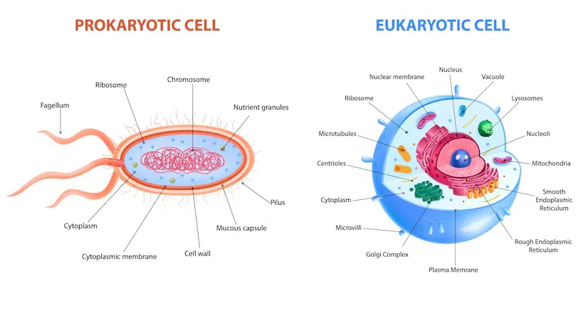
Further research has allowed us to classify cells into two major categories based on their complexity and internal structure: Prokaryotes and Eukaryotes.
Prokaryotes – The First Cells
Cells that lack a nucleus and other membrane-bound organelles.
- Examples:Bacteria and Archaea.
- Complexity:Considered the simplest type of cell.
- Genetic Info:A single, circular chromosome in a nucleoid region.
- Structure:Enclosed by a cell membrane and protected by a cell wall.
- Organelles:Contain ribosomes (non-membrane bound) for protein synthesis.
Eukaryotes
Cells that have a true nucleus and various membrane-bound organelles.
- Examples:Protists, Fungi, Plants, and Animals.
- Complexity:Significantly more complex than prokaryotes.
- Features:Possess a membrane-bound nucleus, organelles, and a cytoskeleton.
- Structures:All have a Nucleus, Cell Membrane, and Cytoplasm.
- Main Types:Plant Cells and Animal Cells.
Cell or Plasma Membrane and Phospholipids
When you go swimming, have you ever wondered why your cells don't fill up with water or why substances don't leak out? The reason is a critical structure called the cell membrane. It protects the cell from the outside environment and determines what can enter and leave—a property we call semi-permeability.
Cell Membrane Structure
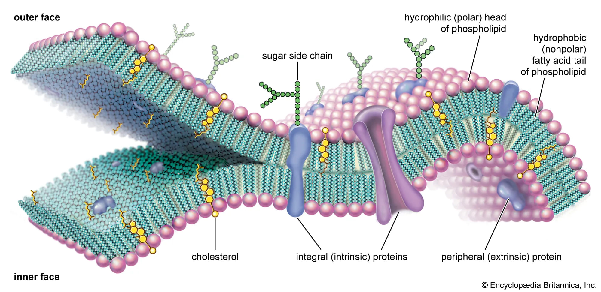
Viewed with an electron microscope, the membrane appears as a double-layered structure about 7.5-10 nanometers thick. It's primarily composed of proteins and special fat-like molecules called phospholipids, which form a structure known as the Lipid Bilayer.
The Phospholipid Molecule
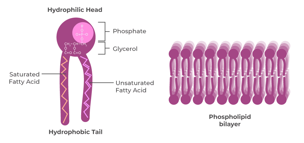
Each phospholipid has two distinct parts:
- Phosphate Head: Hydrophilic (water-loving) and polar. Faces the watery environments inside and outside the cell.
- Fatty Acid Tails: Two hydrophobic (water-fearing), non-polar chains that face inward, forming the core of the membrane.
 Phospholipid Diagram
Chemical Compositions
- Proteins: Embedded in or attached to the bilayer, they act as doors, channels, and communicators for the cell. Integral proteins span the entire membrane, while peripheral proteins are found on the inner or outer surface.
- Carbohydrates: Found only on the outer surface, attached to proteins (glycoproteins) or lipids (glycolipids). They act as the cell's "identification tags."
- Other Lipids: Molecules like
Cholesterolare also present, helping to maintain the membrane's fluidity and stability.
Functions of the Cell Membrane
The cell membrane performs several vital jobs:
Transport Mechanisms: How Cells Move Things
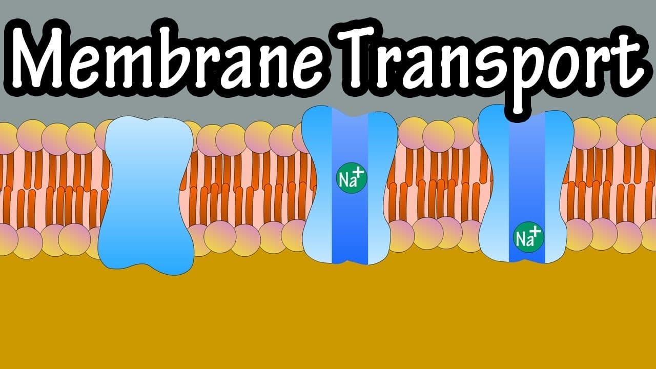
A cell constantly needs to bring in supplies and get rid of waste. The cell membrane acts as a gatekeeper, using different methods to move substances across. How easily a substance crosses often depends on whether it "likes" fats (lipids) or water.
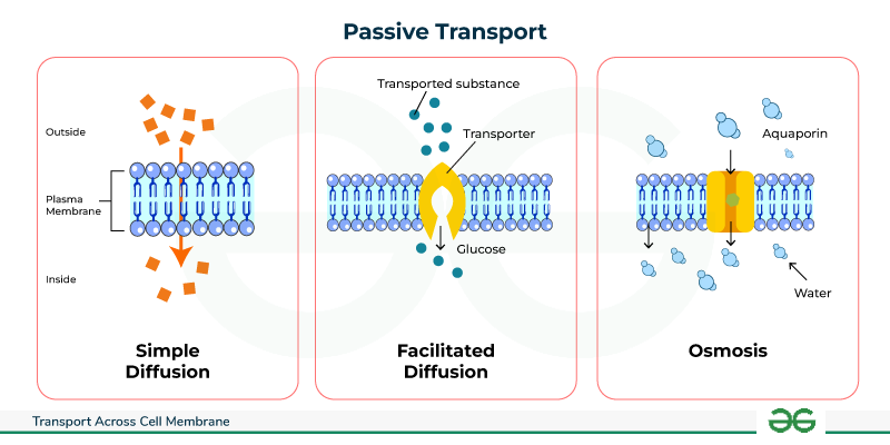
1. Passive Transport: No Energy Needed!
Substances move from an area of high concentration to an area of low concentration (down the concentration gradient).
A. Simple Diffusion
The simplest way small, fat-soluble molecules (like O₂, CO₂) sneak right through the lipid bilayer. It's a slow process driven by the concentration gradient.
B. Facilitated Diffusion
Substances that can't easily cross (like glucose) get a "ride" using special carrier proteins. This is much faster than simple diffusion but still requires no energy.
C. Osmosis
This is specifically the movement of water across a semi-permeable membrane, from an area of more water to an area of less water, to even out solute concentrations.
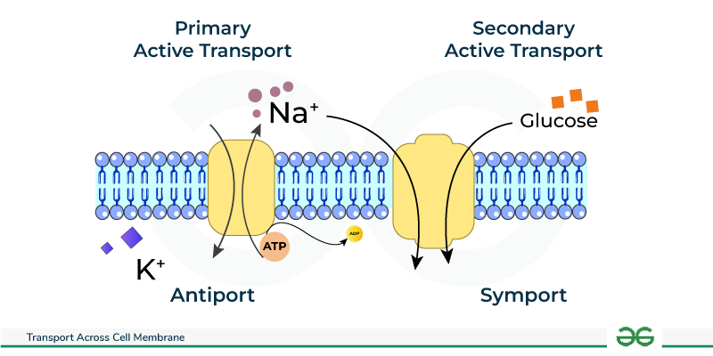
2. Active Transport: Energy Required!
This is like pushing a ball uphill. It moves substances against their concentration gradient (from low to high concentration) and requires energy, usually from ATP.
Key Example: The Sodium-Potassium Pump
This famous pump uses ATP to constantly push 3 sodium ions (Na+) OUT of the cell and pull 2 potassium ions (K+) INTO the cell. This is vital for nerve signals and muscle contractions.

3. Ion Channels & Coupled Transport
Ion Channels are protein "tunnels" that allow charged ions (like Na+, K+) to pass through quickly when a gate opens. Coupled Transport uses a single transporter to move multiple substances, often in the same direction (Symport) or opposite directions (Antiport).
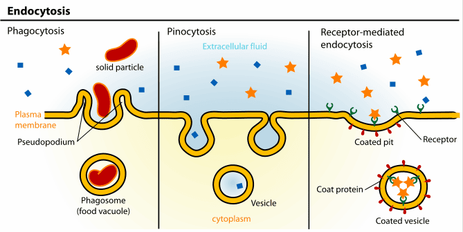
4. Vesicular Transport: For Big Stuff!
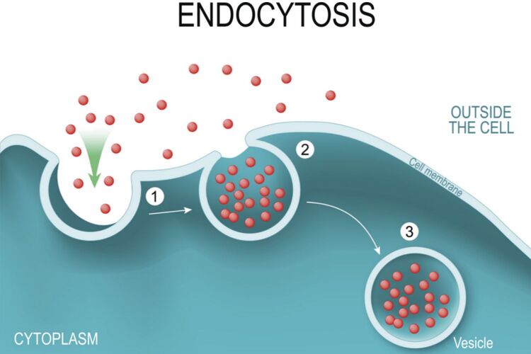
Cells move very large particles by wrapping them in a membrane-bound sac called a vesicle.
Endocytosis ("Bringing In")
The cell membrane engulfs a substance to bring it inside. Includes Phagocytosis ("cell eating" for solids) and Pinocytosis ("cell drinking" for liquids).
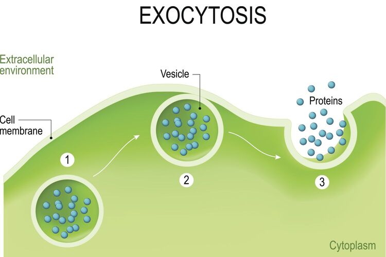
Exocytosis ("Sending Out")
A vesicle inside the cell fuses with the membrane to release its contents outside. Used for hormones, neurotransmitters, and waste.
Organelles: The Cell's Specialized Structures
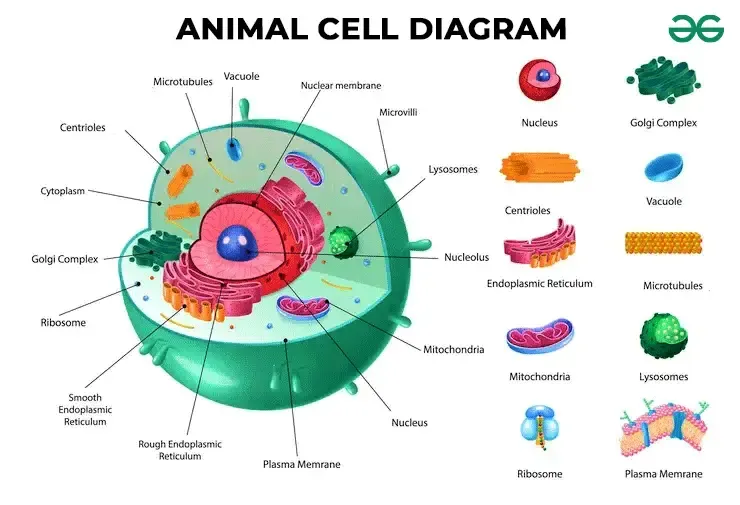
An organelle is a specific, membrane-bound structure within a cell that performs a specialized function. They are often called vesicles and are identified by microscopy. It's important to note that the cell membrane itself is not an organelle, as it is the outer boundary, not a structure contained within the cytoplasm.
The Basic Structure: Boundary and Contents
The two fundamental components of any cell are its outer boundary and its internal contents.
Cell Membrane
The outer boundary, or "bag," that contains all internal components. It's a phospholipid bilayer embedded with proteins.
Cytoplasm
The term for all the material and fluid inside the cell membrane but outside the nucleus.
Function 1: Synthesizing & Transporting Proteins
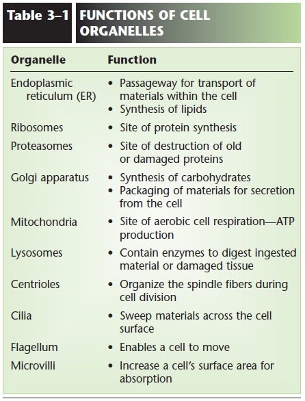
This is the central manufacturing and logistics pathway of the cell, involving a coordinated effort from the nucleus to the Golgi apparatus.
The Nucleus: The Cell's Control Center
Often called the "brain" of the cell, the nucleus houses the genetic blueprint (DNA) and controls all cellular activities. Its primary functions are DNA Replication and Transcription (creating RNA from DNA).
Key Structures of the Nucleus
1. Nuclear Envelope: A double membrane enclosing the nucleus. The outer layer is continuous with the Rough ER.
2. Nuclear Pores: Regulated gateways that control transport between the nucleus and cytoplasm.
3. Nucleolus: The "Ribosome Factory," where ribosomal RNA (rRNA) is synthesized.
4. Chromatin: The complex of DNA and proteins (histones) inside the nucleus. It exists as loosely packed Euchromatin (active genes) or tightly packed Heterochromatin (inactive genes).
Ribosomes: The "Workers"
These small organelles read the messenger RNA (mRNA) blueprint from the nucleus and assemble proteins in a process called Translation. They are found either attached to the Rough ER (membrane-bound) or floating freely in the cytoplasm (cytosolic).
Rough Endoplasmic Reticulum (Rough ER): The "Finishing Department"
A network of folded membranes connected to the nucleus, covered in ribosomes. After a protein is made, it enters the Rough ER to be folded into its 3D shape, checked for quality, and tagged with a molecular "shipping label" (e.g., through glycosylation).
Golgi Apparatus: The "Packaging & Shipping Center"
A stack of flattened sacs that receives proteins from the Rough ER, performs final modifications, sorts them, and packages them into vesicles for delivery to the cell membrane, outside the cell, or to other organelles.
Smooth Endoplasmic Reticulum (Smooth ER)
A tubular network that lacks ribosomes. It does not process proteins but has other vital roles:
- Lipid Synthesis: Makes fats, phospholipids, and steroids.
- Detoxification: Neutralizes drugs, toxins, and alcohol in the liver.
- Calcium Storage: Sequesters and releases Ca²⁺ ions, crucial for muscle contraction.
Function 2: Converting Energy
Mitochondria: The "Powerhouse"
Performs cellular respiration to convert energy from food (glucose) into ATP, the cell's main energy currency. Mitochondria contain their own DNA (mtDNA), inherited from the mother.
Key Structures of the Mitochondrion
Outer Membrane: Smooth and permeable. Inner Membrane: Highly folded into cristae, where the Electron Transport Chain occurs. Matrix: The innermost space containing enzymes for the Krebs Cycle and mtDNA.
Function 3: Storage & Waste Breakdown
Lysosome
The "Recycling Crew." A vesicle filled with digestive enzymes to break down pathogens, old organelles, and initiate programmed cell death (apoptosis).
Peroxisome
The "Detox Center." Contains enzymes to neutralize toxins and break down harmful hydrogen peroxide.
Function 4: Reproduction & Structure
Centrosome
Composed of two centrioles, this is the main organizing center for microtubules and forms the spindle fibers that pull chromosomes apart during cell division.
Cytoskeleton
A dynamic network of protein fibers (microtubules, microfilaments, intermediate filaments) that provides shape, support, and a "highway" system for intracellular transport.
External Structures for Movement & Surface Area
- Microvilli: Finger-like folds that increase surface area for absorption (e.g., in the small intestine). They do not move.
- Cilia: Short, hair-like projections that move in unison to sweep substances across the cell's surface (e.g., in the respiratory tract).
- Flagellum: A long, whip-like tail used to propel the entire cell. In humans, the only flagellated cell is the sperm.
Cell Functions and Functional Specialization
Even though all cells share basic structures and carry out essential life processes, different types of cells in our body are highly specialized to perform specific functions. This functional specialization is what allows us to have complex tissues, organs, and organ systems. Think of the different workers in our factory – some are builders, some are packers, some are security guards, each with a unique role.
Some fundamental functions that most cells perform to stay alive and maintain the organism include:
- Metabolism: The sum of all chemical processes that occur in the body. Cells carry out metabolic reactions to obtain energy (like cellular respiration in mitochondria) and to synthesize or break down molecules needed for their structure and function.
- Responsiveness: The ability to detect and respond to changes in their environment. This can be sensing chemical signals, physical touch, or electrical impulses. For example, nerve cells respond to stimuli by generating electrical signals.
- Movement: Can refer to movement of the entire cell (like white blood cells moving to an infection site) or movement of structures within the cell (like organelles being transported) or movement produced by the cell (like muscle cells contracting).
- Growth: An increase in cell size or an increase in the number of cells through cell division.
- Differentiation: The process by which a less specialized cell becomes a more specialized cell type. This is how a single fertilized egg develops into all the different cell types in the body (nerve cells, muscle cells, skin cells, etc.).
- Reproduction: Can refer to the formation of new cells for growth, repair, or replacement (through mitosis) or the production of a new organism (through meiosis and fertilization).
Now, let's look at how different cells are specialized for particular jobs, often by having more of certain organelles or unique structures:
- Muscle Cells: Specialized for contraction. They are packed with protein filaments (actin and myosin) that slide past each other to shorten the cell, producing force and movement. They also have abundant mitochondria for energy and specialized smooth ER (sarcoplasmic reticulum) for calcium storage, which is crucial for contraction.
- Nerve Cells (Neurons): Specialized for transmitting electrical and chemical signals over long distances. They have long extensions called axons and dendrites. Their plasma membrane is excitable, meaning it can generate and conduct electrical impulses. They have many ribosomes and ER for synthesizing neurotransmitters.
- Red Blood Cells: Specialized for transporting oxygen. They lack a nucleus and most organelles (like mitochondria) in their mature state, which maximizes the space available for hemoglobin, the protein that binds oxygen. Their biconcave shape also increases surface area for gas exchange and allows them to squeeze through narrow blood vessels.
- Epithelial Cells: Specialized for covering surfaces, lining cavities, protection, absorption, and secretion. They are often tightly packed together and may have specialized structures like microvilli (to increase surface area for absorption, like in the intestines) or cilia (to move substances, like in the airways).
- Gland Cells: Specialized for secretion (producing and releasing substances like hormones, enzymes, or mucus). They have abundant ribosomes, ER, and Golgi apparatus to synthesize, process, and package their secretory products into vesicles.
- Bone Cells (Osteocytes): Specialized for maintaining bone tissue. They are embedded in a hard extracellular matrix they helped produce, providing structural support to the body.
- White Blood Cells (e.g., Macrophages): Part of the immune system, specialized for defense. Some can move actively (
amoeboid movement) and engulf foreign particles or debris (phagocytosis), acting like the body's cleanup crew and security. They have abundant lysosomes to break down ingested material.
Understanding cell specialization helps us appreciate how the different parts of the body perform their unique roles and how disruptions at the cellular level can impact the function of entire tissues and organs.
Cell Division: The Process of Life's Replication
Learning Objectives:
- Describe the two types of cell division and their roles.
- Describe each phase of mitosis.
Introduction to Cell Division
Cells reproduce through a fundamental process called cell division, essential for growth, repair, and reproduction. There are two primary types:
Mitotic Cell Division (Mitosis)
- Role: Growth and tissue repair.
- Occurs in: Somatic (body) cells.
- Outcome: Two identical daughter cells.
- Chromosomes: 46 (same as parent).
Meiotic Cell Division (Meiosis)
- Role: Production of sex cells (sperm/ova).
- Occurs in: Reproductive organs only.
- Outcome: Four daughter cells.
- Chromosomes: 23 (half of parent).

The Cell Cycle: A Cell's Life Journey
The cell cycle describes a cell's entire lifespan. Mitosis itself is only a small part (5-10%); most of the cell's time is spent in Interphase.
Interphase: The "Resting" and Preparation Phase
This is the longest phase of the cell cycle, a period of intense growth and preparation for division. Key events include:
- Cell Growth: The cell grows and performs normal metabolic functions.
- Organelle Synthesis: New organelles are assembled.
- Chromosome Replication (DNA Replication): The most critical event. The DNA "unzips," and new nucleotides attach to create two identical DNA molecules, ensuring each daughter cell gets a full set of 46 chromosomes.
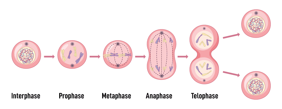
Mitotic Phases: The Stages of Nuclear Division
Once interphase is complete, the cell enters mitosis. It is a continuous process, but we divide it into four sequential phases for easier understanding.
A. Prophase
- • Replicated chromosomes coil and condense, becoming visible.
- • Each chromosome consists of two identical sister chromatids joined at a centromere.
- • The nuclear envelope disappears.
- • The mitotic spindle begins to form.
B. Metaphase
- • Replicated chromosomes line up precisely at the cell's equator (the metaphase plate).
- • Centromeres are attached to the spindle fibers.
C. Anaphase
- • Centromeres divide, and the sister chromatids separate.
- • Each chromatid is now considered an individual chromosome.
- • Spindle fibers pull the chromosomes towards opposite poles of the cell.
D. Telophase
- • The spindle fibers disassemble.
- • A new nuclear envelope forms around each set of chromosomes.
- • Chromosomes uncoil and return to their chromatin form.
Cytokinesis: Division of the Cytoplasm
Usually occurring during late anaphase and telophase, cytokinesis is the final step. A furrow forms in the plasma membrane, deepens, and eventually pinches the parent cell into two separate, genetically identical daughter cells, each with its own nucleus and cytoplasm.
Knowledge Check
Movement of materials in and out of cells is controlled by the plasma membrane.
Molecules of DNA located in chromosomes control the activities of cells.
Aerobic respiration occurs within mitochondria.
The sites of protein synthesis are ribosomes.
The nucleolus assembles protein and RNA to form ribosomes.
The endoplasmic reticulum consists of intracellular membranous channels for material transport.
Movement of molecules from an area of their higher concentration to an area of their lower concentration is known as diffusion.
Movement of molecules across a membrane by carrier proteins without the expenditure of energy is a form of facilitated diffusion.
Breakdown of organic nutrients in cells to release energy and form ATP is called cellular respiration.
Instructions for synthesizing a protein are carried from DNA to ribosomes by messenger RNA (mRNA).
The equal distribution of chromosomes to daughter nuclei occurs by mitosis.
Test Your Knowledge
Check your understanding of the concepts covered in this post.
1. Which of the following is a primary function of the Golgi apparatus in a human cell?
- DNA replication
- Detoxification of drugs
- Modification, sorting, and packaging of proteins and lipids
- ATP synthesis
2. In human cells, which organelle is responsible for generating the majority of ATP through oxidative phosphorylation?
- Nucleus
- Peroxisome
- Mitochondrion
- Ribosome
3. Which type of cellular junction is crucial for preventing the leakage of substances between epithelial cells, such as those lining the digestive tract?
- Gap junction
- Desmosome
- Tight junction
- Hemidesmosome
4. A patient is diagnosed with a lysosomal storage disease. This typically means there is a deficiency in the function of which organelle?
- Endoplasmic Reticulum
- Lysosome
- Golgi Apparatus
- Proteasome
5. Which component of the human cell cytoskeleton is primarily involved in maintaining cell shape, resisting tension, and anchoring organelles?
- Microtubules
- Microfilaments (Actin filaments)
- Intermediate filaments
- Cilia
6. The process of programmed cell death, vital for tissue development and removing damaged cells in humans, is known as:
- Necrosis
- Mitosis
- Apoptosis
- Phagocytosis
7. Which phase of the human cell cycle involves the primary growth of the cell and normal metabolic functions before DNA replication?
- S phase
- M phase
- G1 phase
- G2 phase
8. Which organelle in human cells is responsible for synthesizing lipids, metabolizing carbohydrates, and detoxifying drugs and poisons?
- Rough Endoplasmic Reticulum
- Smooth Endoplasmic Reticulum
- Nucleolus
- Ribosome
9. The process by which a human cell engulfs extracellular fluid containing dissolved solutes is called:
- Phagocytosis
- Receptor-mediated endocytosis
- Pinocytosis
- Exocytosis
10. Which of the following structures is responsible for synthesizing ribosomal RNA (rRNA) and assembling ribosomal subunits within the nucleus of a human cell?
- Nuclear envelope
- Chromatin
- Nucleolus
- Nuclear pore
11. In human cells, the genetic material is found within the __________ in the form of chromatin.
12. The plasma membrane of human cells is a selectively permeable barrier composed primarily of a __________ and associated proteins.
13. The cellular process by which proteins are synthesized from mRNA templates on ribosomes is called __________.
14. Human cells utilize __________ to transport substances out of the cell, often involving vesicles fusing with the plasma membrane.
15. The __________ are small, membrane-bound organelles that contain enzymes involved in various metabolic reactions, including the breakdown of fatty acids and the detoxification of harmful substances, producing hydrogen peroxide as a byproduct.
Quiz Complete!
Your Score:
0%
0 / 0 correct
A Deep Summary into The Living Cell
Must Know
Ribosomes: The Protein Factories
Ribosomes are tiny molecular machines whose only job is to build proteins by linking amino acids together in the order specified by messenger RNA (mRNA). They are made of ribosomal RNA (rRNA) and proteins.
Forms of Ribosomes:
- Free Ribosomes: Float freely in the cytoplasm. They synthesize proteins that will function within the cell itself (e.g., enzymes for metabolism, cytoskeleton proteins).
- Bound Ribosomes: Are attached to the surface of the Rough Endoplasmic Reticulum. They synthesize proteins that are destined to be exported from the cell, inserted into cell membranes, or delivered to specific organelles like lysosomes.
Example: A muscle cell needs the protein actin to contract. This protein stays inside the cell, so it is made by free ribosomes. A pancreatic cell makes the hormone insulin, which must be exported into the blood. Insulin is therefore made by bound ribosomes on the RER.
The Nucleus: The Control Center
The nucleus is the most prominent organelle, serving as the cell's command center. It houses and protects the cell's genetic material (DNA).
- Nuclear Envelope: A double membrane that surrounds the nucleus.
- Nuclear Pores: Channels that regulate the passage of molecules (like RNA and proteins) in and out of the nucleus.
- Nucleolus: A dense region within the nucleus where ribosomes are assembled.
Forms of DNA:
- Chromatin: The normal state of DNA in a non-dividing cell. It's a complex of DNA and proteins (histones), resembling a tangled thread.
Euchromatin:Loosely packed chromatin. It is genetically active, meaning the DNA is accessible for transcription (making RNA).Heterochromatin:Tightly packed chromatin. It is genetically inactive.
- Chromosomes: When a cell is about to divide, the chromatin condenses into these highly organized, visible structures to ensure the DNA is safely and equally distributed to the daughter cells.
Endoplasmic Reticulum (ER): The Production Network
The ER is a vast network of interconnected membranes (cisternae) that is continuous with the nuclear envelope. It comes in two forms.
1. Rough Endoplasmic Reticulum (RER)
Named "rough" because its surface is studded with ribosomes.
Where is extensive RER found and what is its significance?
You find a huge amount of RER in cells that are specialized for protein secretion. The large surface area of the RER allows for massive production of proteins.
Examples:
- Plasma Cells: These immune cells produce and secrete enormous quantities of antibodies (which are proteins).
- Pancreatic Acinar Cells: Produce and secrete digestive enzymes (like trypsinogen) into the small intestine.
Functions of the RER:
- Protein Synthesis and Modification: Synthesizes proteins destined for export, membranes, or specific organelles.
- Protein Folding: Contains chaperone proteins that help newly made proteins fold into their correct 3D shape.
- Glycosylation: Attaches carbohydrate chains to proteins, creating glycoproteins. This is important for protein stability, folding, and cell-cell recognition.
Clinical Condition: Misfolded Proteins
If proteins do not fold correctly in the RER, they can be targeted for destruction. In diseases like Cystic Fibrosis, a mutation causes the CFTR protein to misfold in the RER. It is then degraded instead of being sent to the cell membrane, leading to the disease's symptoms.
2. Smooth Endoplasmic Reticulum (SER)
Named "smooth" because it lacks ribosomes. Its structure is more tubular.
Functions of the SER (More than 7):
- Steroid Synthesis: Synthesizes steroid hormones (which are derived from cholesterol). Examples: Cells in the adrenal cortex and gonads have extensive SER.
- Lipid Synthesis: Synthesizes cholesterol, triglycerides, and phospholipids.
- Detoxification of Drugs and Poisons: Primarily in liver cells. Contains enzymes called
cytochrome P450that modify drugs. - Calcium Storage and Release: In muscle cells, the SER is specialized and called the Sarcoplasmic Reticulum (SR).
- Carbohydrate Metabolism: In the liver, it helps break down glycogen to release glucose.
- Membrane Formation: Provides lipids and proteins for new membranes.
- Metabolism of Fats: Involved in the metabolism of fatty acids.
Detoxification and Drug Tolerance
When the liver is exposed to certain drugs, it responds by increasing the amount of SER (SER hypertrophy). This increases the rate of detoxification, meaning a person will need more of the drug to achieve the same effect. This is the cellular basis for drug tolerance.
Golgi Apparatus: The Post Office
The Golgi is a stack of flattened membrane sacs (cisternae). It receives proteins and lipids from the ER, modifies them, sorts them, and packages them into vesicles for delivery.
Major Roles of the Golgi:
- Modification of Proteins and Lipids
- Post-Translational Modification
- Carbohydrate Synthesis
- Sorting and Packaging
- Formation of Lysosomes
Cleaving Pro-insulin and its Significance
The hormone insulin is first synthesized on the RER as an inactive precursor called pro-insulin. It is then transported to the Golgi. Inside the Golgi, enzymes cleave pro-insulin into active insulin and a fragment called C-peptide. Both are secreted together. This is an essential activation step. A condition called hyperproinsulinemia can be a sign of pancreatic beta-cell stress or tumors.
Diseases Related to the Golgi: I-Cell Disease
I-cell disease (Mucolipidosis II) is a devastating disease caused by a Golgi defect. The Golgi normally "tags" lysosomal enzymes with mannose-6-phosphate. In I-cell disease, this tag is not added. As a result, the enzymes are mistakenly secreted outside the cell, and the lysosomes cannot break down waste products, which accumulate and cause severe developmental problems.
Lysosomes & Peroxisomes: The Cleanup Crew
Lysosomes: The Recycling Center
Lysosomes are vesicles filled with powerful digestive enzymes called acid hydrolases, which function best in an acidic environment (pH ~ 5). This is a crucial safety feature. Phagocytic immune cells, like macrophages, use lysosomes to destroy pathogens.
Peroxisomes: The Detoxification Center
Peroxisomes are small vesicles containing oxidative enzymes. Their functions include:
- Breakdown of Very Long-Chain Fatty Acids (VLCFA)
- Synthesis of Bile Acids and Plasmalogens
- Detoxification of substances like alcohol, creating hydrogen peroxide (H₂O₂).
- Neutralizing H₂O₂ using the enzyme catalase.
Diseases Related to Peroxisomes: Zellweger Syndrome
This is a severe congenital disorder where the body fails to form functional peroxisomes. As a result, VLCFAs accumulate in the blood and tissues, especially the brain, liver, and kidneys, leading to severe neurological defects.
Mitochondria: The Powerhouse
Mitochondria generate most of the cell's ATP through cellular respiration.
- Outer Membrane: Contains channels called porins, allowing small molecules to pass freely.
- Inner Membrane: Highly folded and strictly impermeable. It contains cardiolipin, making it very tight to maintain the proton gradient for ATP synthesis.
- Cristae: These are the folds of the inner membrane, which dramatically increase the surface area for the protein complexes of the electron transport chain and ATP synthase.
Relationship Between Mitochondria Number and Function
The number of mitochondria in a cell directly correlates with its energy demand. Cells like muscle cells, neurons, and sperm have a high number, while cells with lower metabolic activity have fewer.
The Cytoskeleton: Scaffolding and Highways
The cytoskeleton is a network of protein filaments that provides structural support and allows for movement.
Microtubules:
These are hollow cylinders made of tubulin. Their "dynamic instability" (rapid growing and shrinking) is crucial for processes like cell division, where they form the mitotic spindle to pull chromosomes apart. They also form the core of cilia and flagella, where motor proteins called dynein cause bending and movement.
Disease Outcome: Kartagener's Syndrome
Also known as Primary Ciliary Dyskinesia, this is a genetic disorder where the dynein arms in cilia and flagella are defective. This renders the cilia immobile, leading to chronic respiratory infections and infertility.
The Cell Membrane and Its Connections
The Cell Membrane: The Gatekeeper
The cell membrane is a fluid, flexible barrier described by the Fluid Mosaic Model. Its contents include the Phospholipid Bilayer, Cholesterol (for fluidity), Proteins (channels, pumps, receptors), and the Glycocalyx (for cell-cell recognition).
Cellular Connections:
- Tight Junctions: Form a watertight seal between cells, preventing leaks (e.g., in the intestines).
- Gap Junctions: Form channels that directly connect the cytoplasm of adjacent cells, allowing for rapid communication (e.g., in heart muscle).
Exosomes: Intercellular Messengers
Exosomes are very small vesicles released by cells that contain a cargo of proteins, lipids, and RNA, acting as a sophisticated form of cell-to-cell communication.