Introduction to Basic Physiology & The Cell
Physiology is the science of studying the functional activities and its mechanisms in the biological body. For example: why can the heart automatically beat? Physiology derived from two Greek words - physis = nature; logos = study.
Physiology Involves Process and Function
Words, names and terms are very important in any discipline because most often they carry precise meaning in them. Knowing and understanding the relationships of the meanings of these words will help tremendously in remembering and comprehending the information in a much deeper way. This information will also stay with you long after the course is over, and you will recognize important elements in other disciplines when you connect to the deeper meanings.
Physiology
The etymology (word origin) of the term Physiology comes from the 1560’s French which comes directly from Latin physiologia, meaning “The study and description of natural objects, natural philosophy". This is derived from ‘physios’ meaning "nature, natural, physical"; and ‘logia’ meaning "study". This gives us the fuller meaning of Physiology as the "Science of the normal function of living things". When studying physiology, it is imperative that we also understand the basic anatomy involved, as anatomy (structure) and physiology (function) go hand in hand.
Anatomy
The etymology (word origin) of the term Anatomy comes from the Late 1300’s terms in both Latin, anatomia and Greek, anatome. These words are derived from ana which means "up"; and tomos (or temnein) which means "to cut". Together this gives "a cutting up", which is clearly involved in dissection! In general, anatomy is considered the “Study or knowledge of the structure (form) and function of the human body“. Courses and textbooks for anatomy and physiology are different, but are inextricably connected to each other.
Etymology for the Language of Physiology
Another useful concept related to the importance of words in physiology (and anatomy) is knowing the etymology (origin of the word) of the vast array of scientific terms used in the health care field. Since many of these words are derived from Latin and Greek, it is incredibly helpful to know the origins and ‘translations’ of these terms. Becoming aware of the origins of words will greatly help students to: 1) understand what the term means; and 2) assist you in predicting what a brand new term means when you first encounter it.
Here are two examples:
- The solution is hypertonic. Hyper means above normal and tonic means strength. The solution is strong or concentrated.
- The person has hypoglycemia. Hypo is the opposite of hyper and means below normal. The glyc portion means glucose (a type of sugar), and emia means blood. Therefore, this statement means the person has low blood sugar.
One more example:
- A runner has hyponatremia. Hypo still means below normal. The natr portion means natrium which is the Latin word for sodium (hence why the chemical symbol for sodium is Na), and emia still means blood. Therefore, this statement means the person has low sodium levels in their blood.
Along the way in this physiology course we will encounter many of these terms that, once we know the origin and meaning of, will help us figure out newer terms with ease and familiarity. Anyone who has taken a medical terminology course will know the value of understanding the meaning of roots, prefixes, and suffixes.
Now you do this one:
There is a diagnosis of pancytopenia. (Hint: there are 3 terms here: pan, cyto and penia).
Please feel free to use any reference resource available to you, and remember there is a Glossary of Anatomy and Physiology Etymology terms provided in this text (page 649) to help find out what this diagnosis literally means.
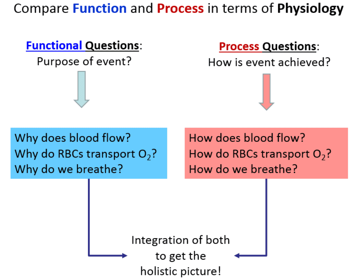
Compare Function and Process in Human Physiology
As we look to understand the central themes of physiology, an important concept is how to ask questions about what’s occurring in the human body. In general, there are two basic approaches to physiology: 1) We can ask Functional Questions; and 2) We can ask Process Questions.
1. Functional Questions (Why)
These are related to Why something occurs. For example, what is the purpose of the heart beating? These can often be answered without much detail.
Q: Why does blood flow?
A: To transport nutrients, wastes and gases around the body.
Q: Why do RBCs transport O₂?
A: To deliver O₂ to the body tissue that need it.
Q: Why do we breathe?
A: To extract the oxygen (O₂) from inhaling atmosphere air and also to release carbon dioxide (CO₂) when exhaling air back out of the body.
2. Process Questions (How)
These are related to How something occurs. For example, how does the heart actually beat? Often these issues are answered in a detailed step-by-step manner.
Q: How does blood flow?
A: The tissue fluid pressures and the ventricles of the heart act in coordination to generate a pressure gradient down which blood flows throughout the body.
Q: How do RBCs transport O₂?
A: Inside the red blood cells (RBCs) the heme portion of the molecule hemoglobin has a high affinity for O₂ when the partial pressure of the surroundings for O₂ is high, and a low affinity for O₂ when the surrounding partial pressure for O₂ is low.
Q: How do we breathe?
A: Changes can be made in the volume of the thoracic cavity by the contraction and relaxation of the skeletal muscles of respiration. This causes inverse changes in the pressure of the thoracic cavity, causing air to move down its pressure gradient.
Things to notice about Function and Process
Notice the How part (process) requires more details and also involves a sort of ‘pathway’ approach. It is more like story telling compared to the less detailed functional aspects. The more arduous component of physiology is the detailed processes. This is the reason we need to take our time and fully understand the fundamentals before we delve into intricate details.
What most students recognize about physiology is that it is more conceptual than anatomy because there is often a process to describe in a step by step manner. There are usually two sides to the functions discussed in physiology. This is because at the center of the human body is balance, which provides the equilibrium necessary to function properly. When we explain the mechanism of how we breathe in, we must also explain how we breathe out. Often once you master one side of the story, the other side falls into place more easily.
Basic Functions of a Complex Organism
Holistically, we will examine Human Physiology as it relates the foundational basics of how a multi-system living organism functions as a single coordinated entity. The basic functions are listed below:
- Differentiation
- Responsiveness
- Metabolism
- Growth and Repair
- Movement
- Excretion
- Reproduction
What we will find is that all of the systems we will study in this course will contain many if not all of these functions embedded in them.
Levels of Organization & Body Systems
A body system (also called an organ system) is an integrated collection of organs in the body that work together to perform a specific vital function. The truth is that all systems are intimately connected, but it is useful to study them separately, even though they are not separate at all. With all of our body systems operating constantly, it is necessary to have a system in place to maintain stability and equilibrium across the integrated systems. This unifying element in physiology is called homeostasis.
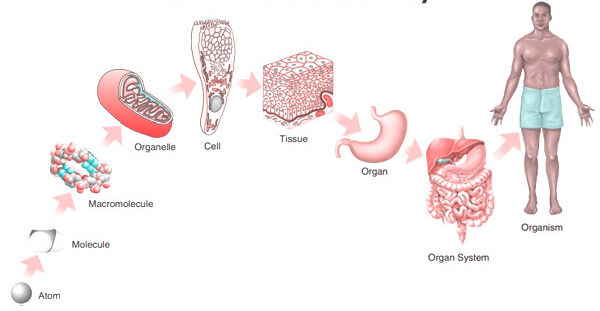
The Cell: The Fundamental Unit of Life
A. The Outer Boundary: The City Wall and Gates
The Cell (Plasma) Membrane
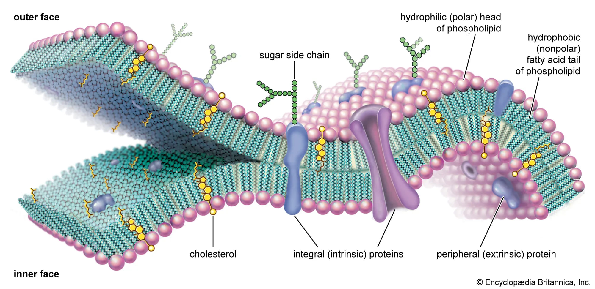
This is the outer boundary of the cell, a thin, flexible, and selectively permeable (or semipermeable) barrier. It's primarily composed of a phospholipid bilayer, with embedded proteins, carbohydrates, and cholesterol.
- Phospholipid Bilayer: Two layers of phospholipids. Each has a hydrophilic ("water-loving") head facing the watery environments inside and outside the cell, and two hydrophobic ("water-fearing") tails facing inward, forming the core of the membrane.
- Proteins: These are crucial for function. Integral (transmembrane) proteins span the entire membrane, forming channels and receptors. Peripheral proteins are loosely attached to the surface, often involved in signaling or anchoring.
- Cholesterol: Found within the hydrophobic core, it helps stabilize the membrane's fluidity.
- Glycocalyx (Carbohydrates): Chains of carbohydrates attached to proteins (glycoproteins) or lipids (glycolipids) on the outer surface, forming a unique "sugar coat" or cellular "ID tag."
Physiological Functions of the Cell Membrane:
- Selective Permeability: Controls what enters and leaves the cell, maintaining homeostasis. (The city's border control).
- Cell Recognition: The glycocalyx allows cells to recognize each other.
- Communication/Signaling: Receptor proteins bind to chemical messengers like hormones.
- Cell Adhesion: Proteins allow cells to stick together to form tissues.
- Protection: Provides a physical barrier.
B. The Cell's Internal Environment: The City Hall and Workers
Cytoplasm
The cytoplasm is everything inside the cell membrane but outside the nucleus. It consists of:
- Cytosol: The jelly-like, semi-fluid portion where organelles are suspended. It's mostly water with dissolved solutes (ions, glucose, amino acids, ATP, etc.).
- Organelles: The "little organs" with specific functions (discussed next).
- Inclusions: Temporary storage bodies, such as glycogen granules, lipid droplets, and pigment granules.
Physiological Functions of the Cytoplasm:
- Site of Many Metabolic Reactions: Key pathways like glycolysis (the first step of glucose breakdown) occur in the cytosol.
- Suspension of Organelles: Provides the medium for all organelles to exist and function.
E. Energy Production: The Power Plant
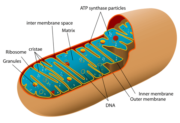
Mitochondria
Oval-shaped organelles enclosed by a double membrane: a smooth outer membrane and an inner membrane highly folded into cristae to increase surface area. The fluid-filled space within is the matrix.
Physiological Function (The "Powerhouses of the Cell"):
The primary site of aerobic cellular respiration, converting fuel molecules like glucose into ATP (adenosine triphosphate), the main energy currency of the cell.
F. Waste Management and Recycling: The Cleaning Crew
Lysosomes
Spherical sacs containing powerful hydrolytic (digestive) enzymes. They act as the "Recycling Centers," breaking down ingested substances, worn-out organelles (autophagy), and cellular debris.
Peroxisomes
Smaller sacs containing oxidative enzymes like catalase. They act as the "Detoxification Squad," neutralizing harmful free radicals and alcohol, and also break down fatty acids.
G. The Cell's Internal Support and Movement: The Infrastructure
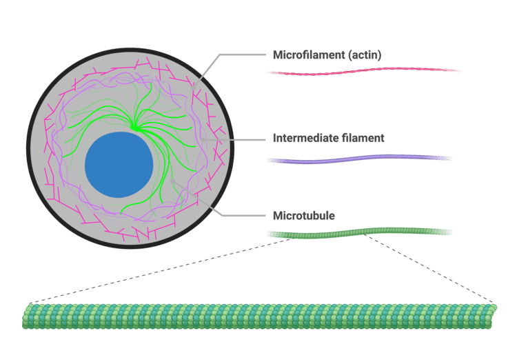
The Cytoskeleton
An intricate network of protein filaments extending throughout the cytoplasm, providing shape, support, and pathways for transport. It consists of three main types:
- Microfilaments (Actin): Thinnest; involved in cell movement, shape changes, and muscle contraction.
- Intermediate Filaments: Provide structural stability and resist mechanical stress.
- Microtubules: Largest, hollow tubes; form tracks for organelle movement and are the structural core of cilia, flagella, and the mitotic spindle.
Centrosomes, Cilia, and Flagella
- Centrosomes and Centrioles: Located near the nucleus, the centrosome contains two centrioles. It acts as the main Microtubule-Organizing Center (MTOC), organizing the mitotic spindle during cell division.
- Cilia and Flagella: Hair-like projections made of microtubules. Cilia are short and numerous, moving substances across the cell surface (e.g., mucus). Flagella are long and singular, propelling the entire cell (e.g., sperm).
Summary Table of Organelles
| Organelle | Key Functions |
|---|---|
| Plasma Membrane | Selective barrier, cell recognition, communication |
| Nucleus | Genetic control, DNA replication, transcription |
| Ribosomes | Protein synthesis (translation) |
| Rough ER (RER) | Synthesis & modification of proteins for export/membranes |
| Smooth ER (SER) | Lipid synthesis, detoxification, Ca²⁺ storage |
| Golgi Apparatus | Modifies, sorts, and packages proteins and lipids |
| Mitochondria | Cellular respiration, ATP synthesis (powerhouse) |
| Lysosomes | Intracellular digestion, waste removal |
| Peroxisomes | Detoxification (free radicals), fatty acid breakdown |
| Cytoskeleton | Cell shape, support, intracellular transport, motility |
| Centrosomes | Organize mitotic spindle during cell division |
| Cilia / Flagella | Move substances across cell surface or propel the cell |
Biological Membranes
Biological membranes are dynamic, fluid structures that define the boundaries of cells (plasma membrane) and organelles. They are essential for maintaining cellular integrity, regulating transport, facilitating communication, and housing vital enzymatic reactions. The most widely accepted model describing membrane structure is the Fluid Mosaic Model.
The Fluid Mosaic Model
Proposed by Singer and Nicolson in 1972, this model describes the cell membrane as a fluid lipid bilayer where proteins are embedded or attached, much like a mosaic.
- "Fluid": Refers to the constant movement of individual phospholipid molecules and proteins within the plane of the membrane. Lipids and many proteins can drift laterally, rotate, and flex.
- "Mosaic": Refers to the diverse "patchwork" of proteins and other molecules (like cholesterol and carbohydrates) embedded within the lipid bilayer.
A. Lipids of the Cell Membrane
The central, structural framework of the membrane is a fluid lipid bilayer, predominantly made of phospholipids and cholesterol.
1. Phospholipids
Phospholipids are the most abundant lipids in the membrane. They are amphipathic, meaning they have a hydrophilic (water-loving) polar head and two hydrophobic (water-fearing) non-polar fatty acid tails. In water, they spontaneously form a bilayer where the hydrophobic tails face inward, away from the water, and the hydrophilic heads face the watery environments inside and outside the cell.
2. Cholesterol
Cholesterol molecules are rigid, ring-shaped lipids inserted between the phospholipids. They act as a membrane buffer, regulating fluidity. At body temperature, cholesterol reduces fluidity, making the membrane stronger. At low temperatures, it increases fluidity by preventing phospholipids from packing too tightly and solidifying.
Lipid Functions in the Cell Membrane:
- Forms the fundamental bilayer structure.
- Provides a selectively permeable barrier, primarily allowing fat-soluble substances (O₂, CO₂, steroids) to pass through directly.
- Acts as a barrier for water-soluble substances (glucose, ions), which require assistance from proteins to cross.
B. Membrane Proteins
Proteins are the workhorses of the membrane, performing most of its specific functions.
1. Integral (Transmembrane) Proteins
Tightly bound proteins that span the entire membrane. They can only be removed by disrupting the bilayer. They function as channels, carriers, pumps, receptors, and enzymes.
2. Peripheral Proteins
Loosely bound to the membrane's surface (either inside or outside). They do not penetrate the core and are easily detached. They often function as enzymes or cytoskeletal anchors.
Functions of Membrane Proteins:
- Transport: Facilitating the movement of specific substances (channels, carriers, pumps).
- Enzymatic Activity: Catalyzing metabolic reactions.
- Signal Transduction: Acting as receptors for chemical messengers.
- Cell-Cell Recognition: Acting as identification tags (glycoproteins).
- Intercellular Joining: Forming junctions between cells.
- Attachment to Cytoskeleton & ECM: Providing structural stability.
C. Carbohydrates of the Cell Membrane
Carbohydrates are always found on the external surface of the plasma membrane. They are attached to lipids (forming glycolipids) or proteins (forming glycoproteins). This entire "sugar coat" is called the glycocalyx, which serves as a unique molecular signature for each cell type.
Functions of Membrane Carbohydrates (Glycocalyx):
- Cell-Cell Recognition: Crucial for distinguishing "self" from "non-self" (e.g., immune responses, blood types).
- Cell Adhesion: Helps cells bind to one another.
- Receptors: Can act as receptors for hormones or toxins.
- Protection: Provides a protective barrier against damage.
Properties of the Cell Membrane
The composition and arrangement of lipids, proteins, and carbohydrates give the cell membrane its essential properties:
1. Selectively Permeable (or Semi-permeable)
This is the most important property. The membrane precisely regulates which substances can enter or leave the cell. The hydrophobic core acts as the primary barrier. Small, nonpolar molecules (O₂, CO₂) and lipid-soluble molecules pass directly, while ions and large polar molecules (glucose) require specific transport proteins.
2. Fluidity
The membrane is not rigid; its components are in constant motion. Fluidity is influenced by temperature, cholesterol (which acts as a buffer), and the saturation of fatty acid tails. This property is essential for membrane fusion, cell division, and protein function.
3. Asymmetry
The two faces (inner and outer leaflets) of the plasma membrane are structurally and functionally different. For example, carbohydrates are only on the outer surface (glycocalyx), and specific lipids and proteins are oriented in a particular direction. This is vital for directional signaling and cell recognition.
4. Self-Sealing Capability
Due to hydrophobic interactions, if the membrane is punctured, it has a natural tendency to re-seal itself, preventing leakage of cytoplasmic contents. This is crucial for maintaining cell integrity.
Functions of the Cell Membrane
Summary of Key Membrane Functions:
- Protective Barrier: Encloses the cell's contents, separating the intracellular from the extracellular environment.
- Selective Transport: Regulates the passage of substances into and out of the cell.
- Cell-Cell Communication: Contains receptors for hormones and neurotransmitters.
- Cell Recognition & Adhesion: Facilitates cell identification and the formation of tissues.
- Enzymatic Activity: Houses enzymes that catalyze specific biochemical reactions.
- Maintenance of Cell Shape: Provides structural support in conjunction with the cytoskeleton.
- Generates Membrane Potential: Crucial for nerve and muscle cell function.
- Endocytosis & Exocytosis: Manages bulk transport into and out of the cell.
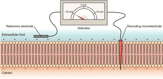
Membrane Potential: The Electrical Voltage Across the Membrane
Before looking at how things move across the membrane, it's essential to understand that there's an electrical difference, or voltage, across the cell membrane. This is called the membrane potential.
Membrane Potential is the difference in electrical charge (or potential energy) between the inside and outside of a cell. By convention, the inside of the cell is measured as being negative relative to the outside.
How is the Membrane Potential Established?
1. Unequal Distribution of Ions
There are different concentrations of ions (charged particles) inside and outside the cell.
- Outside the cell (ECF): High concentration of Na⁺ (sodium) and Cl⁻ (chloride).
- Inside the cell (ICF): High concentration of K⁺ (potassium) and negatively charged proteins/phosphates (which are too large to leave the cell).
2. Selective Permeability of the Membrane
The cell membrane is not equally permeable to all ions. At rest, it is much more permeable to K⁺ than to Na⁺, allowing K⁺ to leak out down its concentration gradient, which makes the inside of the cell more negative.
3. Sodium-Potassium Pump (Na⁺/K⁺ ATPase)
This active transport pump constantly ejects 3 Na⁺ ions out of the cell for every 2 K⁺ ions it pumps in. Since it pumps out more positive charge than it brings in, this pump is electrogenic and contributes directly to the negative charge inside the cell.
Resting Membrane Potential
In a resting (non-stimulated) neuron or muscle cell, the steady-state potential established by these factors is called the Resting Membrane Potential. It is typically around -70 mV (millivolts).
Physiological Significance
The resting membrane potential is not just a passive state; it's a form of stored energy crucial for:
- Excitability: It allows excitable cells (like neurons and muscle cells) to generate rapid electrical signals (action potentials) for communication and contraction.
- Secondary Active Transport: The energy stored in the Na⁺ and K⁺ ion gradients can be harnessed to power the transport of other substances across the membrane.
Membrane Transport
Membrane transport is a fundamental physiological process that governs the movement of substances across biological membranes. It's essential for maintaining cellular homeostasis, acquiring nutrients, expelling waste products, and facilitating cell-to-cell communication. Substances cross the membrane via two general mechanisms: Passive Transport and Active Transport.
1. Passive Transport: Moving Downhill
Passive transport is the movement of substances across a cell membrane without the direct expenditure of cellular metabolic energy (ATP). This movement is always down the electrochemical gradient of the substance. The energy for this movement comes from the inherent kinetic energy of the molecules and the potential energy stored in the concentration gradient.
1.1. Simple Diffusion: Through the Lipid Bilayer
In simple diffusion, substances move directly through the lipid bilayer without the help of membrane proteins.
- Highly Permeable: Small, nonpolar (lipophilic) molecules like O₂, CO₂, and steroid hormones readily dissolve in the hydrophobic core and pass through.
- Moderately Permeable: Small, uncharged polar molecules like water and ethanol can pass to a limited degree.
- Impermeable: Large polar molecules (glucose) and all charged ions (Na⁺, K⁺) cannot pass through on their own.
The driving force is the concentration gradient. Random molecular motion (kinetic energy) results in a net movement from an area of higher concentration to an area of lower concentration until equilibrium is reached.
Key Characteristics of Simple Diffusion:
- No membrane proteins are involved.
- Does not exhibit saturation kinetics: The rate increases linearly with the concentration gradient and does not have a maximum transport rate (Vmax).
- The rate is directly proportional to the gradient magnitude, lipid solubility, and surface area, and inversely proportional to molecular size and membrane thickness.
1.2. Facilitated Diffusion: Protein-Assisted Passage
This process uses integral membrane proteins (channels or carriers) to facilitate the movement of specific substances down their electrochemical gradient. It is still passive as no ATP is directly consumed.
A. Channel Proteins (Pores)These proteins form a water-filled pore across the membrane, allowing incredibly rapid passage of specific ions or water. Most channels are gated, meaning they open or close in response to specific stimuli:
- Voltage-Gated Channels: Respond to changes in membrane potential (e.g., Na⁺/K⁺ channels in neurons).
- Ligand-Gated Channels: Respond to the binding of a chemical messenger (e.g., neurotransmitter receptors).
- Mechanically-Gated Channels: Respond to physical deformation (e.g., touch receptors).
- Leak Channels: Are generally always open and contribute to the resting membrane potential.
Examples include ion channels (Na⁺, K⁺, Cl⁻, Ca²⁺) and aquaporins, which are specialized water channels.
B. Carrier Proteins (Transporters)These proteins bind to a specific molecule, undergo a conformational (shape) change, and release the molecule on the other side. This process is much slower than channel-mediated transport.
- Saturation Kinetics: Because there are a finite number of carriers, the transport rate has a maximum (Vmax) when all carriers are occupied.
- Specificity: Carriers are highly specific for the molecule(s) they transport.
- Competition: Structurally similar molecules can compete for the same binding site.
Examples include Glucose Transporters (GLUT proteins) and amino acid transporters.
Key Characteristics of Facilitated Diffusion:
- Involves specific membrane proteins (channels or carriers).
- Exhibits saturation kinetics (Vmax) due to the limited number of transporters.
- Can be subject to competition.
- Can be regulated by the cell (e.g., by gating channels or inserting/removing carriers from the membrane).
1.3. Osmosis: The Grand Movement of Water
Osmosis is the net movement of water across a selectively permeable membrane, from an area of higher water concentration (lower solute concentration) to an area of lower water concentration (higher solute concentration). The driving force is the water potential gradient, determined by the difference in solute concentration.
Osmotic pressure is the "pulling" force a solution with a higher solute concentration exerts on water. Tonicity refers to the effect of a solution on cell volume:
- Isotonic: No net water movement; cell volume remains normal.
- Hypotonic: Water moves into the cell, causing it to swell and potentially burst (lysis).
- Hypertonic: Water moves out of the cell, causing it to shrink (crenation).
2. Active Transport: Against the Current, with Energy
Active transport is the process of moving substances across a cell membrane against their electrochemical gradient (i.e., from a region of lower concentration to a region of higher concentration). This "uphill" movement necessitates the direct or indirect expenditure of cellular metabolic energy, almost invariably derived from the hydrolysis of ATP.
2.1. Primary Active Transport: Direct ATP Expenditure
Primary active transporters are integral membrane proteins that function as ATPases, directly binding and hydrolyzing ATP to power the movement of solutes. These transporters are often called "pumps."
Na⁺/K⁺ ATPase (Sodium-Potassium Pump)
Found in virtually all animal cells, this vital pump moves 3 Na⁺ ions out of the cell and 2 K⁺ ions into the cell for every ATP hydrolyzed. It is electrogenic (creates a charge imbalance) and is fundamental for maintaining Na⁺/K⁺ gradients, establishing the resting membrane potential, regulating cell volume, and driving secondary active transport.
Ca²⁺ ATPases (e.g., SERCA, PMCA)
These pumps maintain the extremely low intracellular Ca²⁺ concentration. SERCA pumps Ca²⁺ into the sarcoplasmic/endoplasmic reticulum for storage (crucial for muscle relaxation), while PMCA pumps Ca²⁺ directly out of the cell.
H⁺/K⁺ ATPase (Gastric Proton Pump)
Located in the parietal cells of the stomach, this pump secretes H⁺ into the stomach lumen, creating the highly acidic environment (pH 1-2) necessary for digestion. It is the target of Proton Pump Inhibitor (PPI) drugs.
ABC Transporters (ATP-Binding Cassette)
A huge superfamily of transporters that move a vast array of substrates. Examples include MDR1 (P-glycoprotein), which causes multidrug resistance in cancer cells by pumping out chemotherapy drugs, and the CFTR protein, a Cl⁻ channel whose mutation causes Cystic Fibrosis.
2.2. Secondary Active Transport (Co-transport)
Secondary active transport does not directly hydrolyze ATP. Instead, it uses the potential energy stored in an existing electrochemical gradient (typically the Na⁺ gradient created by the Na⁺/K⁺ pump) to drive the transport of a second substance against its own gradient.
1. Symporters (Cotransporters)
Both the driving ion (e.g., Na⁺) and the transported solute move in the same direction. Examples include the Na⁺-Glucose Symporter (SGLT) in the intestine and kidneys, which absorbs glucose against its gradient.
2. Antiporters (Exchangers)
The driving ion and the transported solute move in opposite directions. Examples include the Na⁺-Ca²⁺ Exchanger (NCX), crucial for removing Ca²⁺ from cardiac muscle cells, and the Na⁺-H⁺ Exchanger (NHE) for regulating intracellular pH.
3. Vesicular Transport (Bulk Transport): For the Heavy Lifting
Vesicular transport is used for moving large molecules, macromolecules, and particulate matter into or out of the cell. It involves the formation and fusion of membrane-bound sacs called vesicles and always requires energy (ATP).
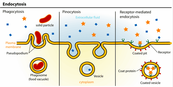
3.1. Endocytosis: Bringing the Outside In
Endocytosis is the process by which cells internalize substances. The plasma membrane invaginates and pinches off to form an intracellular vesicle.
- Phagocytosis ("Cell Eating"): The ingestion of large particles like bacteria or cellular debris by specialized immune cells (e.g., macrophages). The cell extends pseudopods to engulf the target, forming a phagosome.
- Pinocytosis ("Cell Drinking"): The non-specific uptake of extracellular fluid and dissolved solutes.
- Receptor-Mediated Endocytosis: A highly specific process where extracellular ligands (e.g., LDL-cholesterol, iron) bind to complementary receptors, which then cluster in clathrin-coated pits and are brought into the cell in a vesicle.
3.2. Exocytosis: Releasing to the Outside
Exocytosis is the process by which cells release substances. Intracellular vesicles fuse with the plasma membrane, releasing their contents to the outside.
- Constitutive Secretion: A continuous, unregulated process that delivers new lipids/proteins to the plasma membrane and secretes components of the extracellular matrix.
- Regulated Secretion: Occurs only in specialized secretory cells (e.g., neurons, endocrine cells). Secretory vesicles containing products like neurotransmitters or hormones are stored and only released in response to a specific signal (often a rise in intracellular Ca²⁺).
The "Why": Importance and Functions of Membrane Transport
The precise control over what enters and exits a cell underlies virtually every physiological process.
- Maintenance of Cellular Homeostasis: Strict ion gradients (e.g., low intracellular Na⁺, high K⁺) and pH are maintained at great energy cost to prevent cell death and ensure proper enzyme function.
- Nutrient Acquisition: Transport systems enable cells to efficiently scavenge and concentrate essential molecules like glucose and amino acids.
- Waste Removal: Active transporters expel harmful metabolic waste products, preventing their toxic accumulation.
- Generation of Electrical Signals: In neurons and muscle cells, the controlled movement of ions through channels generates action potentials, the basis of thought and movement.
- Cell-to-Cell Communication: Exocytosis releases neurotransmitters and hormones, while endocytosis regulates receptor sensitivity.
- Regulation of Cell Volume: Ion pumps, especially the Na⁺/K⁺ ATPase, control intracellular osmolarity, preventing cells from swelling or shrinking.
- Absorption and Reabsorption: Coordinated transport processes in the GI tract and kidneys are essential for absorbing nutrients and regulating the body's water, electrolyte, and acid-base balance.