Muscles of the Lower Limb: From Pelvis to Toe.
Anatomy of the Lower Extremities
The Hip Joint
The hip joint is one of the most important joints in the body for movement, like walking or dancing.
Part 1: The Bony Pelvis & The Hip Bone
The bony pelvis is a basin-shaped ring of bones connecting the vertebral column to the femurs, formed by the sacrum, coccyx, and the two hip bones (Os coxae).
The Hip Bone (Os Coxa)
Each large, irregularly shaped hip bone is a fusion of three primary bones that completes by the end of puberty:
- Ilium: The largest, most superior part, forming the prominent "wings" of the pelvis.
- Ischium: Forms the posteroinferior (lower-back) part of the hip bone.
- Pubic Bone (Pubis): Forms the anterior part of the hip bone.
The Acetabulum
The deep, cup-shaped socket on the lateral surface of the hip bone, formed by the union of all three bones. It articulates with the head of the femur. Key features include the crescent-shaped Lunate Surface (articular), the central Acetabular Fossa, and the fibrocartilaginous Acetabular Labrum that deepens the socket for increased stability.
Detailed Anatomy of the Hip Bone
- Ilium:
- Iliac Crest: The palpable superior border, terminating anteriorly as the Anterior Superior Iliac Spine (ASIS) and posteriorly as the Posterior Superior Iliac Spine (PSIS).
- Other Spines: Anterior Inferior Iliac Spine (AIIS) and Posterior Inferior Iliac Spine (PIIS).
- Surfaces: The large, concave internal Iliac Fossa; the rough outer Gluteal Surface for gluteal muscle attachment; and the medial Auricular Surface for articulation with the sacrum.
- Notches: The Greater Sciatic Notch, a large indentation for passage of the sciatic nerve.
- Ischium:
- Ischial Tuberosity: The large, roughened "sitting bone" that supports body weight when seated.
- Ischial Spine: A pointed projection posterior to the acetabulum, separating the Greater and Lesser Sciatic Notches.
- Ramus of the Ischium: Projects forward to join with the pubis.
- Pubis:
- Body of Pubis: The central part that meets the other pubic bone at the Pubic Symphysis.
- Superior & Inferior Rami: Bars of bone that help form the acetabulum and obturator foramen.
- Key Markings: Includes the Pubic Tubercle and Obturator Crest for ligament and muscle attachments.
Obturator Foramen
The large opening created by the ischium and pubis. It is mostly closed by the obturator membrane but allows the obturator nerve and vessels to pass through the obturator canal into the thigh.
The Femur (Thigh Bone)
The femur is the longest, strongest, and heaviest bone in the body, transmitting weight from the hip to the tibia.
Key Features of the Femur:
- Proximal End: Features the spherical Head (with its Fovea Capitis for the ligament of the head of the femur), the constricted Neck (a common fracture site), and the large Greater and Lesser Trochanters for muscle attachment. The Intertrochanteric Line (anterior) and Crest (posterior) connect the trochanters.
- Shaft: Includes the prominent posterior ridge, the Linea Aspera, for attachment of many thigh muscles. Proximally, it gives rise to the Pectineal Line and Gluteal Tuberosity.
- Distal End: Forms the knee joint with the large Medial and Lateral Condyles. The deep posterior notch between them is the Intercondylar Fossa. It also features the Medial and Lateral Epicondyles for ligament attachment and the anterior Patellar Surface.
Key Ligaments of the Hip Joint
- Iliofemoral Ligament (Y-ligament of Bigelow): The strongest ligament in the body, located anteriorly. It prevents hyperextension of the hip.
- Pubofemoral Ligament: Located anteroinferiorly, it limits excessive abduction and extension.
- Ischiofemoral Ligament: Located posteriorly, it limits internal rotation and adduction.
- Ligament of the Head of the Femur (Ligamentum Teres): Located inside the joint, connecting the fovea capitis to the acetabulum.
- Transverse Acetabular Ligament: Bridges the acetabular notch, completing the socket.
Muscles of the Lower Limb
The powerful muscles of the lower limb are designed for stability, locomotion, and maintaining an upright posture. We will cover them regionally, starting with the hip and gluteal region.
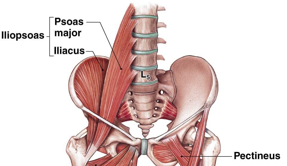
Hip Muscles: The Iliopsoas Group
The Iliopsoas is the strongest hip flexor in the body. It's a composite muscle formed by the Psoas Major and Iliacus, which merge to insert on the lesser trochanter of the femur.
- Psoas Major: Originates from the lumbar vertebrae.
- Iliacus: Originates from the iliac fossa.
- Main Actions: As the main flexor of the hip, it is essential for walking, running, and lifting the leg.
1. Muscles of the Gluteal Region (Buttocks)
These muscles are essential for hip movement, stability, and posture, divided into superficial and deep layers.
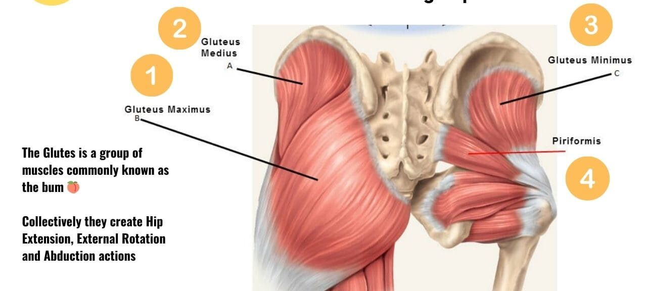
Superficial Gluteal Muscles
Gluteus Maximus
The largest and most superficial gluteal muscle. It is the main extensor of the thigh (crucial for climbing stairs or standing up) and a lateral rotator.
Gluteus Medius
Lies deep to Gluteus Maximus. It is the main abductor and a medial rotator of the thigh. It is crucial for stabilizing the pelvis during walking to prevent the hip from dropping on the unsupported side (Trendelenburg sign).
Gluteus Minimus
The smallest and deepest gluteal muscle. It works with the Gluteus Medius to abduct and medially rotate the thigh and stabilize the pelvis.
Tensor Fasciae Latae (TFL)
A small anterolateral muscle that flexes, abducts, and medially rotates the thigh. It tenses the iliotibial (IT) tract, which helps to stabilize the knee in extension.
Deep Gluteal Muscles (Short External Rotators)
This group of six smaller muscles lies deep to the gluteus maximus. They collectively function as powerful lateral rotators of the thigh and help stabilize the head of the femur in the acetabulum.
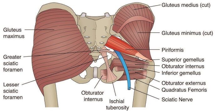
Piriformis
- Origin: Anterior surface of sacrum.
- Insertion: Superior border of greater trochanter.
- Innervation: Nerve to Piriformis (S1, S2).
- Actions: Laterally rotates, abducts (when hip is flexed), and extends the thigh.
Superior Gemellus
- Origin: Ischial spine.
- Insertion: Medial surface of greater trochanter (with Obturator Internus tendon).
- Innervation: Nerve to Obturator Internus (L5, S1).
- Actions: Laterally rotates and abducts the thigh.
Obturator Internus
- Origin: Pelvic surface of obturator membrane.
- Insertion: Medial surface of greater trochanter.
- Innervation: Nerve to Obturator Internus (L5, S1).
- Actions: Laterally rotates and abducts the thigh.
Inferior Gemellus
- Origin: Ischial tuberosity.
- Insertion: Medial surface of greater trochanter (with Obturator Internus tendon).
- Innervation: Nerve to Quadratus Femoris (L4, L5, S1).
- Actions: Laterally rotates and abducts the thigh.
Obturator Externus
- Origin: External surface of obturator membrane.
- Insertion: Trochanteric fossa of femur.
- Innervation: Obturator Nerve (L3, L4).
- Actions: Laterally rotates and adducts the thigh.
Quadratus Femoris
- Origin: Lateral border of ischial tuberosity.
- Insertion: Quadrate tubercle on intertrochanteric crest.
- Innervation: Nerve to Quadratus Femoris (L4, L5, S1).
- Actions: A powerful lateral rotator and adductor of the thigh.
Summary Table of Gluteal Muscles
| Muscle | Origin | Insertion | Innervation | Main Actions |
|---|---|---|---|---|
| Gluteus Maximus | Ilium, sacrum, coccyx | IT tract, gluteal tuberosity | Inferior Gluteal N. | Extends & laterally rotates thigh |
| Gluteus Medius | External surface of ilium | Greater trochanter | Superior Gluteal N. | Abducts & medially rotates thigh; stabilizes pelvis |
| Gluteus Minimus | External surface of ilium | Greater trochanter | Superior Gluteal N. | Abducts & medially rotates thigh; stabilizes pelvis |
| Tensor Fasciae Latae | ASIS, iliac crest | IT tract | Superior Gluteal N. | Flexes, abducts, medially rotates thigh |
| Piriformis | Anterior sacrum | Greater trochanter | N. to Piriformis | Laterally rotates & abducts thigh |
| Obturator Internus | Obturator membrane | Greater trochanter | N. to Obturator Internus | Laterally rotates & abducts thigh |
| Gemelli (Sup & Inf) | Ischial spine/tuberosity | Greater trochanter | Varies | Laterally rotate & abduct thigh |
| Quadratus Femoris | Ischial tuberosity | Intertrochanteric crest | N. to Quadratus Femoris | Powerful lateral rotator of thigh |
2. Muscles of the Thigh
The powerful muscles of the thigh are divided into three compartments: anterior (extensors), medial (adductors), and posterior (flexors/hamstrings).
Anterior Compartment of the Thigh (Extensors)
- Innervation: Femoral Nerve (L2, L3, L4)
- Main Actions: Primarily extension of the knee; some flexion of the hip.
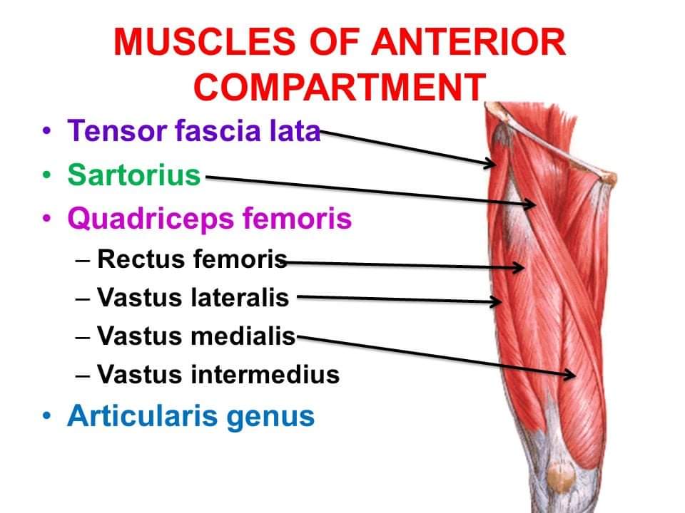
Quadriceps Femoris
A group of four muscles (Rectus Femoris, Vastus Lateralis, Vastus Medialis, Vastus Intermedius) that converge on the patellar tendon. It is the powerful extensor of the knee. The Rectus Femoris is unique as it also flexes the hip.
Sartorius
The longest muscle in the body. It flexes, abducts, and laterally rotates the thigh, and also flexes the knee (the "tailor's muscle" for crossing legs).
Medial Compartment of the Thigh (Adductors)
- Innervation: Mostly Obturator Nerve (L2, L3, L4).
- Main Actions: Primarily adduction of the thigh.
This group includes the Pectineus, Adductor Longus, Adductor Brevis, the powerful Adductor Magnus (which has both an adductor and a hamstring part), and the long, strap-like Gracilis.
Posterior Compartment of the Thigh (Hamstrings)
- Innervation: Sciatic Nerve (Tibial portion), except for the short head of Biceps Femoris.
- Main Actions: Primarily flexion of the knee and extension of the hip.
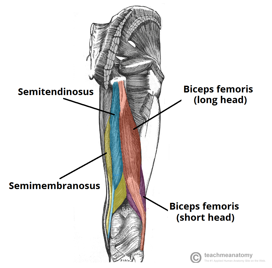
Biceps Femoris
The lateral hamstring muscle, with a long and a short head. It flexes the knee and laterally rotates the leg.
Semitendinosus
A medial hamstring muscle. It flexes the knee and medially rotates the leg.
Semimembranosus
A medial hamstring muscle, deep to the Semitendinosus. It flexes the knee and medially rotates the leg.
Summary Table of Thigh Muscles
| Muscle | Origin | Insertion | Innervation | Main Actions |
|---|---|---|---|---|
| ANTERIOR COMPARTMENT | ||||
| Rectus Femoris | AIIS | Patella & Tibial Tuberosity | Femoral N. | Extends knee, flexes hip |
| Vastus Lateralis | Greater trochanter, linea aspera | Extends knee | ||
| Vastus Medialis | Intertrochanteric line, linea aspera | Extends knee | ||
| Vastus Intermedius | Femoral shaft | Extends knee | ||
| Sartorius | ASIS | Medial tibia (Pes Anserinus) | Femoral N. | Flexes, abducts, lat. rotates thigh; flexes knee |
| MEDIAL COMPARTMENT | ||||
| Adductor Longus/Brevis/Magnus | Pubis, Ischial ramus | Femur (linea aspera) | Obturator N. (Magnus also Sciatic N.) | Adduct thigh; Magnus also extends thigh |
| Gracilis | Pubic symphysis | Medial tibia (Pes Anserinus) | Obturator N. | Adducts thigh, flexes knee |
| POSTERIOR COMPARTMENT (HAMSTRINGS) | ||||
| Biceps Femoris | Long: Ischial tuberosity; Short: Linea aspera | Head of fibula | Sciatic N. (Tibial & Common Fibular) | Flexes knee, extends hip, lat. rotates leg |
| Semitendinosus | Ischial tuberosity | Medial tibia (Pes Anserinus) | Sciatic N. (Tibial) | Flexes knee, extends hip, med. rotates leg |
| Semimembranosus | Ischial tuberosity | Medial condyle of tibia | Sciatic N. (Tibial) | Flexes knee, extends hip, med. rotates leg |
3. Muscles of the Leg
The muscles of the leg are divided into four compartments by interosseous membrane and fascial septa: anterior, lateral, posterior superficial, and posterior deep.
Anterior Compartment of the Leg
- Innervation: Deep Fibular (Peroneal) Nerve (L4, L5, S1)
- Main Actions: Primarily dorsiflexion of the ankle and extension of the toes.
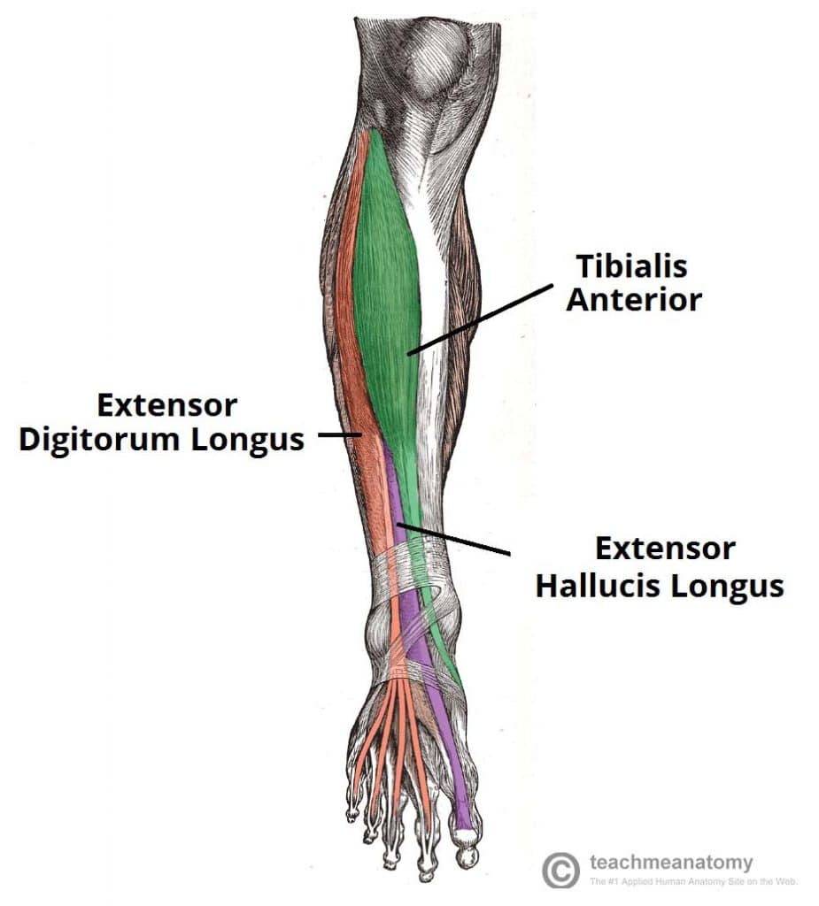
This compartment includes the Tibialis Anterior (main dorsiflexor and invertor), Extensor Digitorum Longus (extends lateral four toes), Extensor Hallucis Longus (extends great toe), and Fibularis Tertius (dorsiflexes and everts).
Lateral Compartment of the Leg
- Innervation: Superficial Fibular (Peroneal) Nerve (L5, S1, S2)
- Main Actions: Primarily eversion of the foot; some plantarflexion.
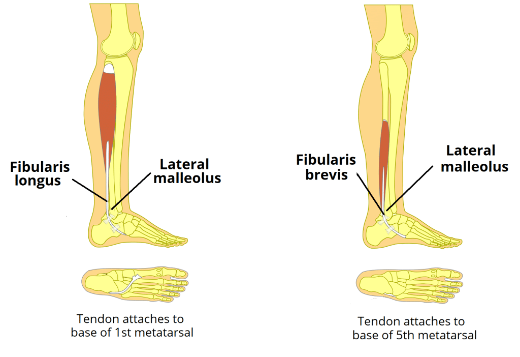
This compartment contains two muscles: Fibularis (Peroneus) Longus and Fibularis (Peroneus) Brevis. Together, they are the main everters of the foot.
Posterior Compartment of the Leg
- Innervation: Tibial Nerve (L4-S2)
- Main Actions: Primarily plantarflexion of the ankle, inversion of the foot, and flexion of the toes.
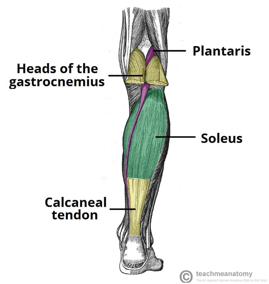
Superficial Group
This group forms the bulk of the calf and inserts via the calcaneal (Achilles) tendon. It includes the two-headed Gastrocnemius, the powerful underlying Soleus, and the small Plantaris. Together, Gastrocnemius and Soleus are known as the Triceps Surae and are powerful plantarflexors.
Deep Group
These muscles lie deep to the superficial group. They include the Popliteus (unlocks the knee), Flexor Digitorum Longus (flexes lateral four toes), Flexor Hallucis Longus (flexes the great toe), and the Tibialis Posterior (the main invertor of the foot).

Summary Table of Leg Muscles
| Muscle | Origin | Insertion | Innervation | Main Actions |
|---|---|---|---|---|
| ANTERIOR COMPARTMENT | ||||
| Tibialis Anterior | Lateral tibia | Medial cuneiform, 1st metatarsal | Deep Fibular N. | Main dorsiflexor; inverts foot |
| Extensor Digitorum Longus | Tibia, fibula | Distal phalanges of digits 2-5 | Deep Fibular N. | Extends lateral four toes |
| Extensor Hallucis Longus | Fibula | Distal phalanx of great toe | Deep Fibular N. | Extends great toe |
| LATERAL COMPARTMENT | ||||
| Fibularis (Peroneus) Longus | Head of fibula | 1st metatarsal, medial cuneiform | Superficial Fibular N. | Everts foot; plantarflexes ankle |
| Fibularis (Peroneus) Brevis | Lateral fibula | Base of 5th metatarsal | Superficial Fibular N. | Everts foot; plantarflexes ankle |
| POSTERIOR COMPARTMENT (SUPERFICIAL) | ||||
| Gastrocnemius | Femoral condyles | Calcaneus via Achilles tendon | Tibial N. | Plantarflexes ankle, flexes knee |
| Soleus | Tibia, fibula | Powerful plantarflexor | ||
| POSTERIOR COMPARTMENT (DEEP) | ||||
| Tibialis Posterior | Tibia, fibula | Navicular, cuneiforms, etc. | Tibial N. | Main invertor of foot |
| Flexor Digitorum Longus | Posterior tibia | Distal phalanges of digits 2-5 | Flexes lateral four toes | |
| Flexor Hallucis Longus | Posterior fibula | Distal phalanx of great toe | Flexes great toe | |
4. Muscles of the Foot
The intrinsic muscles of the foot are divided into dorsal (top) and plantar (sole) groups, responsible for fine motor control, supporting the arches, and assisting in locomotion.
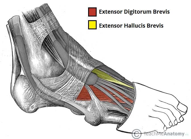
Dorsal Muscles of the Foot
Extensor Digitorum Brevis
Originates from the calcaneus and helps extend toes 2-4.
Extensor Hallucis Brevis
Originates from the calcaneus and helps extend the great toe.
Plantar Muscles of the Foot
These muscles are organized into four layers from superficial to deep. They are primarily innervated by the Medial and Lateral Plantar Nerves (branches of the Tibial Nerve) and are crucial for supporting the arches and controlling fine movements of the toes.
Layer 1 (Superficial)

Abductor Hallucis
Abducts and flexes the great toe. (Innervated by Medial Plantar N.)
Flexor Digitorum Brevis
Flexes the lateral four toes at the PIP joints. (Innervated by Medial Plantar N.)
Abductor Digiti Minimi
Abducts and flexes the little toe. (Innervated by Lateral Plantar N.)
Layer 2

Quadratus Plantae
Assists the Flexor Digitorum Longus (FDL) tendon in flexing the toes by straightening its line of pull. (Innervated by Lateral Plantar N.)
Lumbricals (4)
Flex the MTP joints and extend the IP joints of the lateral four toes. (Innervated by both Medial and Lateral Plantar Nerves).
Layer 3
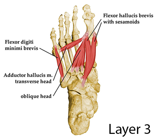
Flexor Hallucis Brevis
Flexes the great toe at the MTP joint. (Innervated by Medial Plantar N.)
Adductor Hallucis
Has two heads (oblique and transverse); adducts the great toe. (Innervated by Lateral Plantar N.)
Flexor Digiti Minimi Brevis
Flexes the little toe. (Innervated by Lateral Plantar N.)
Layer 4 (Deepest)

Plantar Interossei (3)
Adduct toes 3-5 (PAD - Plantar Adduct). (Innervated by Lateral Plantar N.)
Dorsal Interossei (4)
Abduct toes 2-4 (DAB - Dorsal Abduct). (Innervated by Lateral Plantar N.)
Summary Table of Foot Muscles
| Layer/Group | Muscle | Origin | Insertion | Innervation | Main Actions |
|---|---|---|---|---|---|
| DORSAL MUSCLES | |||||
| Dorsal | Extensor Digitorum Brevis | Calcaneus | Extensor expansions 2-4 | Deep Fibular N. | Extends toes 2-4 |
| Extensor Hallucis Brevis | Calcaneus | Prox. phalanx of great toe | Extends great toe | ||
| PLANTAR MUSCLES | |||||
| Layer 1 | Abductor Hallucis | Calcaneus | Prox. phalanx of great toe | Medial Plantar N. | Abducts & flexes great toe |
| Flexor Digitorum Brevis | Calcaneus | Middle phalanges 2-5 | Medial Plantar N. | Flexes toes 2-5 (PIP) | |
| Abductor Digiti Minimi | Calcaneus | Prox. phalanx of 5th toe | Lateral Plantar N. | Abducts & flexes 5th toe | |
| Layer 2 | Quadratus Plantae | Calcaneus | Tendon of FDL | Lateral Plantar N. | Assists FDL in flexing |
| Lumbricals (4) | Tendons of FDL | Extensor expansions 2-5 | Medial & Lateral Plantar N. | Flex MTPs, Extend IPs | |
| Layer 3 | Flexor Hallucis Brevis | Cuboid, cuneiforms | Prox. phalanx of great toe | Medial Plantar N. | Flexes great toe |
| Adductor Hallucis | Metatarsals 2-4 | Prox. phalanx of great toe | Lateral Plantar N. | Adducts great toe | |
| Flexor Digiti Minimi Brevis | Base of 5th metatarsal | Prox. phalanx of 5th toe | Lateral Plantar N. | Flexes little toe | |
| Layer 4 | Plantar Interossei (3) | Metatarsals 3-5 | Prox. phalanges 3-5 | Lateral Plantar N. | Adduct toes (PAD) |
| Dorsal Interossei (4) | Adjacent metatarsals | Prox. phalanges 2-4 | Lateral Plantar N. | Abduct toes (DAB) | |
Test Your Knowledge
A quiz on the Muscles of the Lower Limb (Pelvis to Toe).
1. Which muscle is the most powerful extensor of the hip, especially when climbing stairs or rising from a seated position?
- Gluteus Medius
- Gluteus Minimus
- Gluteus Maximus
- Piriformis
Correct (c): The Gluteus Maximus is the largest and most powerful muscle for hip extension, especially against resistance.
Incorrect (a, b): Gluteus Medius and Minimus are primary hip abductors.
Incorrect (d): Piriformis is an external rotator of the hip.
2. Damage to the superior gluteal nerve would most likely result in weakness in which primary action of the hip?
- Extension
- Adduction
- Abduction
- Flexion
Correct (c): The Superior Gluteal Nerve innervates the Gluteus Medius and Minimus, primary hip abductors. Damage leads to the "Trendelenburg gait."
Incorrect (a): Hip extension is by the Gluteus Maximus (inferior gluteal nerve).
Incorrect (b): Hip adduction is by the adductor group (obturator nerve).
Incorrect (d): Hip flexion is primarily by the Iliopsoas.
3. The "pes anserinus" is the common insertion for which three muscles on the medial tibia?
- Semitendinosus, Semimembranosus, Biceps Femoris
- Sartorius, Gracilis, Semitendinosus
- Vastus Medialis, Vastus Lateralis, Rectus Femoris
- Adductor Longus, Adductor Brevis, Adductor Magnus
Correct (b): The Sartorius, Gracilis, and Semitendinosus muscles insert together via a common tendon onto the superomedial surface of the tibia, forming the "pes anserinus" or goose's foot.
4. All anterior thigh muscles are innervated by the Femoral Nerve, EXCEPT for which muscle?
- Rectus Femoris
- Iliacus
- Sartorius
- Psoas Major
Correct (d): The Psoas Major is innervated by anterior rami of lumbar nerves (L1-L3) directly from the lumbar plexus.
Incorrect (a, b, c): Rectus Femoris, Iliacus, and Sartorius are all innervated by the Femoral Nerve.
5. Which of the following muscles is NOT a component of the quadriceps femoris group?
- Vastus Lateralis
- Rectus Femoris
- Semitendinosus
- Vastus Medialis
Correct (c): Semitendinosus is one of the hamstring muscles, located in the posterior compartment of the thigh.
Incorrect (a, b, d): The other three muscles are all part of the quadriceps femoris group, along with the Vastus Intermedius.
6. The primary action of the muscles in the lateral compartment of the leg is:
- Dorsiflexion and inversion
- Plantarflexion and eversion
- Dorsiflexion and eversion
- Plantarflexion and inversion
Correct (b): The muscles of the lateral compartment (Fibularis/Peroneus Longus and Brevis) are strong evertors of the foot and also assist in plantarflexion.
7. A patient presents with "foot drop" and an inability to dorsiflex the ankle. Which nerve is most likely damaged?
- Tibial Nerve
- Common Fibular (Peroneal) Nerve
- Saphenous Nerve
- Obturator Nerve
Correct (b): The Common Fibular Nerve (specifically its deep branch) innervates the anterior compartment of the leg, responsible for dorsiflexion. Damage leads to "foot drop."
Incorrect (a): The Tibial Nerve innervates the plantarflexors.
Incorrect (c): The Saphenous Nerve is sensory.
Incorrect (d): The Obturator Nerve innervates thigh adductors.
8. Which muscle's tendon acts like a "stirrup" to support the longitudinal arches of the foot?
- Tibialis Anterior
- Tibialis Posterior
- Fibularis (Peroneus) Longus
- Extensor Digitorum Longus
Correct (c): The Fibularis Longus tendon passes under the foot, acting like a "stirrup" to support the longitudinal and transverse arches.
9. The muscles of the deep posterior compartment of the leg include all of the following EXCEPT:
- Popliteus
- Flexor Digitorum Longus
- Soleus
- Tibialis Posterior
Correct (c): The Soleus muscle is part of the superficial posterior compartment, along with the Gastrocnemius and Plantaris.
Incorrect (a, b, d): The others are all deep posterior compartment muscles.
10. Which group of muscles contributes to maintaining the transverse arch of the foot?
- Dorsal Interossei
- Lumbricals
- Adductor Hallucis (transverse and oblique heads)
- Flexor Digiti Minimi Brevis
Correct (c): The Adductor Hallucis, particularly its transverse head, plays a significant role in maintaining the transverse arch of the foot by pulling the metatarsal heads together.
11. The powerful hip flexor formed by the fusion of the Iliacus and Psoas Major is known as the __________ group.
12. The primary action of the Quadriceps Femoris group is extension of the __________.
13. The common origin point for the hamstring muscles is the __________.
14. The three muscles that form the triceps surae are the Gastrocnemius, Soleus, and __________.
15. The intrinsic foot muscles responsible for flexing the MTP joints and extending the IP joints are the __________ and Interossei.
Quiz Complete!
Your Score:
0%
0 / 0 correct