
The Brachial Plexus
The brachial plexus is a complex network of nerves formed by the anterior rami of the lower four cervical nerves (C5, C6, C7, C8) and the first thoracic nerve (T1). It is responsible for the motor and sensory innervation of the entire upper limb.
Understanding the plexus is best done by following its five main divisions, remembered by the mnemonic: "Real Texans Drink Cold Beer" (Roots, Trunks, Divisions, Cords, Branches).
1. Roots (C5, C6, C7, C8, T1)
The five roots are the anterior primary rami of the spinal nerves, emerging between the anterior and middle scalene muscles in the neck.
Key Branches from Roots:
- Dorsal Scapular Nerve (C5): Innervates Rhomboids and Levator Scapulae.
- Long Thoracic Nerve (C5, C6, C7): Innervates Serratus Anterior.
2. Trunks (Superior, Middle, Inferior)
The five roots unite to form three trunks, which pass over the first rib.
- Upper Trunk: Formed by the union of C5 and C6 roots.
- Middle Trunk: A continuation of the C7 root.
- Lower Trunk: Formed by the union of C8 and T1 roots.
Key Branches from Trunks:
- Suprascapular Nerve (C5, C6): From the Upper Trunk; innervates Supraspinatus and Infraspinatus.
3. Divisions (Anterior and Posterior)
Each of the three trunks divides into an anterior and a posterior division, passing under the clavicle. The posterior divisions supply future extensors, while the anterior divisions supply future flexors.
4. Cords (Lateral, Posterior, Medial)
The six divisions regroup to form three cords, named for their position relative to the axillary artery.
- Lateral Cord (C5-C7): From the anterior divisions of the upper and middle trunks.
- Posterior Cord (C5-T1): From the posterior divisions of all three trunks.
- Medial Cord (C8-T1): From the anterior division of the lower trunk.
Key Branches from Cords:
- Lateral Pectoral Nerve: From the Lateral Cord.
- Upper & Lower Subscapular Nerves, Thoracodorsal Nerve: From the Posterior Cord.
- Medial Pectoral Nerve, Medial Cutaneous Nerves: From the Medial Cord.
5. Branches (The 5 Major Terminal Nerves)
The three cords give rise to the five major terminal nerves that innervate the entire upper limb.
Musculocutaneous Nerve (C5-C7)
Motor: Anterior arm compartment (Biceps Brachii, Brachialis, Coracobrachialis).
Sensory: Skin of the lateral forearm.
Axillary Nerve (C5-C6)
Motor: Deltoid and Teres Minor.
Sensory: Skin over the lower deltoid ("regimental badge area").
Radial Nerve (C5-T1)
Motor: All muscles of the posterior compartments of the arm and forearm (all extensors).
Sensory: Posterior skin of arm and forearm, dorsal aspect of lateral 2.5 digits.
Median Nerve (C5-T1)
Motor: Most anterior forearm muscles (flexors/pronators), and thenar muscles of the thumb.
Sensory: Skin of the lateral palm and palmar aspect of the lateral 3.5 digits.
Ulnar Nerve (C8-T1)
Motor: Two anterior forearm muscles (Flexor Carpi Ulnaris, medial half of FDP) and most intrinsic muscles of the hand.
Sensory: Skin of the medial 1.5 digits (palmar and dorsal).
Brachial Plexus Summary Table
| Level | Components | Key Nerve Branches | Clinical Notes |
|---|---|---|---|
| ROOTS | Anterior Rami of C5, C6, C7, C8, T1 | Dorsal Scapular N (C5): Rhomboids, Levator Scapulae Long Thoracic N (C5-C7): Serratus Anterior | Emerge between Scalenes. Injury to Long Thoracic N. → Winged Scapula. |
| TRUNKS | Upper: C5 + C6 Middle: C7 Lower: C8 + T1 | Suprascapular N (C5, C6): Supraspinatus, Infraspinatus N. to Subclavius (C5, C6): Subclavius | Pass over 1st rib. Erb-Duchenne palsy is an upper trunk injury. |
| DIVISIONS | Each trunk divides into an Anterior & Posterior Division | No direct named branches. | Posterior divisions supply extensors; Anterior supply flexors. |
| CORDS | Lateral: Ant. divisions of Upper & Middle Posterior: Post. divisions of all 3 Medial: Ant. division of Lower | Lateral Pectoral N. Upper & Lower Subscapular N., Thoracodorsal N. Medial Pectoral N., Medial Cutaneous Nerves | Named for position around axillary artery. |
| BRANCHES | Terminal Nerves | Musculocutaneous N. Axillary N. Radial N. Median N. Ulnar N. | Major nerves of the upper limb. Injuries lead to distinct motor & sensory deficits. |
Brachial Plexus Injuries and Clinical Correlates
Upper Plexus Injury (Erb-Duchenne Palsy)

Affects C5-C6 roots. Caused by an excessive angle between the neck and shoulder. Results in the classic "Waiter's Tip" position (adducted shoulder, medially rotated arm, extended elbow).
Lower Plexus Injury (Klumpke's Palsy)
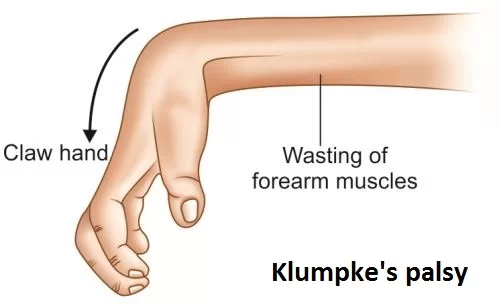
Affects C8-T1 roots. Caused by excessive abduction of the arm. Affects intrinsic hand muscles, leading to a "Claw Hand" of the 4th and 5th digits.
Radial Nerve Injury (Wrist Drop)
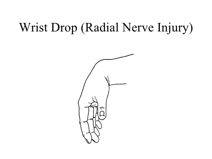
Commonly caused by mid-shaft humeral fractures or compression in the axilla ("Saturday night palsy"). Results in an inability to extend the wrist and fingers.
Median Nerve Injury (Carpal Tunnel Syndrome)
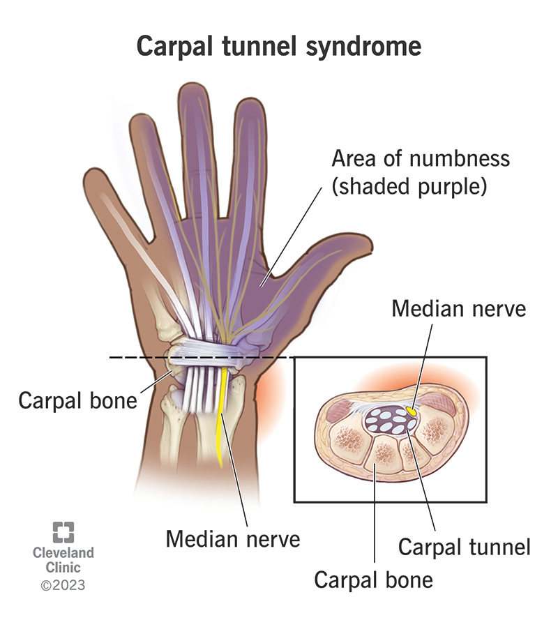
Compression of the median nerve at the wrist. Causes numbness and tingling in the lateral 3.5 digits and weakness/atrophy of the thenar (thumb) muscles.
Ulnar Nerve Injury ("Claw Hand")
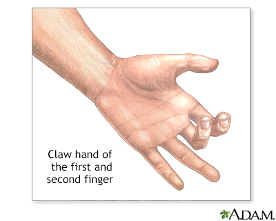
Injury at the elbow ("funny bone") or wrist. Affects intrinsic hand muscles, leading to "clawing" of the 4th and 5th digits and sensory loss over the medial hand.
Muscles of the Chest (Pectoral Region)
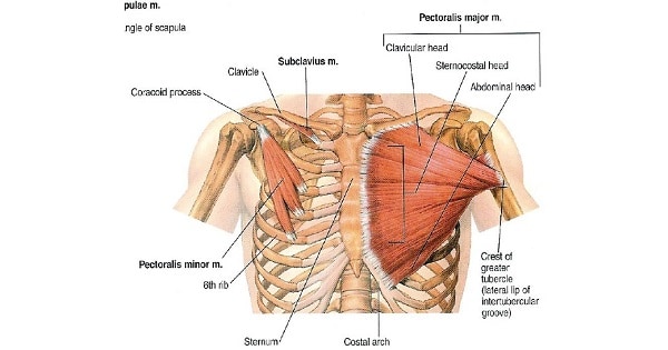
1. Superficial Muscles of the Pectoral Region
These muscles connect the upper limb to the anterior and lateral thoracic wall.
a. Pectoralis Major
A large, fan-shaped muscle covering the upper chest. It is a powerful adductor and medial rotator of the arm. Its clavicular head also flexes the arm, while the sternocostal head helps extend it from a flexed position.
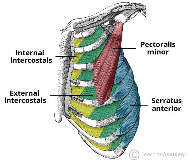
b. Pectoralis Minor
A thin, triangular muscle lying deep to Pectoralis Major. It depresses the shoulder and protracts the scapula (pulls it forward and downward).
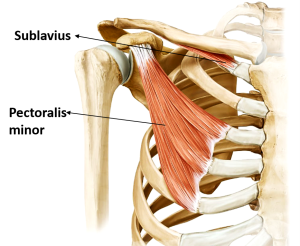
c. Subclavius
A small muscle located inferior to the clavicle. It anchors and depresses the clavicle, and helps protect the underlying subclavian vessels and brachial plexus.
d. Serratus Anterior
The "boxer's muscle" on the lateral thoracic wall. It is the prime mover for protracting the scapula (punching/pushing) and is essential for rotating the scapula to allow for full arm elevation. Paralysis leads to "winged scapula".
2. Deep Muscles of the Thorax (Associated with Respiration)
These muscles are primarily involved in the mechanics of breathing.
a. Intercostal Muscles (External, Internal, Innermost)
Three layers of muscles in the intercostal spaces. The External Intercostals elevate the ribs for forced inspiration. The Internal and Innermost Intercostals depress the ribs for forced expiration.
b. Transversus Thoracis
A thin muscle on the inner anterior thoracic wall that weakly depresses the ribs.
Summary Table of Chest Muscles
| Muscle | Origin | Insertion | Innervation | Main Actions |
|---|---|---|---|---|
| SUPERFICIAL PECTORAL MUSCLES | ||||
| Pectoralis Major | Clavicle, Sternum, Costal Cartilages 1-6 | Intertubercular groove of humerus | Lat & Med Pectoral N. | Adducts & medially rotates arm; flexes & extends arm |
| Pectoralis Minor | Ribs 3-5 | Coracoid process of scapula | Medial Pectoral N. | Depresses shoulder; protracts scapula |
| Subclavius | 1st rib | Inferior surface of clavicle | N. to Subclavius | Depresses & anchors clavicle |
| Serratus Anterior | Ribs 1-9 | Medial border of scapula | Long Thoracic N. | Protracts & rotates scapula (prevents winging) |
| DEEP THORACIC (RESPIRATORY) MUSCLES | ||||
| External Intercostals | Rib above | Rib below | Intercostal Nerves | Elevate ribs (forced inspiration) |
| Internal Intercostals | Rib above | Rib below | Intercostal Nerves | Depress ribs (forced expiration) |
Muscles of the Upper Limbs
The muscles of the upper limb enable a remarkable range of movements, from powerful lifting to delicate fine motor skills. We will cover them regionally: shoulder, arm, forearm, and hand.
1. Muscles of the Shoulder
These muscles act primarily on the glenohumeral (shoulder) joint, providing movement and stability.
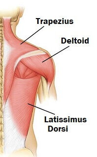
a. Deltoid
The large, triangular muscle forming the rounded contour of the shoulder. Its three parts (anterior, middle, posterior) allow it to perform a wide range of actions. The entire muscle is the prime mover of arm abduction (after the first 15 degrees). The anterior part flexes and medially rotates the arm, while the posterior part extends and laterally rotates it.
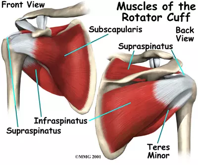
b. Rotator Cuff Muscles (SITS)
A group of four muscles that surround the shoulder joint, providing crucial stability. Their tendons blend with the joint capsule. Remembered by the mnemonic SITS.
Supraspinatus
Action: Initiates arm abduction (first 15 degrees). Most commonly torn rotator cuff muscle.
Infraspinatus
Action: Laterally rotates the arm.
Teres Minor
Action: Laterally rotates the arm.
Subscapularis
Action: Medially rotates the arm.
c. Teres Major
A thick muscle inferior to Teres Minor, often called "Lat's Little Helper." It is not part of the rotator cuff. Its main actions are to extend, adduct, and medially rotate the arm, similar to the Latissimus Dorsi.
Summary Table of Shoulder Muscles
| Muscle | Origin | Insertion | Innervation | Main Actions |
|---|---|---|---|---|
| Deltoid | Clavicle, acromion, spine of scapula | Deltoid tuberosity of humerus | Axillary N. (C5, C6) | Abducts arm; flexes & medially rotates; extends & laterally rotates |
| Supraspinatus | Supraspinous fossa | Greater tubercle of humerus | Suprascapular N. (C5, C6) | Initiates arm abduction (first 15°) |
| Infraspinatus | Infraspinous fossa | Greater tubercle of humerus | Suprascapular N. (C5, C6) | Laterally rotates arm |
| Teres Minor | Lateral border of scapula | Greater tubercle of humerus | Axillary N. (C5, C6) | Laterally rotates arm |
| Subscapularis | Subscapular fossa | Lesser tubercle of humerus | Upper & Lower Subscapular N. | Medially rotates arm |
| Teres Major | Inferior angle of scapula | Intertubercular groove of humerus | Lower Subscapular N. | Extends, adducts, medially rotates arm |
2. Muscles of the Arm
The muscles of the arm are divided into anterior (flexor) and posterior (extensor) compartments by intermuscular septa.

Anterior (Flexor) Compartment of the Arm
- Innervation: Musculocutaneous Nerve (C5, C6, C7)
- Arterial Supply: Brachial artery
- Main Actions: Flexion at the elbow and shoulder; supination of the forearm.
Biceps Brachii
A prominent two-headed muscle. It is a powerful supinator of the forearm and a strong flexor of the forearm at the elbow.
Brachialis
Lies deep to the biceps. It is the "workhorse" and primary flexor of the forearm at the elbow.
Coracobrachialis
The smallest of the three. It assists in flexion and adduction of the arm at the shoulder.
Posterior (Extensor) Compartment of the Arm
- Innervation: Radial Nerve (C6, C7, C8, T1)
- Arterial Supply: Deep brachial artery
- Main Actions: Extension at the elbow.
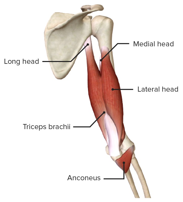
Triceps Brachii
The sole muscle of the posterior arm, with three heads (long, lateral, medial). It is the powerful extensor of the forearm at the elbow. The long head also assists in extending and adducting the arm at the shoulder.
Anconeus
A small muscle at the posterior elbow. It assists the triceps in forearm extension and helps stabilize the elbow joint.
Summary Table of Arm Muscles
| Muscle | Origin | Insertion | Innervation | Main Actions |
|---|---|---|---|---|
| ANTERIOR COMPARTMENT | ||||
| Biceps Brachii | Long: Supraglenoid tubercle; Short: Coracoid process | Radial tuberosity | Musculocutaneous N. | Supinates forearm, flexes forearm |
| Brachialis | Anterior humerus | Coronoid process of ulna | Musculocutaneous N. | Primary flexor of forearm |
| Coracobrachialis | Coracoid process | Medial surface of humerus | Musculocutaneous N. | Flexes and adducts arm |
| POSTERIOR COMPARTMENT | ||||
| Triceps Brachii | Long: Infraglenoid tubercle; Lat/Med: Posterior humerus | Olecranon process of ulna | Radial N. | Powerful extensor of forearm |
| Anconeus | Lateral epicondyle of humerus | Lateral olecranon | Radial N. | Assists triceps in extension |
3. Muscles of the Forearm
The numerous muscles of the forearm are complexly arranged in layers and are divided into anterior (flexor/pronator) and posterior (extensor/supinator) compartments.
Anterior (Flexor-Pronator) Compartment
- Innervation: Mostly Median Nerve; Flexor Carpi Ulnaris & medial half of FDP by Ulnar Nerve.
- Main Actions: Flexion of wrist and fingers; pronation of forearm.
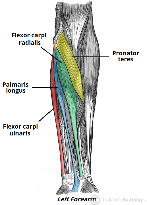
 Superficial Layer
Pronator Teres
Pronates and flexes forearm.
Flexor Carpi Radialis
Flexes and abducts wrist.
Palmaris Longus
Flexes wrist (often absent).
Flexor Carpi Ulnaris
Flexes and adducts wrist.
Intermediate Layer
Flexor Digitorum Superficialis
Flexes the middle phalanges of digits 2-5.
Deep Layer
Flexor Digitorum Profundus
Flexes the distal phalanges of digits 2-5.
Flexor Pollicis Longus
Flexes the thumb.
Pronator Quadratus
Primary pronator of the forearm.
Posterior (Extensor-Supinator) Compartment
- Innervation: Radial Nerve and its deep branch (Posterior Interosseous Nerve).
- Main Actions: Extension of wrist and fingers; supination of forearm.

Superficial Layer
Includes wrist extensors (ECRL, ECRB, ECU), finger extensors (Extensor Digitorum, Extensor Digiti Minimi), and the unique Brachioradialis, which flexes the elbow.
Deep Layer
Includes the Supinator muscle, and the "outcropping" muscles of the thumb: Abductor Pollicis Longus (APL), Extensor Pollicis Brevis (EPB), and Extensor Pollicis Longus (EPL), which form the anatomical snuffbox. Also includes the Extensor Indicis for independent index finger extension.
Summary Table of Forearm Muscles
| Muscle | Origin | Insertion | Innervation | Main Actions |
|---|---|---|---|---|
| ANTERIOR COMPARTMENT | ||||
| Pronator Teres | Medial epicondyle, coronoid process | Lateral radius | Median N. | Pronates & flexes forearm |
| Flexor Carpi Radialis | Medial epicondyle | Base of 2nd & 3rd metacarpals | Median N. | Flexes & abducts wrist |
| Palmaris Longus | Medial epicondyle | Palmar aponeurosis | Median N. | Flexes wrist |
| Flexor Carpi Ulnaris | Medial epicondyle, olecranon | Pisiform, hamate, 5th metacarpal | Ulnar N. | Flexes & adducts wrist |
| Flexor Digitorum Superficialis | Medial epicondyle, coronoid, radius | Middle phalanges of digits 2-5 | Median N. | Flexes middle phalanges |
| Flexor Digitorum Profundus | Ulna, interosseous membrane | Distal phalanges of digits 2-5 | Median N. (lat), Ulnar N. (med) | Flexes distal phalanges |
| Flexor Pollicis Longus | Radius, interosseous membrane | Distal phalanx of thumb | Median N. (AIN) | Flexes thumb |
| Pronator Quadratus | Distal ulna | Distal radius | Median N. (AIN) | Primary pronator of forearm |
| POSTERIOR COMPARTMENT | ||||
| Brachioradialis | Lateral supracondylar ridge | Styloid process of radius | Radial N. | Flexes forearm |
| Extensor Carpi Radialis Longus | Lateral supracondylar ridge | Base of 2nd metacarpal | Radial N. | Extends & abducts wrist |
| Extensor Carpi Ulnaris | Lateral epicondyle, posterior ulna | Base of 5th metacarpal | Radial N. (PIN) | Extends & adducts wrist |
| Supinator | Lateral epicondyle, ulna | Proximal radius | Radial N. (Deep br.) | Primary supinator of forearm |
4. Muscles of the Hand
The intrinsic muscles of the hand are responsible for the fine motor control and dexterity required for complex movements. They are divided into three main groups.
a. Thenar Muscles (Ball of the Thumb)
This group of muscles acts on the thumb (pollux). All are innervated by the Recurrent Branch of the Median Nerve, except for the Adductor Pollicis.
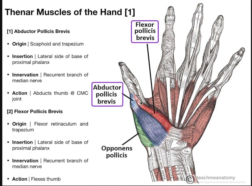
Abductor Pollicis Brevis (APB)
Abducts the thumb.
Flexor Pollicis Brevis (FPB)
Flexes the thumb.
Opponens Pollicis (OP)
Opposes the thumb (brings it across the palm).
Adductor Pollicis
Adducts the thumb (innervated by Ulnar Nerve).
b. Hypothenar Muscles (Ball of the Little Finger)
This group acts on the little finger (digiti minimi). All are innervated by the Deep Branch of the Ulnar Nerve.
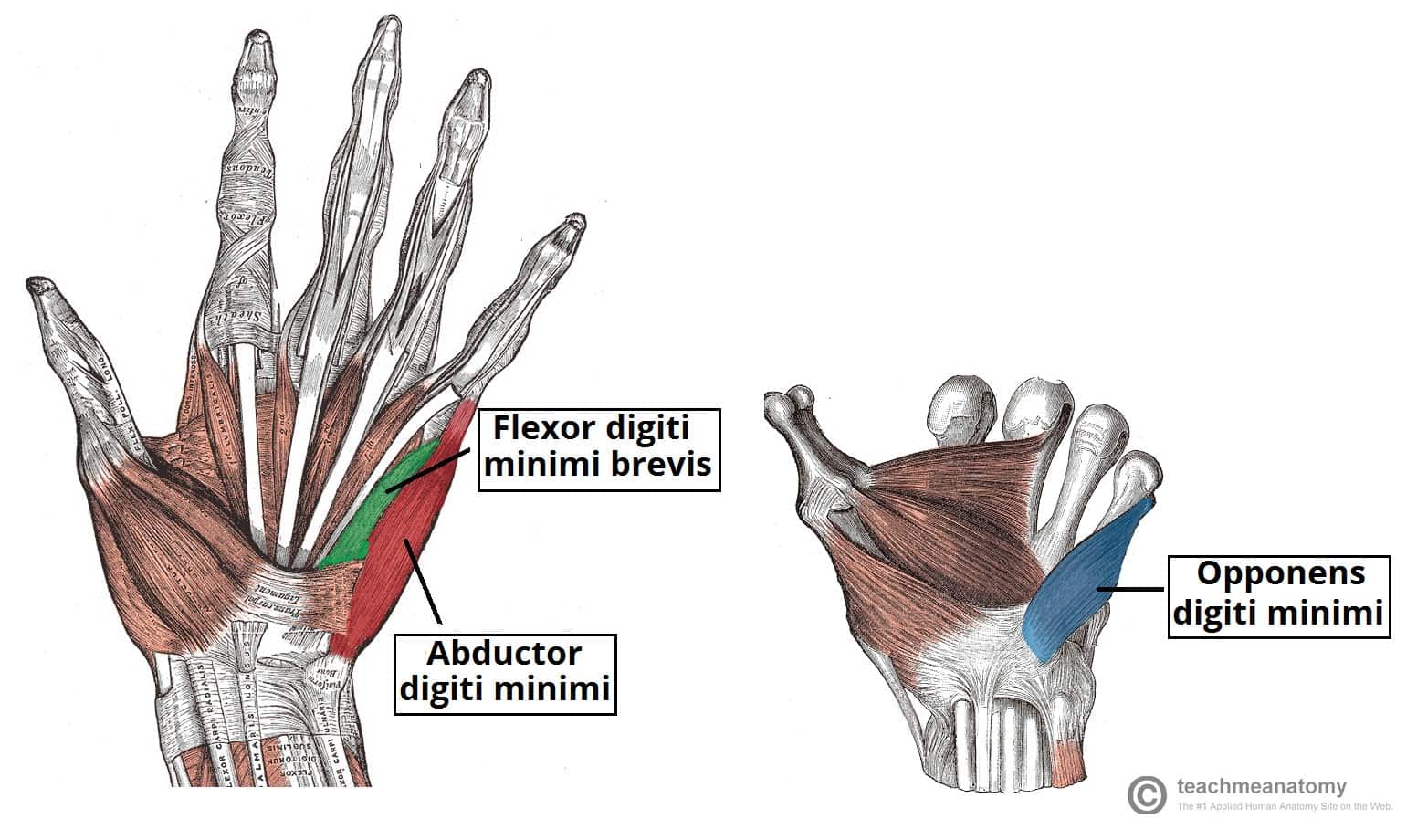
Abductor Digiti Minimi (ADM)
Abducts the little finger.
Flexor Digiti Minimi Brevis (FDMB)
Flexes the little finger.
Opponens Digiti Minimi (ODM)
Opposes the little finger (cups the palm).
c. Intrinsic Muscles of the Hand

Lumbricals (4 muscles)
Small, worm-shaped muscles that originate from the tendons of Flexor Digitorum Profundus. They flex the MCP joints and extend the IP joints. The lateral two are innervated by the Median Nerve, and the medial two by the Ulnar Nerve.
Interossei (7 muscles)
Muscles located between the metacarpals, all innervated by the Ulnar Nerve. The 4 Dorsal Interossei Abduct the fingers (DAB), and the 3 Palmar Interossei Adduct the fingers (PAD).
Summary Table of Hand Muscles
| Group | Muscle | Origin | Insertion | Innervation | Action |
|---|---|---|---|---|---|
| Thenar | Abductor Pollicis Brevis | Flexor retinaculum, scaphoid, trapezium | Proximal phalanx of thumb | Median N. (Recurrent br.) | Abducts thumb |
| Flexor Pollicis Brevis | Flexor retinaculum, trapezium | Proximal phalanx of thumb | Median N. (Recurrent br.) | Flexes thumb | |
| Opponens Pollicis | Flexor retinaculum, trapezium | 1st metacarpal | Median N. (Recurrent br.) | Opposes thumb | |
| Adductor Pollicis | Capitate, 2nd & 3rd metacarpals | Proximal phalanx of thumb | Ulnar N. (Deep br.) | Adducts thumb | |
| Hypothenar | Abductor Digiti Minimi | Pisiform | Proximal phalanx of digit 5 | Ulnar N. (Deep br.) | Abducts little finger |
| Flexor Digiti Minimi Brevis | Hook of hamate | Proximal phalanx of digit 5 | Ulnar N. (Deep br.) | Flexes little finger | |
| Opponens Digiti Minimi | Hook of hamate | 5th metacarpal | Ulnar N. (Deep br.) | Opposes little finger | |
| Intrinsic | Lumbricals (4) | Tendons of FDP | Extensor expansions | Lat 2: Median; Med 2: Ulnar | Flex MCPs, Extend IPs |
| Dorsal Interossei (4) | Adjacent metacarpals | Proximal phalanges | Ulnar N. (Deep br.) | Abduct fingers (DAB) | |
| Palmar Interossei (3) | Single metacarpal | Proximal phalanges | Ulnar N. (Deep br.) | Adduct fingers (PAD) |